. 2025 Jun 24;44(6):115737.
doi: 10.1016/j.celrep.2025.115737. Epub 2025 May 20.

ADP-ribosylation of NuMA promotes DNA single-strand break repair and transcription

Affiliations

ADP-ribosylation of NuMA promotes DNA single-strand break repair and transcription

Arwa A Abugable et al. Cell Rep. .

Abstract

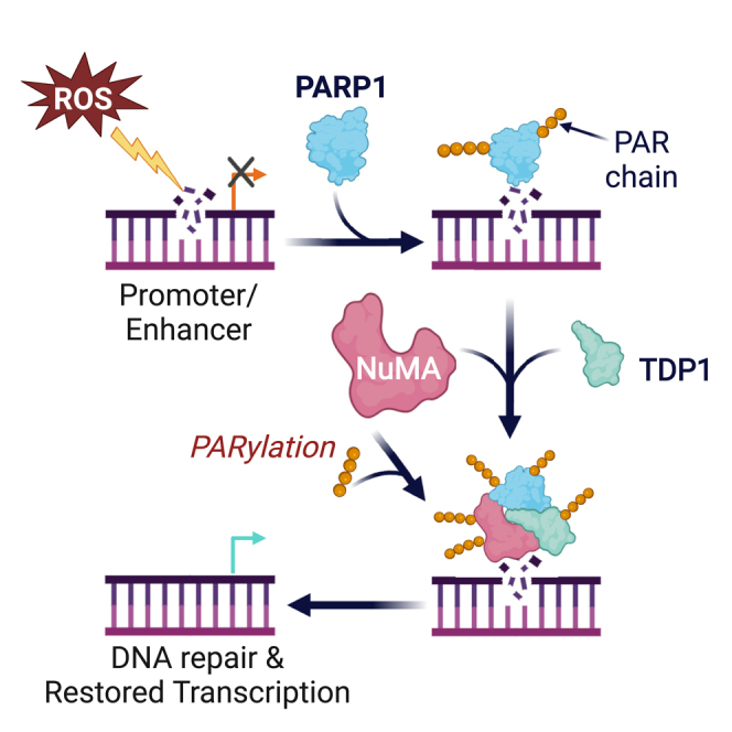
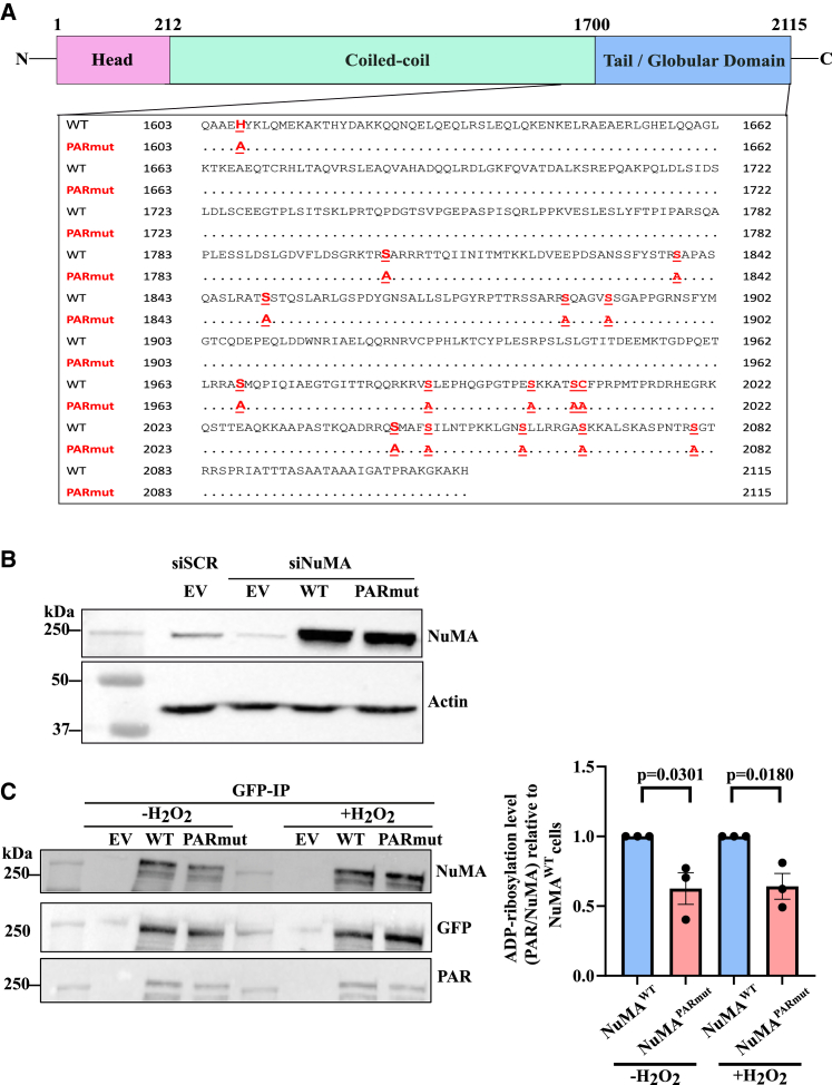
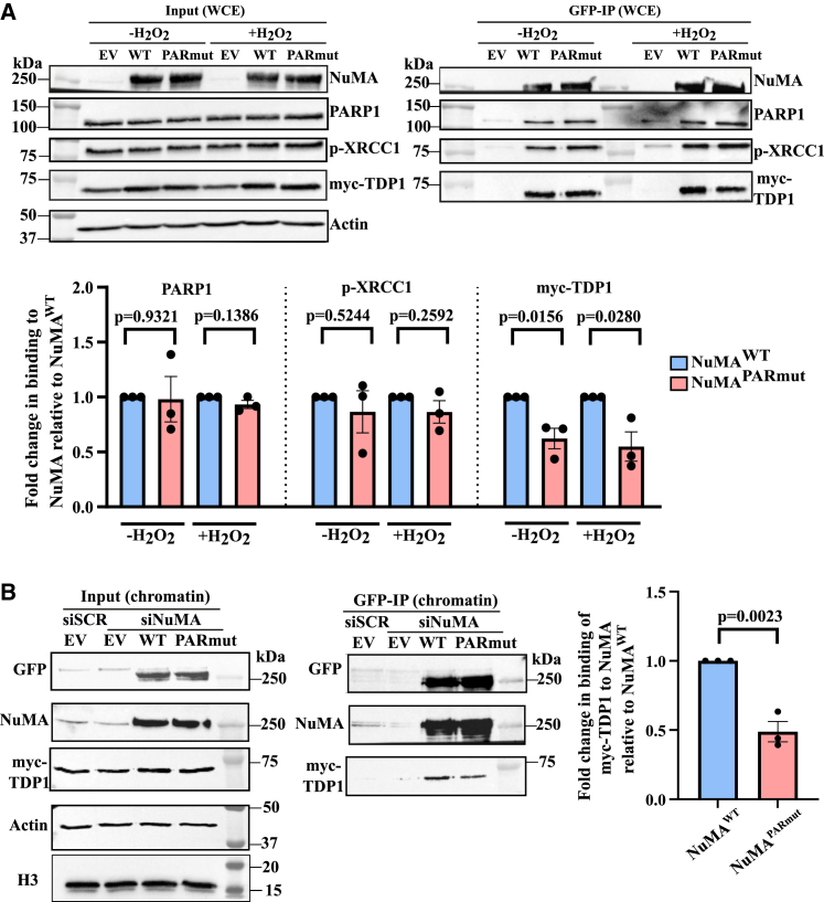
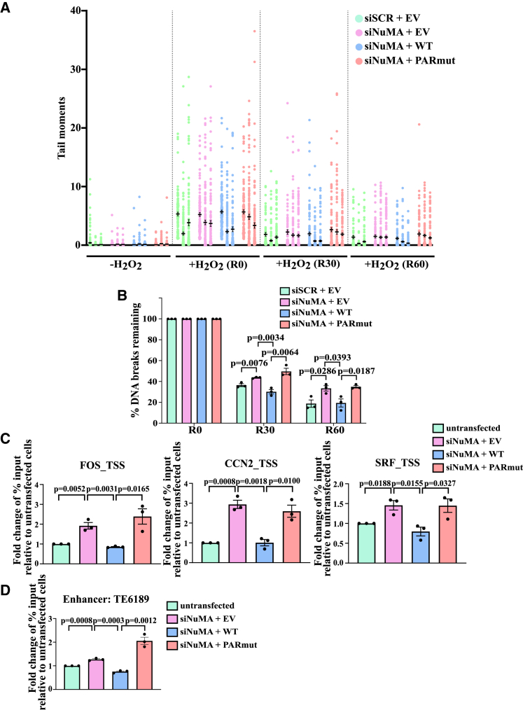
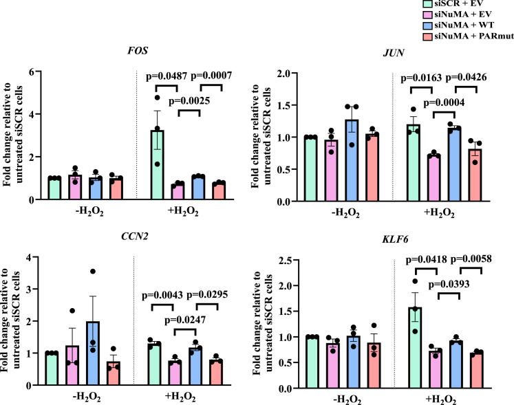
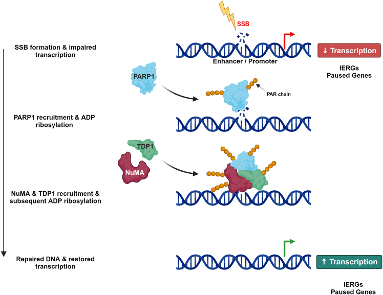
Single-strand breaks (SSBs) are prevalent DNA lesions implicated in genome instability. The nuclear mitotic apparatus protein (NuMA) has been reported to promote SSB repair (SSBR) and regulate transcription following oxidative stress. ADP-ribosylation, an important post-translational modification, regulates several processes, including chromatin remodeling, transcription, and DNA repair. To investigate its role in NuMA-dependent functions, we generated an ADP-ribosylation-deficient NuMA construct and report that NuMA ADP-ribosylation is required for its interaction with tyrosyl DNA phosphodiesterase 1 (TDP1), an SSBR player. Cells expressing ADP-ribosylation-deficient NuMA exhibit delayed SSBR kinetics following oxidative stress and reduced repair at promoter and enhancer regions, consistent with a role of NuMA in protecting non-coding regulatory regions from DNA damage. Furthermore, the expression of NuMA-regulated genes following oxidative stress requires ADP-ribosylation. Our findings demonstrate that ADP-ribosylation of NuMA promotes SSBR and transcription following oxidative stress, underscoring the importance of ADP-ribosylation in modulating DNA repair and gene expression.

Keywords: ADP-ribosylation; CP: Molecular biology; DDR; DNA damage response; DNA repair; IEGs; NuMA; brain health; cancer; dementia; gene regulatory elements; immediate early genes; oxidative DNA damage; oxidative stress; single-strand DNA breaks; transcription.


Conflict of interest statement

Declaration of interests The authors declare no competing interests.

Figures

None
Graphical abstract
Figure 1
Figure 1
Generation and validation of ADP-ribosylation-deficient construct (A) Schematic illustration highlighting the head, coiled-coil, and tail (C terminus globular domain) of NuMA. The sites susceptible to ADP-ribosylation are shown and highlighted in bold and red for NuMAWT, and the subsequent mutated residue, alanine, is highlighted in red for NuMAPARmut. (B) Representative immunoblotting from whole-cell extracts showing the successful knockdown of endogenous NuMA with siRNA and transfection of the empty vector (EV), NuMAWT, and NuMAPARmut constructs. (C) Representative immunoblotting following immunoprecipitation with GFP-trap beads of GFP-NuMAWT and GFP-NuMAPARmut in cells that were either untreated or treated with 10 μM H2O2 for 10 min on ice in the dark. Bar chart shows the fold change in relative ADP-ribosylation levels (PAR/NuMA) in NuMAPARmut relative to NuMAWT. The bar chart represents data collected from three biological replicates, with error bars representing the standard error of the mean. Two-sided unpaired Student’s t test was conducted.
Figure 2
Figure 2
NuMAWT and NuMAPARmut exhibit highly disordered structures (A) Predicted structure of NuMAWT and NuMAPARmut as generated by AlphaFold2. The colors used correspond to AlphaFold2’s predicted local distance difference test (pLDDT) values of the AlphaFold2 structures. The green and black triangles represent the positions of the serine and corresponding alanine residues in NuMAWT and NuMAPARmut that lie within helices. (B) The amino acid sequences of NuMAWT and NuMAPARmut. The ADP-ribosylation sites are shown in red, bold, and underlined. The amino acids corresponding to the helical structures are highlighted in yellow.
Figure 3
Figure 3
ADP-ribosylation-deficient NuMA shows decreased TDP1 interaction (A) Representative immunoblotting from immunoprecipitation with GFP-trap beads of the GFP-NuMAWT and GFP-NuMAPARmut in whole-cell extracts of untreated and H2O2-treated (10 μM H2O2 for 10 min on ice in the dark) cells that were also co-transfected with myc-TDP1. Actin was used as a loading control. Bar charts show the fold change in binding of PARP1, XRCC1, and myc-TDP1 to NuMA in NuMAPARmut relative to NuMAWT. The bar chart represents data from three biological replicates, with error bars representing the standard error of the mean. Two-sided unpaired Student’s t test was conducted. (B) Representative immunoblotting from immunoprecipitation with GFP-trap beads of the GFP-NuMAWT and GFP-NuMAPARmut in the chromatin fraction of cells co-transfected with myc-TDP1 and treated with 10 μM H2O2 for 10 min on ice in the dark. H3 was used as a loading control. Bar chart shows the fold change in binding of myc-TDP1 to NuMA in NuMAPARmut relative to NuMAWT. The bar chart represents data collected from three biological replicates with error bars representing the standard error of the mean. Two-sided unpaired Student’s t test was conducted.
Figure 4
Figure 4
ADP-ribosylation-deficient NuMA demonstrates delayed SSBR kinetics and increased AP sites at promoters and enhancers Cells were transfected with siSCR or siNuMA and then complemented with either EV, NuMAWT, or NuMAPARmut constructs. The cells were left untreated or treated with 20 μM H2O2 for 10 min on ice in the dark, followed by recovery in H2O2-free media for 0, 30, and 60 min, denoted as R0, R30, and R60, respectively, before being subjected to alkaline comet assay. (A) Violin plot showing the distribution of the comet tail moments at the indicated time points. The data shown are from three biological replicates. (B) Bar plot showing the percentage of DNA breaks remaining during the recovery time points. The bar chart represents data from three biological replicates, with error bars representing the standard error of the mean. Two-sided unpaired Student’s t test was conducted. (C and D) Cells were transfected with siSCR or siNuMA and then complemented with either EV, NuMAWT, or NuMAPARmut constructs. OGG1-AP-qPCR was conducted at (C) promoters of FOS, CCN2, and SRF and (D) the TE6189 enhancer. Bar charts show the fold change in percentage of input relative to the untransfected cells. The bar chart represents data from three biological replicates, with error bars representing the standard error of the mean. Two-sided unpaired Student’s t test was conducted.
Figure 5
Figure 5
ADP-ribosylation-deficient NuMA leads to decreased expression of NuMA-regulated genes Cells were transfected with siSCR or siNuMA and then complemented with either EV, NuMAWT, or NuMAPARmut constructs. They were serum starved for 48 h and left either untreated (-H2O2) or treated with 10 μM H2O2 (+H2O2) for 10 min on ice in the dark and recovered in serum-containing media for 90 min. qPCR measuring the expression of transcripts was conducted and represented as a fold change, where untreated and siSCR H2O2-treated cells were normalized to siSCR-untreated cells, while all other conditions were normalized to siSCR H2O2-treated cells. The bar chart represents data from three biological replicates, with error bars representing the standard error of the mean. Two-sided unpaired Student’s t test was conducted.
Figure 6
Figure 6
Model depicting the role of ADP-ribosylation in promoting the roles of NuMA in repair and transcription Upon the formation of an SSB, transcription of immediate-early response genes (IERGs) and paused genes is impaired. PARP1 is then recruited to the break site, where it is ADP-ribosylated. It then ADP-ribosylates other repair proteins such as NuMA and TDP1, facilitating their recruitment to the SSB site to repair the SSB. Once repaired, transcription is restored.

Similar articles

References

    1. Lindahl T., Barnes D.E. Repair of Endogenous DNA Damage. Cold Spring Harb. Symp. Quant. Biol. 2000;65:127–133. doi: 10.1101/sqb.2000.65.127. - DOI - PubMed
    1. Jackson S.P., Bartek J. The DNA-damage response in human biology and disease. Nature. 2009;461:1071–1078. doi: 10.1038/nature08467. - DOI - PMC - PubMed
    1. Caldecott K.W. DNA single-strand break repair and human genetic disease. Trends Cell Biol. 2022;32:733–745. doi: 10.1016/j.tcb.2022.04.010. - DOI - PubMed
    1. Lindahl T. Instability and decay of the primary structure of DNA. Nature. 1993;362:709–715. doi: 10.1038/362709a0. - DOI - PubMed
    1. Demple B., Harrison L. REPAIR OF OXIDATIVE DAMAGE TO DNA: Enzymology and Biology. Annu. Rev. Biochem. 1994;63:915–948. doi: 10.1146/annurev.bi.63.070194.004411. - DOI - PubMed

MeSH terms

LinkOut - more resources