. 2025 May 27;122(21):e2417224122.
doi: 10.1073/pnas.2417224122. Epub 2025 May 21.

Inhibiting 15-PGDH blocks blood-brain barrier deterioration and protects mice from Alzheimer's disease and traumatic brain injury

Yeojung Koh #  1   2   3   4   5 Edwin Vázquez-Rosa #  1   2   3   4   5 Farrah Gao #  6 Hongyun Li  7 Suwarna Chakraborty  8 Sunil Jamuna Tripathi  8 Sarah Barker  1   2   3   4   5 Zea Bud  1   2   3   4   9 Anusha Bangalore  1   2   4   10 Uapingena P Kandjoze  1   2   4   11 Rose A León-Alvarado  1   2   4   11 Preethy S Sridharan  1   2   3   4   10 Brittany A Cordova  7 Youngmin Yu  1   2   3   4   12 Jiwon Hyung  1   2   4 Hua Fang  1   2   4   13   14 Salendra Singh  7   15 Ramachandra Katabathula  7 Thomas LaFramboise  6   7 Lakshmi Kasturi  16 James Lutterbaugh  16 Lydia Beard  16 Erika Cordova  16 Coral J Cintrón-Pérez  1   2   3   4 Kathryn Franke  1   2   3   4 Mariana Franco Fragoso  7 Emiko Miller  1   2   3   4   10 Vidya Indrakumar  1   2   10 Kamryn L Noel  1   2   10 Matasha Dhar  1   2   3   4 Kaouther Ajroud  17   18 Carlos Zamudio  18 Filipa Blasco Tavares Pereira Lopes  19   20 Evangeline Bambakidis  1   2   3   4   21 Xiongwei Zhu  5 Brigid Wilson  3   22 Margaret E Flanagan  17   23 Tamar Gefen  17   24 Hisashi Fujioka  25 Stephen P Fink  7 Amar B Desai  5   7 Dawn Dawson  5   7 Noelle S Williams  26 Young-Kwang Kim  27 Joseph M Ready  26 Bindu D Paul  8   28   29   30 Min-Kyoo Shin  1   2   3   4   27   31 Sanford D Markowitz  7   16   32 Andrew A Pieper  1   2   3   4   5   10
Affiliations

Inhibiting 15-PGDH blocks blood-brain barrier deterioration and protects mice from Alzheimer's disease and traumatic brain injury

Yeojung Koh et al. Proc Natl Acad Sci U S A. .

Abstract

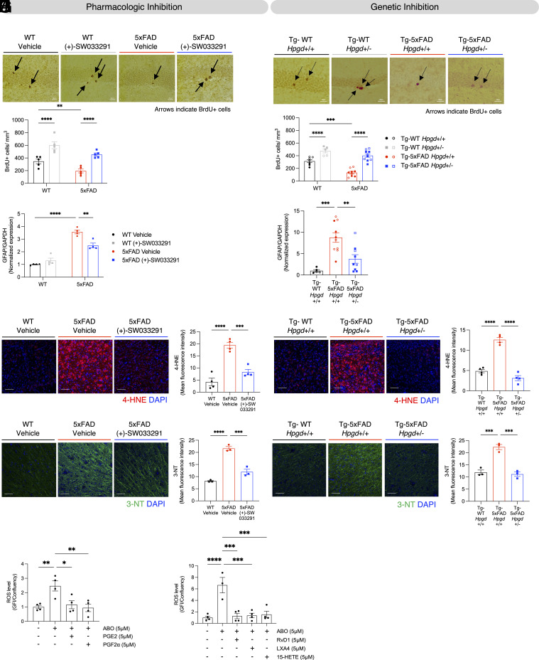
Alzheimer's disease (AD) and traumatic brain injury (TBI) are currently untreatable neurodegenerative disorders afflicting millions of people worldwide. These conditions are pathologically related, and TBI is one of the greatest risk factors for AD. Although blood-brain barrier (BBB) disruption drives progression of both AD and TBI, strategies to preserve BBB integrity have been hindered by lack of actionable targets. Here, we identify 15-hydroxyprostaglandin dehydrogenase (15-PGDH), an enzyme that catabolizes eicosanoids and other anti-inflammatory mediators, as a therapeutic candidate that protects the BBB. We demonstrate that 15-PGDH is enriched in BBB-associated myeloid cells and becomes markedly elevated in human and mouse models of AD and TBI, as well as aging, another major risk factor for AD. Pathological increase in 15-PGDH correlates with pronounced oxidative stress, neuroinflammation, and neurodegeneration, alongside profound BBB structural degeneration characterized by astrocytic endfeet swelling and functional impairment. Pharmacologic inhibition or genetic reduction of 15-PGDH in AD and TBI models strikingly mitigates oxidative damage, suppresses neuroinflammation, and restores BBB integrity. Most notably, inhibiting 15-PGDH not only halts neurodegeneration but also preserves cognitive function at levels indistinguishable from healthy controls. Remarkably, these neuroprotective effects in AD are achieved without affecting amyloid pathology, underscoring a noncanonical mechanism for treating AD. In a murine microglia cell line exposed to amyloid beta oligomer, major protection was demonstrated by multiple anti-inflammatory substrates that 15-PGDH degrades. Thus, our findings position 15-PGDH inhibition as a broad-spectrum strategy to protect the BBB and thereby preserve brain health and cognition in AD and TBI.

Keywords: 15-PGDH; Alzheimer’s disease; blood–brain barrier; neuroprotection; traumatic brain injury.


Conflict of interest statement

Competing interests statement:Y.K., E.V.-R., M.-K.S., J.M.R., N.S.W., S.D.M., and A.A.P. hold (+)-SW033291-related patents, some of which have been licensed and/or optioned to Amgen.

Figures

Fig. 1.
Fig. 1.
Brain 15-PGDH increases in human and mouse AD, TBI, and aging and is enriched in brain myeloid cells. (A) qPCR data from the human cortex reveal that 15-PGDH mRNA (HPGD expression) is increased in subjects with AD, compared to control subjects without dementia. HPGD was normalized to the geometric mean of ACTB, POLR1B, and LDHA (*P < 0.05, unpaired t test). (B) Mouse whole-brain qPCR data reveal that 15-PGDH mRNA (Hpgd expression) is increased in symptomatic (6 mo old) 5xFAD mice, compared to WT littermates. Hpgd was normalized to Actb (**P < 0.01, unpaired t test). (C) Mouse 15-PGDH enzymatic activity is increased in symptomatic (6 and 12 mo old) 5xFAD mice, compared to WT littermates (5xFAD versus WT genotype effect P < 0.001, 6 versus 12 mo time effect P = 0.0533, ***P < 0.001, two-way ANOVA and Tukey’s post hoc analysis). (D) Human brain RNA seq data from Seo et al. (ERP015139) (41) reveals that 15-PGDH mRNA (HPGD expression) is increased in subjects with chronic traumatic encephalopathy (CTE), compared to control subjects (*P < 0.05, unpaired t test). Data are shown from both the superior parietal cortex and posterior visual cortex. (E) Mouse hippocampal 15-PGDH enzymatic activity is increased 3 wk after TBI, relative to sham-injury (****P < 0.0001, unpaired t test). (F) Fluorescent in situ hybridization (FISH) for Hpgd mRNA (red) in the mouse brain shows more 15-PGDH positive cells in the cortex and hippocampus of symptomatic (6 mo old) 5xFAD mice, compared to WT littermates (*P < 0.05, ***P < 0.001, unpaired t test; 2 to 7 brain sections/mouse). (G) CD11b+ myeloid cells show increased 15-PGDH protein expression in the brains of 6-mo-old 5xFAD mice, compared to WT littermates (Interaction**P < 0.01, ****P < 0.0001, two-way ANOVA). (H) Single-cell RNA sequencing data analyzed from Yao et al. (42) reveals that Hpgd-expressing myeloid cells are composed of Mrc1 positive PVMs and Tmem119 positive microglia. A heatmap of cortical myeloid cells depicting expression [log2(SCT)] of 15-PGDH mRNA (Hpgd), Mrc1 [perivascular macrophages (PVMs)], Lyve1 (PVMs), Aif1 (microglia and macrophages), and Tmem119 (microglia) is shown. (I) RNA FISH in the mouse brain shows an increase in Hpgd-expressing microglia [double positive for 15-PGDH mRNA, Hpgd (red) and Tmem119 mRNA (green)], in symptomatic 5xFAD mice (6 mo old), compared to WT littermates, in the cortex and hippocampus (**P < 0.01, unpaired t test; 4 to 8 brain sections/mouse). (J) RNA FISH shows an increase in Hpgd-expressing PVMs [double positive for 15-PGDH mRNA, Hpgd (red) and Mrc1 mRNA (green)] in symptomatic 5xFAD mice (6 mo old), compared to WT littermates, in the cortex and hippocampus (*P < 0.05, **P < 0.01, unpaired t test; 2 to 7 brain sections/mouse). Expanded view of the boxed region is shown at Lower Right. In all graphs (AJ), data points indicate values of individual human subjects or individual mice.
Fig. 2.
Fig. 2.
15-PGDH inhibition protects 5xFAD mice from cognitive impairment and BBB deterioration without reducing amyloid pathology. (A) Schematic diagram of experimental procedure for evaluating the protective efficacy of pharmacologic inhibition of 15-PGDH. Mice were intraperitoneally (IP) injected with (+)-SW033291 twice daily (5 mg/kg × 2 = 10 mg/kg/day) from 2 to 6 mo of age. At 5 mo of age, a bolus of bromodeoxyuridine (BrdU, 150 mg/kg) was administered to label newborn hippocampal neurons. Morris water maze (MWM) testing of cognition was conducted 1 wk before brain tissue was harvested for analysis at 6 mo of age. Males (shown as filled circles or filled squares in subsequent panels) and females (shown as open circles or open squares in subsequent panels) of all four combinations of 5xFAD genotype and treatment groups were housed under normal conditions. (B) Treatment with (+)-SW033291 for 4 mo markedly reduced brain 15-PGDH activity in both 5xFAD mice and WT littermates, as compared to vehicle-treated mice (Interaction P < 0.0001, **P < 0.01, ****P < 0.0001, two-way ANOVA and Tukey’s post hoc analysis). 15-PGDH activity was also higher in vehicle-treated 5xFAD mice than vehicle-treated WT littermates, consistent with the results shown in Fig. 1C. (C) (+) SW033291 fully prevents cognitive deficits in 5xFAD mice in the MWM test. In vehicle-treated animals, 5xFAD mice performed significantly worse than WT littermates, as shown by a significantly longer latency time to first cross the platform area. However, in 5xFAD mice treated with (+)-SW033291, performance (latency time) was preserved at the normal WT littermate vehicle levels. Performance was identical between WT mice treated with either vehicle or (+)-SW033291 (Interaction P < 0.05, **P < 0.01, ***P < 0.001, two-way ANOVA and Tukey’s post hoc analysis). (D) Schematic diagram of experimental procedure for evaluating the protective efficacy of genetic haploinsufficiency of 15-PGDH (Hpgd+/-) in 5xFAD mice. Tg-WT and Tg-5xFAD refer to 5xFAD genotypes, respectively, designating the absence or presence of the 5xFAD transgene, and Hpgd+/+ and Hpgd+/- denote Hpgd genotype. Mice were injected with a bolus of BrdU (150 mg/kg) 1 mo before brains were harvested to label newborn hippocampal neurons. MWM testing of cognition was conducted 1 wk before brain tissue was harvested for analysis at 9 mo of age. Males (shown as filled circles or filled squares in subsequent panels) and females (shown as open circles or open squares in subsequent panels) of all four combinations of 5xFAD and 15-PGDH genotypes were housed under normal conditions. (E) Hpgd haploinsufficiency fully blocks the increase in 15-PGDH enzyme activity in symptomatic 5xFAD mice. Brain 15-PGDH enzyme activity is highly elevated in Tg-5xFAD Hpgd+/+ mice compared to control Tg-WT Hpgd+/+ mice and is preserved at control levels in Tg-5xFAD Hpgd+/- mice, with no difference detected between Tg-WT Hpgd+/+ and Tg-5xFAD Hpgd+/- mice (**P < 0.01, one-way ANOVA and Tukey’s post hoc analysis). (F) Hpgd haploinsufficiency prevents cognitive impairment in 5xFAD mice in the MWM. In the MWM probe test of memory, Tg-5xFAD Hpgd+/+ mice performed significantly worse than control Tg-WT Hpgd+/+ mice, as shown by significantly longer latency to first cross the platform area. Tg-5xFAD Hpgd+/- mice were protected from memory deficits, as evidenced by latency to first cross the platform area in the memory test being significantly lower than Tg-5xFAD Hpgd+/+ mice and not significantly different from Tg-WT Hpgd+/+ mice (Interaction P < 0.05, *P < 0.05, ****P < 0.0001, two-way ANOVA and Tukey’s post hoc analysis). (G) Aβ 4G8 staining shows that (+)-SW033291-mediated inhibition of 15-PGDH does not affect amyloid deposition in the 5xFAD mice hippocampus (**P < 0.001, one-way ANOVA and Tukey’s post hoc analysis, (Scale bar, 50 µm) 3 to 4 images/mouse). (H) Aβ 4G8 staining shows that genetic inhibition of 15-PGDH does not affect amyloid deposition in the Tg-5xFAD mice hippocampus (**P < 0.01, one-way ANOVA and Tukey’s post hoc analysis, (Scale bar, 50 µm) n = 3 to 4 images/mouse). (I) Transmission electron microscopy (TEM) shows structural damage of the BBB, as measured by the percentage of brain capillaries affected by astrocyte endfeet swelling in 5xFAD mice as compared to WT littermates. This damage was completely prevented by (+)-SW033291 treatment (****P < 0.0001, one-way ANOVA and Tukey’s post hoc analysis, (Scale bar, 2 µm). Representative pictures are shown on the top (each graphed data point represents the average value from one mouse, with 50 images/mouse). (J) TEM shows structural damage of the BBB, as measured by the percentage of brain capillaries affected by astrocyte endfeet swelling in Tg-5xFAD Hpgd+/+ mice as compared to control Tg-WT Hpgd+/+ mice. Hpgd haploinsufficiency completely prevented this BBB damage (Tg-5xFAD Hpgd+/- versus Tg-5xFAD Hpgd+/+ comparison) (****P < 0.0001, one-way ANOVA and Tukey’s post hoc analysis, (Scale bar, 2 µm). Representative pictures are shown on the Top (each graphed data point represents the average value from one mouse, with 50 images/mouse). (K) IgG staining shows functional impairment and increased permeability of the BBB as evidenced by IgG infiltration into the brain parenchyma in 5xFAD mice, compared to WT littermates. (+)-SW033291 treatment protects 5xFAD mice from this functional impairment of the BBB. (*P < 0.05, ***P < 0.001, one-way ANOVA and Tukey’s post hoc analysis, (Scale bar, 50 µm) 2 to 3 brain sections/mouse). (L) IgG staining shows functional impairment and increased permeability of the BBB as evidenced by IgG infiltration into the brain parenchyma in Tg-5xFAD Hpgd+/+ mice, compared to control Tg-WT Hpgd+/+ mice. Hpgd haploinsufficiency protects Tg-5xFAD Hpgd+/- mice from this impairment (Tg-5xFAD Hpgd+/+ compared to Tg-5xFAD Hpgd+/-) (*P < 0.05, **P < 0.01, one-way ANOVA and Tukey’s post hoc analysis, (Scale bar, 50 µm) 2 to 3 brain sections/mouse). In all graphs (A–L), data points indicate values of individual mice.
Fig. 3.
Fig. 3.
15-PGDH inhibition protects 5xFAD mice from decreased survival of newborn hippocampal neurons, neuroinflammation, and oxidative stress. (A) 6-mo-old symptomatic 5xFAD mice have impaired survival of BrdU-labeled newborn hippocampal neurons (black arrows), compared to WT littermates, as assessed 28 d post–BrdU injection. (+)-SW033291 treatment preserves normal survival of newborn hippocampal neurons in 5xFAD mice (genotype P < 0.001, treatment P < 0.0001, **P < 0.01, ****P < 0.0001, two-way ANOVA and Tukey’s post hoc analysis, (Scale bar, 20 µm). Each graphed dot represents the average value of BrdU-labeled cells from an individual mouse as determined from 5 to 6 sections spaced at least 400 µm apart across the hippocampus. (B) Tg-5xFAD Hpgd+/+ mice have impaired survival of BrdU-labeled newborn hippocampal neurons (black arrows), versus Tg-WT Hpgd+/+ littermates, as assessed 28 d post–BrdU injection. Genetic reduction of 15-PGDH in Tg-5xFAD Hpgd+/- mice restores newborn hippocampal neuron survival. (Interaction P < 0.05, genotype (Tg-WT vs Tg-5xFAD) P < 0.0001, genotype (Hpgd+/+ vs Hpgd+/-) P < 0.0001, ***P < 0.001, ****P < 0.0001, two-way ANOVA and Tukey’s post hoc analysis, (Scale bar, 20 µm). Each dot represents the average value of BrdU-labeled cells from an individual mouse determined from 5 to 6 sections each spaced at least 400 µm apart across the hippocampus. (C) 5xFAD mice show increased hippocampal GFAP expression, which is significantly reduced by treatment with (+)-SW033291 (Interaction P < 0.001, **P < 0.01, ****P < 0.0001, two-way ANOVA and Tukey’s post hoc analysis). (D) Tg-5xFAD Hpgd+/+ mice show increased whole-brain GFAP expression compared to Tg-WT Hpgd+/+ mice, which is significantly prevented in Tg-5xFAD Hpgd+/- mice (**P < 0.01, ***P < 0.001, one-way ANOVA and Tukey’s post hoc analysis). (E) 4-HNE staining analysis of the brain cortex and its quantification show increased 4-HNE in 5xFAD mice, relative to WT littermates, which is significantly prevented by (+)-SW033291 treatment (***P < 0.001, ****P < 0.0001, one-way ANOVA and Tukey’s post hoc analysis; 2 to 3 brain sections/mouse). (F) 4-HNE staining analysis of the brain cortex and its quantification show increased 4-HNE in Tg-5xFAD Hpgd+/+ mice compared to Tg-WT Hpgd+/+ mice, which is fully prevented in Tg-5xFAD Hpgd+/- mice (****P < 0.0001, one-way ANOVA and Tukey’s post hoc analysis; 2 to 3 brain sections/mouse). (G) 3-NT staining analysis of the brain cortex and its quantification show increased 3-NT in 5xFAD mice, relative to WT littermates, which is significantly prevented by (+)-SW033291 treatment (***P < 0.001, ****P < 0.0001, one-way ANOVA and Tukey’s post hoc analysis; 2 to 3 brain sections/mouse). (H) 3-NT staining analysis of the brain cortex and its quantification show increased 3-NT in Tg-5xFAD Hpgd+/+ mice compared to Tg-WT Hpgd+/+ mice, which is completely prevented in Tg-5xFAD Hpgd+/- mice (***P < 0.001, one-way ANOVA and Tukey’s post hoc analysis; 2 to 3 brain sections/mouse). (I) Pretreatment with prostanoid substrates of 15-PGDH, either 5 µM PGE2 or 5 µM PGF2α, suppressed ROS induction assayed 30 min after 5 µM Aβ oligomer treatment (*P < 0.05, **P < 0.01, one-way ANOVA and Tukey’s post hoc analysis). (J) Pretreatment with 15-PGDH substrates, RvD1, LXA4, 15-HETE suppressed ROS induction assayed 30 min after 5 µM Aβ oligomer treatment (***P < 0.001, ****P < 0.0001, one-way ANOVA and Tukey’s post hoc analysis). In all graphs (A–J), data points indicate the value for an individual mouse or replicate of tested cells.
Fig. 4.
Fig. 4.
15-PGDH inhibition protects mice from TBI-induced cognitive impairment, oxidative stress, neurodegeneration, and BBB damage. (A) Schematic diagram of experimental procedure for evaluating the protective efficacy of (+)-SW033921-mediated 15-PGDH inhibition in TBI. 8-wk-old mice were administered multimodal TBI on day 0, and 24 h later started on a 21 d regimen of twice daily (+)-SW03391 (5 mg/kg × 2 = 10 mg/kg/day). MWM testing was conducted between days 14 and 18, and brain tissue was harvested for analysis on day 21. (B) Post–TBI administration of (+)-SW033291, initiated 24 h after injury and continued for 2 wk, inhibited brain 15-PGDH activity (****P < 0.0001, unpaired t test). (C) Vehicle-treated TBI-injured mice show significantly worse memory than sham-injury mice, as evidenced by significantly greater latency time to cross the platform area in the MWM probe test of memory. Treatment of TBI mice with (+)-SW033921 completely prevented post–TBI memory impairment (Interaction P < 0.01, ****P < 0.0001, two-way ANOVA and Tukey’s post hoc analysis). (D) Vehicle-treated TBI-injured mice showed significantly worse memory than sham-injury mice, as shown by a significantly lower number of platform area crossings on the MWM probe test of memory. Treatment of TBI mice with (+)-SW033921 prevented post–TBI memory impairment. (Interaction P < 0.05, **P < 0.01, two-way ANOVA and Tukey’s post hoc analysis). (E) TBI induces axonal degeneration, which was reversed by 15-PGDH pharmacologic inhibition with (+)-SW03329. Axonal degeneration was visualized and quantified by silver staining analysis, using percent positive area. (**P < 0.01, one-way ANOVA and Tukey’s post hoc analysis, (Scale bar, 5 µm). (Each dot represents average value from one mouse as determined from 6 images/mouse). (F) TEM analysis and its quantification show that TBI-induced structural BBB damage, as evidenced by swelling of astrocyte endfeet, was blocked by 15-PGDH pharmacologic inhibition with (+)-SW033291 (****P < 0.0001, one-way ANOVA and Tukey’s post hoc analysis). (G) 3-NT staining and its quantification show that TBI-induced oxidative stress, as evidenced by increased 3-NT, was blocked by 15-PGDH pharmacologic inhibition with (+)-SW033291 (***P < 0.001, ****P < 0.0001, one-way ANOVA and Tukey’s post hoc analysis; 2 to 3 brain sections/mouse). (H) Schematic diagram of experimental procedure for evaluating the protective efficacy of genetic 15-PGDH inhibition in TBI. Control 8-wk-old Hpgd+/+ (WT) and Hpgd−/− (KO) mice were subjected to multimodal TBI. Brain tissue was harvested for analysis 3 wk after TBI. (I) TBI induces axonal degeneration, which was reversed by biallelic Hpgd gene deletion (Hpgd−/−). Axonal degeneration was visualized and quantified by silver staining analysis, using percent positive area. (**P < 0.01, ***P < 0.001, one-way ANOVA and Tukey’s post hoc analysis, (Scale bar, 5 µm). (Each dot represents average value from one mouse as determined from 6 images/mouse). (J) Electron microscopy analysis and its quantification show that TBI-induced structural BBB damage, as evidenced by swelling of astrocyte endfeet, was blocked by biallelic Hpgd gene deletion (Hpgd−/−) (****P < 0.0001, one-way ANOVA and Tukey’s post hoc analysis). (K) 3-NT staining and its quantification show that TBI-induced oxidative stress, as evidenced by increased 3-NT, was blocked by biallelic Hpgd gene deletion (Hpgd−/−) (****P < 0.0001, one-way ANOVA and Tukey’s post hoc analysis; 2 to 3 brain sections/mouse). In all graphs (AK), data points indicate values of individual mice.

Comment in

Similar articles

Cited by

References

    1. Geneva: World Health Organization, Global Action Plan on The Public Health Response to Dementia 2017–2025 (World Health Organization, 2017).
    1. Montagne A., et al. , Blood-brain barrier breakdown in the aging human hippocampus. Neuron 85, 296–302 (2015). - PMC - PubMed
    1. van de Haar H. J., et al. , Blood-brain barrier leakage in patients with early alzheimer disease. Radiology 281, 527–535 (2016). - PubMed
    1. van de Haar H. J., et al. , Neurovascular unit impairment in early Alzheimer’s disease measured with magnetic resonance imaging. Neurobiol. Aging. 45, 190–196 (2016). - PubMed
    1. Montagne A., et al. , Brain imaging of neurovascular dysfunction in Alzheimer’s disease. Acta Neuropathol. 131, 687–707 (2016). - PMC - PubMed

MeSH terms

LinkOut - more resources