. 2025 May 23;11(21):eadv6445.
doi: 10.1126/sciadv.adv6445. Epub 2025 May 21.

Immunopeptidomics-guided discovery and characterization of neoantigens for personalized cancer immunotherapy

Affiliations

Immunopeptidomics-guided discovery and characterization of neoantigens for personalized cancer immunotherapy

Yangyang Cai et al. Sci Adv. .

Abstract

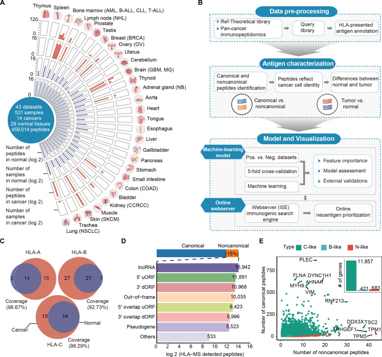
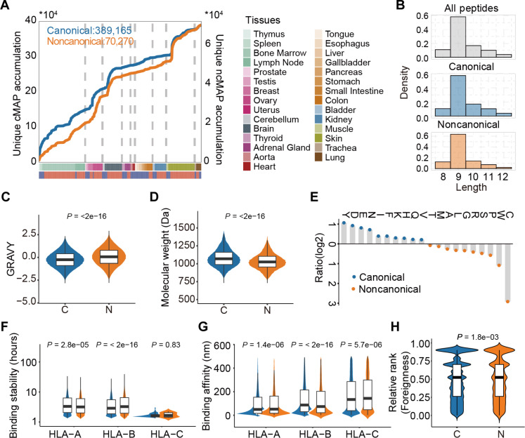
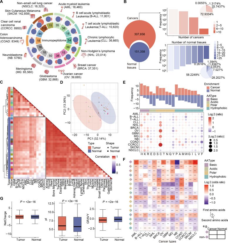
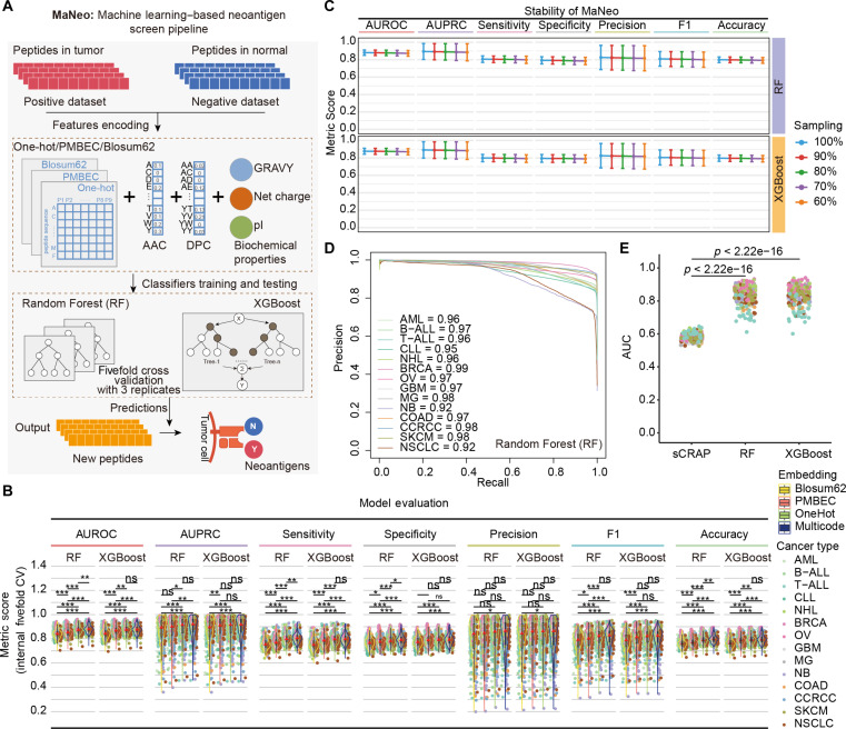
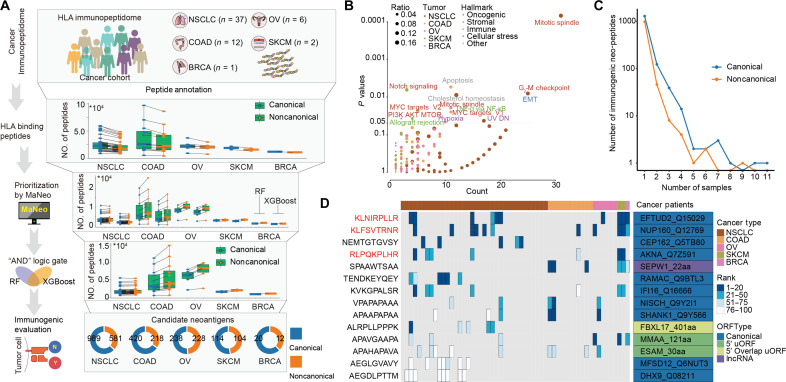
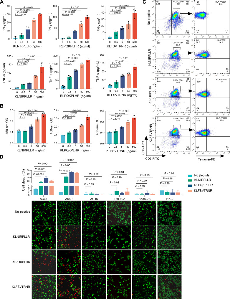
Neoantigens have emerged as ideal targets for personalized cancer immunotherapy. We depict the pan-cancer peptide atlas by comprehensively collecting immunopeptidomics from 531 samples across 14 cancer and 29 normal tissues, and identify 389,165 canonical and 70,270 noncanonical peptides. We reveal that noncanonical peptides exhibit comparable presentation levels as canonical peptides across cancer types. Tumor-specific peptides exhibit significantly distinct biochemical characteristics compared with those observed in normal tissues. We further propose an immunopeptidomic-guided machine learning-based neoantigen screening pipeline (MaNeo) to prioritize neo-peptides as immunotherapy targets. Benchmark analysis reveals MaNeo results in the accurate identification of shared and tumor-specific canonical and noncanonical neo-peptides. Last, we use MaNeo to detect and validate three neo-peptides in cancer cell lines, which can effectively induce increased proliferation of active T cells and T cell responses to kill cancer cells but not damage healthy cells. The pan-cancer peptide atlas and proposed MaNeo pipeline hold great promise for the discovery of canonical and noncanonical neoantigens for cancer immunotherapies.


Figures

Fig. 1.
Fig. 1.. Overview of the HLA-presented tumor-normal immunopeptidome atlas.
(A) Overview of the immunopeptidome cohort across the 14 cancer types and 29 normal tissues. AML: Acute myeloid leukemia, B-ALL: B cell acute lymphoblastic leukemia, T-ALL: T cell acute lymphoblastic leukemia, CLL: Chronic lymphocytic leukemia, NHL: Non-Hodgkin’s lymphoma, BRCA: Breast cancer, OV: Ovarian cancer, GBM: Glioblastoma, M.G.: Meningioma, NB: Neuroblastoma, COAD: Colon adenocarcinoma, CCRCC: Clear cell renal carcinoma, SKCM: Skin cutaneous melanoma, NSCLC: Non–small cell lung cancer. (B) Workflow for the construction of tumor-normal immunopeptidome atlas. (C) Coverage of HLA-A, B, and C alleles among the worldwide population, respectively. (D) Number of peptides among diverse ORF types. (E) Number of gene-encoded canonical and non-canonical peptides. C-like represents the gene deriving canonical peptides more than noncanonical ones. B-like represents the gene deriving canonical peptides equal to noncanonical ones. N-like represents the gene deriving canonical peptides less than noncanonical ones. lncRNA, long noncoding RNA.
Fig. 2.
Fig. 2.. Comparison of distinct aspects of canonical and noncanonical peptides.
(A) Accumulated unique canonical and non-canonical peptide distributions from each tissue. (B) Comparison of the length distributions of the canonical and noncanonical peptides. (C) GRAVY is shown for the canonical (n = 389,165) and noncanonical peptides (n = 70,270) with the Wilcoxon rank sum test. (D) Molecular weights of the peptides were compared via the Wilcoxon rank sum test (389,165 canonical versus 70,270 noncanonical peptides). (E) Usage tendency of different amino acids in canonical and noncanonical peptides. The orange color indicates that the amino acid is enriched in noncanonical peptides, whereas the blue color indicates that the amino acid is enriched in canonical peptides. (F to H) Presentation levels between canonical and noncanonical peptides are from (F) binding stability (HLA-A: 101,200 canonical versus 10,455 noncanonical, HLA-B: canonical: 102,232 versus 7235 noncanonical, HLA-C: 163 canonical versus 7 noncanonical), (G) binding affinity (HLA-A: 160,290 canonical versus 17,459 noncanonical, HLA-B: canonical: 166,184 versus 12,116 noncanonical, HLA-C: 71,012 canonical versus 5742 noncanonical) and (H) the relative rank of foreignness (TCR recognition probability homologous to known pathogen-derived peptides in the IEDB) (149,130 canonical versus 24,852 noncanonical). Wilcoxon rank sum test was used to compare the differences between groups.
Fig. 3.
Fig. 3.. Differences in the features of tumor and normal peptides.
(A) Peptides were classified exclusively into tumor samples as tumor peptides, whereas the rest were classified as normal peptides. The size of the circle indicates the number of peptides. The red color represents the tumor peptides, whereas the blue color represents the normal peptides. (B) The Venn diagram displaying the number of all the tumor and normal peptides. The bar and pie plots show the frequency and proportion of peptides found in several tumors or normal tissues. (C) Heatmap with hierarchically clustered correlations of position frequency vectors across tumors and normal tissues. (D) PCA based on the matrix of the AAC at each position to distinguish tumors from normal tissues. (E) Stacked bar plot at the top illustrates the amino acid enrichment across various types of tumors. The dot plot at the bottom indicates enriched amino acids in each tumor. (F) Connection of amino acids displays different degrees of enrichment among tumors, *P < 0.05, **P < 0.01, and ***P < 0.001. (G) Comparisons of biochemical features—such as net charge, pI, and GRAVY—between tumor (n = 307,656) and normal peptides (n = 151,358) with the Wilcoxon rank sum test.
Fig. 4.
Fig. 4.. Predicting neo-peptides within the HLA-presented peptides.
(A) Schematic representation of MaNeo for the training and testing procedures used to develop the predictive models. For each machine learning model, the hyperparameter was tuned with 5-fold cross-validation with three replicates. (B) Model performance was assessed via various metrics (n = 406, Wilcoxon rank sum test), *P < 0.05, **P < 0.01, and ***P < 0.001. (C) Performance of the models when a smaller amount of training data is used. (D) AUPRC curves for the different groups of tumors generated via the RF classifier. (E) Comparison of performance between the MaNeo and sCRAP algorithms (n = 377, Wilcoxon rank sum test). ns, not significant.
Fig. 5.
Fig. 5.. Validation of MaNeo in independent cancer cohorts.
(A) Schematic of the computational platform for the identification of immunogenic neo-peptides. (B) Gene set enrichment with source genes of immunogenic neo-peptides for each tumor. (C) Number of samples in which the immunogenic neo-peptides were detected (1 to 11). (D) Immunogenic neo-peptides that were presented in at least five samples. The colors of the heatmap represent the rank of the peptides in the samples. PI3K, phosphatidylinositol 3-kinase; UV, ultraviolet; MTOR, mammalian target of rapamycin; NF-κB, nuclear factor κB.
Fig. 6.
Fig. 6.. In vitro validation of neoantigens for tumors.
(A) Activation assay (IFN-γ and TNF-α enzyme-linked immunosorbent assay) for NP/DC-CD8+ T cells and no peptide. n = 3 [means ± SD; ordinary one-way analysis of variance (ANOVA) followed by Dunnett’s multiple comparisons test]. (B) CCK-8 assay for NP/DC-CD8+ T, n = 3 (means ± SD; ordinary one-way ANOVA followed by Dunnett’s multiple comparisons test). (C) CD8+ T cells from healthy individuals stained with KLNIRPLLR, RLPQKPLHR, and KLFSVTRNR tetramers. (D) Cytotoxicity assay for tumor cells (A375: human malignant melanoma line, A549: human NSCLC cancer cell line) and other tissue cells (AC16, THLE-2, Beas-2B, and HK-2). n = 3 (means ± SD; ordinary two-way ANOVA followed by Tukey’s multiple comparisons test).

Similar articles

References

    1. Lang F., Schrörs B., Löwer M., Türeci Ö., Sahin U., Identification of neoantigens for individualized therapeutic cancer vaccines. Nat. Rev. Drug Discov. 21, 261–282 (2022). - PMC - PubMed
    1. Katsikis P. D., Ishii K. J., Schliehe C., Challenges in developing personalized neoantigen cancer vaccines. Nat. Rev. Immunol. 24, 213–227 (2024). - PMC - PubMed
    1. Camarena M. E., Theunissen P., Ruiz M., Ruiz-Orera J., Calvo-Serra B., Castelo R., Castro C., Sarobe P., Fortes P., Perera-Bel J., Albà M. M., Microproteins encoded by noncanonical ORFs are a major source of tumor-specific antigens in a liver cancer patient meta-cohort. Sci. Adv. 10, eadn3628 (2024). - PMC - PubMed
    1. Cai Y., Li D., Lv D., Yu J., Ma Y., Jiang T., Ding N., Liu Z., Li Y., Xu J., MHC-I-presented non-canonical antigens expand the cancer immunotherapy targets in acute myeloid leukemia. Sci. Data 11, 831 (2024). - PMC - PubMed
    1. Chong C., Coukos G., Bassani-Sternberg M., Identification of tumor antigens with immunopeptidomics. Nat. Biotechnol. 40, 175–188 (2022). - PubMed