. 2025 May 23;11(21):eadt6632.
doi: 10.1126/sciadv.adt6632. Epub 2025 May 21.

Astrocyte-derived CCL5-mediated CCR5+ neutrophil infiltration drives depression pathogenesis

Affiliations

Astrocyte-derived CCL5-mediated CCR5+ neutrophil infiltration drives depression pathogenesis

Hang Yao et al. Sci Adv. .

Abstract

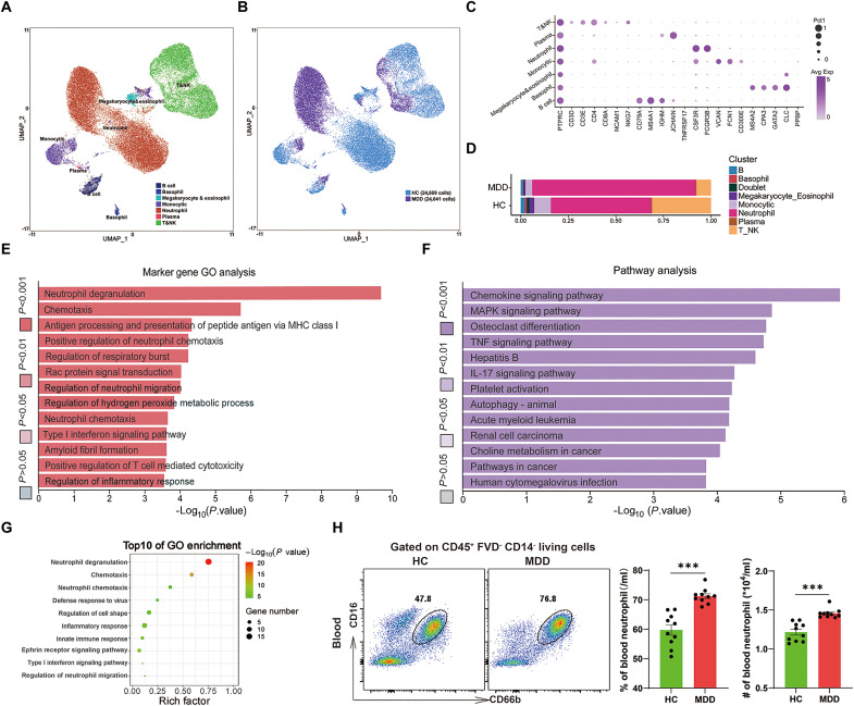
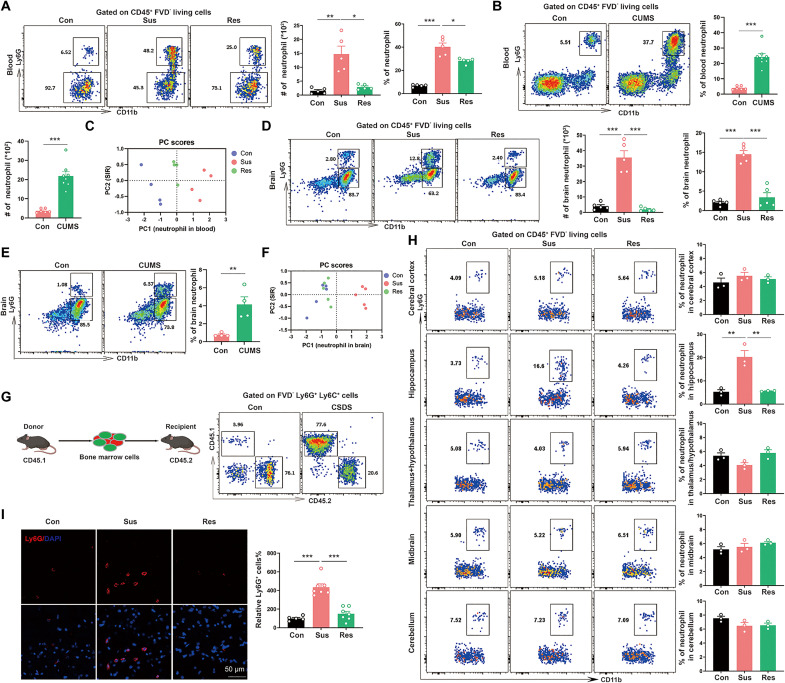
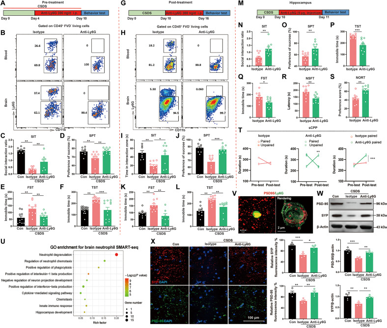
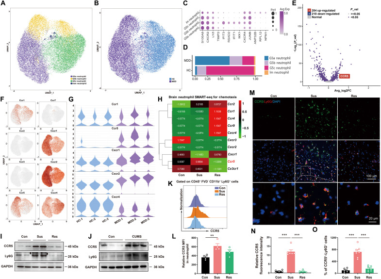
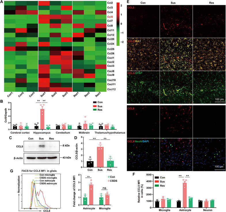
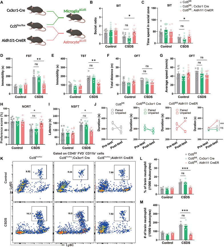
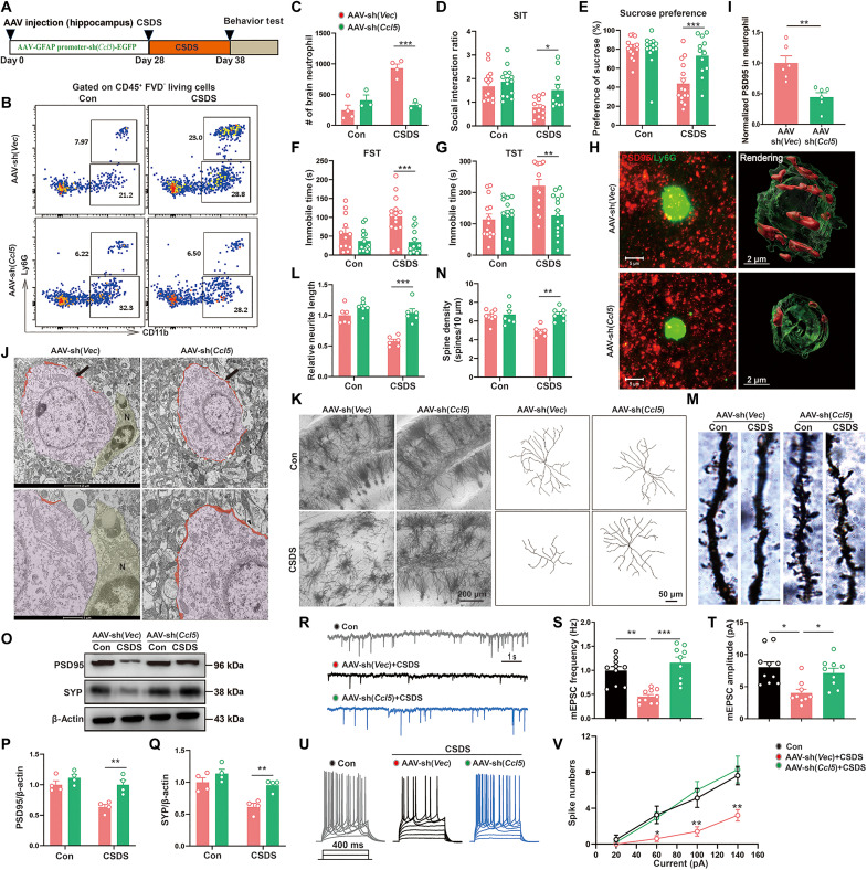
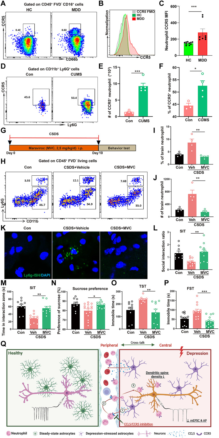
Cross-talk between the nervous and immune systems is involved in neurological diseases. However, their potential interplay in depression has yet to be elucidated. Here, using single-cell RNA and neutrophil SMART RNA sequencing, we showed that CCR5+ neutrophils were significantly increased in patients with depression and preferentially migrated to the hippocampus in a mouse model of depression. Infiltrated neutrophils engulf neuronal spines and subsequently promote depressive symptoms in male mice. Furthermore, by genetic or pharmacologic disruption, we identified a chemotactic effect of the astrocyte-derived chemokine CCL5 on mediating the infiltration of CCR5+ neutrophils and behavioral disorders in male depressed mice. Our findings therefore highlight the critical role of neutrophils in depression pathogenesis and astrocytes in mediating the dysregulation of innate immune responses and suggest that inhibition of CCL5/CCR5-mediated neutrophil infiltration represents a potential therapeutic strategy for noninfectious brain diseases such as depression.


Figures

Fig. 1.
Fig. 1.. Single-cell RNA sequencing reveals an increase in the number of neutrophils in depressive patients.
(A) UMAP embedding plot from three patients with MDD and three HCs showing seven major clusters. (B) UMAP embedding plot showing cell distribution of patients (n = 24,641) and HC (n = 24,609). (C) Dot plot showing the scaled expression of selected signature genes for each cluster, colored by the average expression of each gene in each cluster scaled across all clusters. (D) Proportions of the eight clusters in six samples. (E) GO analysis for marker gene of neutrophil clusters. (F) KEGG analysis for marker gene of neutrophil clusters. (G) GO analysis for DEG of neutrophil clusters between MDD and HC. (H) Flow cytometry analysis of neutrophils in blood from patients with MDD and HC (n = 10). The data shown are the mean ± SEM. Student’s t test was used. ***P < 0.001.
Fig. 2.
Fig. 2.. Neutrophils migrate into the brain in a mouse model of depression.
(A and B) Flow cytometry analysis of neutrophils in blood from mice following CSDS procedure [(A), n = 5 animals] and CUMS procedure [(B), n = 6 to 8 animals]. (C) PCA plot of circulated neutrophil and social interaction ratio (SIR) in control, susceptible mice, and resilient mice. (D and E) Flow cytometry analysis of neutrophils in brain from mice following CSDS procedure [(D), n = 5 animals] and CUMS procedure [(E), n = 4 animals]. (F) PCA plot of brain neutrophil and SIR in control, susceptible, mice and resilient mice. (G) Schematic diagram depicts the tracing of neutrophils. Flow cytometry plots show CD45.1-expressing neutrophils in brain following CSDS procedure. (H) Flow cytometry analysis of neutrophils in the cerebral cortex, hippocampus, thalamus/hypothalamus, midbrain, or cerebellum (n = 4 to 5 animals mixed). (I) Immunofluorescence staining showed the infiltration of neutrophils in hippocampus (n = 5 to 7 animals). Con, nonstressed mice; Sus, susceptible mice; Res, resilient mice. The data shown are the mean ± SEM. One-way analysis of variance (ANOVA) with Tukey’s post hoc tests was used [(A), (D), (H), and (I)]. Student’s t test was used [(B) and (E)]. *P < 0.05, **P < 0.01, ***P < 0.001.
Fig. 3.
Fig. 3.. Neutrophil depletion improves CSDS-induced depressive-like behaviors and reduces the engulfment of neuronal synapses.
(A) Schematic for Ly6G neutralizing antibody treatment in mice during CSDS procedure. (B) Flow cytometry plots show neutrophils in the blood and brain (n = 5 animals). (C) SI ratio (n = 8 to 10 animals). (D) Sucrose preference (n = 9 to 11 animals). (E and F) Total immobility time in FST [(E), n = 10 to 11 animals] and in TST [(F), n = 12 to 16 animals]. (G) Schematic for Ly6G neutralizing antibody treatment in mice after CSDS procedure. (H) Flow cytometry plots show neutrophils in the blood and brain (n = 5 animals). (I) Time spent in the interaction zone from SIT (n = 8 to 10 animals). (J) Sucrose preference (n = 8 animals). (K and L) Total immobility time in FST [(K), n = 7 to 11 animals] and in TST [(L), n = 8 to 12 animals]. (M) Experimental outline. (N) SI ratio (n = 10 animals). (O) Sucrose preference (n = 10 animals). (P and Q) Total immobility time in TST [(P), n = 10 animals] and in FST [(O), n = 10 animals]. (R) Latency in NSFT (n = 10 animals). (S) Preference score in NORT (n = 10 animals). (T) sCPP of isotype or anti-Ly6G–injected mice (n = 10 animals). (U) GO analysis for DEG of brain-infiltrating neutrophil. (V) Representative images and three-dimensional (3D) surface rendering of Ly6G+ neutrophils (green) containing PSD95+ puncta (red) in the hippocampus after CSDS procedure. (W) WB analysis of PSD95 and SYP expression in the hippocampus (n = 4 to 5 animals). (X) Representative images and quantitative analyses of immunostaining for PSD95 (green), SYP (red), and DAPI (blue) in the hippocampus (n = 4 animals). Isotype, mouse IgG1 isotype control; Anti-Ly6G, Ly6G neutralizing antibody. The data shown are the mean ± SEM. One-way ANOVA followed by Tukey’s post hoc test [(C) to (F), (I) to (L), (W), and (X)]. Two-way ANOVA followed by Tukey’s post hoc tests (T). Student’s t test [(N) to (S)]. *P < 0.05, **P < 0.01, ***P < 0.001.
Fig. 4.
Fig. 4.. Neutrophil CCR5 signal is up-regulated in depressive patients and mice.
(A) Seurat-Recluster analyzed the UMAP embedding plot showing four major neutrophil clusters. (B) UMAP embedding plot showing neutrophil distribution of MDD and HC. (C) Dot plot showing the scaled expression of selected signature genes for neutrophil cluster. (D) Proportions of the four clusters in six samples. (E) Volcano plot showing differentially expressed genes in G5a neutrophil cluster between MDD and HC. (F) UMAP embedding plot showing the expression of chemokine receptors in neutrophil clusters. (G) Violin plots showing the expression of chemokine receptors in neutrophil clusters between MDD and HC. (H) Heatmap of the chemokine receptor–related genes determined by SMART-seq of brain neutrophils following CSDS procedure. (I and J) WB analysis of CCR5 and Ly6G expression in the hippocampus following CSDS and CUMS procedure. (K and L) Flow cytometry plots and bar graph showing up-regulation of CCR5 in brain neutrophils (n = 4 animals). (M to O) Representative images and quantitative analyses of immunostaining for CCR5 (green), Ly6G (red), and DAPI (blue) in the hippocampus (n = 5 to 7 animals). The data shown are the mean ± SEM. One-way ANOVA with Tukey’s post hoc tests was used. **P < 0.01, ***P < 0.001.
Fig. 5.
Fig. 5.. The chemokine CCL5 is increased in astrocytes in a mouse model of depression.
(A) Heatmap showing the expression of chemokine ligand in the hippocampus by qPCR analysis. (B) qPCR analysis showing the expression of CCL5 in the cerebral cortex, hippocampus, thalamus/hypothalamus, midbrain, or cerebellum (n = 4 animals). (C and D) WB analysis of CCL5 expression in the hippocampus (n = 3 animals). (E) Representative micrographs of CCL5 (red) costained with IBA1 (orange), GFAP (green), or NeuN (green) in LMol. (F) Quantification of CCL5 fluorescence intensity in (E) (n = 3 to 5 animals). (G) Flow cytometry plots and bar graph showing up-regulation of CCL5 in astrocytes following CSDS procedure (n = 3 to 4 animals). The data shown are the mean ± SEM. One-way ANOVA with Tukey’s post hoc tests was used [(B), (D), and (F)]. Student’s t test (G). **P < 0.01, ***P < 0.001.
Fig. 6.
Fig. 6.. Astrocytic Ccl5 deletion prevents neutrophil brain infiltration and stress susceptibility.
(A) Schematic for astrocytic Ccl5 deletion or microglial Ccl5 deletion mice. For Cre-lox experiments, Ccl5fl/flmice were crossbred with Cx3cr1Cre or Aldh1l1CreER mice. (B and C) SI ratio and time spent in the interaction zone from SIT in Ccl5 fl/fl mice, Aldh1l1 CreER; Ccl5 fl/fl mice, or Cx3cr1 Cre; Ccl5 fl/fl mice following CSDS (n = 9 animals). (D and E) Total immobility time in FST (n = 9 animals) and in TST (n = 8 animals). (F and G) Movement distance and speed within 5 min was recorded in open field test (OFT) (n = 9 animals). (H) Preference score in NORT (n = 8 animals). (I) Latency in NSFT (n = 8 animals). (J) sCPP of Ccl5 fl/fl mice, Aldh1l1 CreER; Ccl5 fl/fl mice, or Cx3cr1 Cre; Ccl5 fl/fl mice (n = 8 animals). (K to M) Flow cytometry analysis of neutrophils in brain from Ccl5 fl/fl mice, Aldh1l1 CreER; Ccl5 fl/fl mice, or Cx3cr1 Cre; Ccl5 fl/fl mice following CSDS procedure (n = 5 animals). The data shown are the mean ± SEM. Two-way ANOVA with Tukey’s post hoc tests was used. *P < 0.05, **P < 0.01, ***P < 0.001. ns, not significant.
Fig. 7.
Fig. 7.. Astrocyte-expressing CCL5 mediates neutrophil brain infiltration and stress susceptibility.
(A) Diagram of the experimental design. (B and C) Flow cytometry analysis of neutrophils in brain from AAV-vector– or AAV-sh(Ccl5)–injected mice (n = 3 to 4 animals). (D) SI ratio (n = 10 to 15 animals). (E) Sucrose preference (n = 12 to 15 animals). (F and G) Total immobility time in FST [(F), n = 12 to 15 animals] and in TST [(G), n = 14 to 15 animals]. (H) Representative images and 3D surface rendering of Ly6G+ neutrophils (green) containing PSD95+ puncta (red) in the hippocampus after CSDS procedure (n = 6 animals). (I) Quantification of PSD95+ puncta in neutrophils as indicated in (H). (J) Electron micrograph showing a region of hippocampus. In AAV-vector–injected mice, a phagocyte [(N), neutrophils] closely apposing part of a neuronal soma in close vicinity can be observed. Red indicates axosomatic synapses. (K) Representative images of neuronal dendrites and 3D reconstruction of neuronal dendrites in mice. (L) Summarized data for relative neuronal dendrite length (n = 6 animals). (M) Representative images of neuronal dendrites by Golgi-Cox staining in mice. (N) Summarized data for spine numbers per 10 μm (n = 6 animals). Scale bar, 5 μm. (O to Q) WB analysis of PSD95 and SYP expression in the hippocampus (n = 4 animals). (R to T) Representative traces (R) and summarized data of mEPSC frequency (S) and amplitude (T) from hippocampal pyramidal neurons in mice (n = 5 animals). (U and V) Representative traces (U) and statistical data of step current injection–induced spike numbers (V) from hippocampal pyramidal neurons in mice (n = 5 animals). The data shown are the mean ± SEM. One way [(S), (T), and (V)] or two-way ANOVA [(C) to (G), (L), (N), (P), and (Q)] with Tukey’s post hoc tests was used. Student’s t test was used (I). *P < 0.05, **P < 0.01, ***P < 0.001.
Fig. 8.
Fig. 8.. Inhibition of neutrophil CCR5 alleviates brain neutrophil infiltration and depressive-like behavior in male mice.
(A) Schematic diagram shows the experimental design. Bone marrow cells were isolated from femur bone marrow of donor Ccr5+/+ or Ccr5−/− mice and then injected into femur bone marrow of recipient mice that were subjected to lethal irradiation. (B and C) Flow cytometry analysis of neutrophils in brain from Ccr5+/+ or Ccr5−/− bone marrow cell chimeric mice following CSDS (n = 3 animals). (D and E) SI ratio and time spent in the interaction zone from SIT (n = 8 to 14 animals). (F) Sucrose preference (n = 7 to 14 animals). (G and H) Total immobility time in FST [(G), n = 10 to 11 animals] and TST [(H), n = 9 to 14 animals]. (I) Representative images and 3D surface rendering of Ly6G+ neutrophils (green) containing PSD95+ puncta (red) in the hippocampus (n = 6 to 7 animals). (J) Quantification of PSD95+ puncta in neutrophils as indicated in (I). (K) Representative images of neuronal dendrites and 3D reconstruction of neuronal dendrites in the indicated groups (n = 6 animals). (L) Summarized data for relative neuronal dendrite length (n = 6 animals). (M and N) Representative images of neuronal dendrites by Golgi-Cox staining and summarized data for spine numbers per 10 μm in Ccr5+/+ or Ccr5−/− bone marrow cell chimeric mice (n = 6 animals). Scale bar, 5 μm. (O to Q) Representative traces (O) and summarized data of mEPSC frequency (P) and amplitude (Q) from hippocampal pyramidal neurons in mice following CSDS (n = 5 animals). (R and S) Representative traces (R) and statistical data of step current injection–induced spike numbers (S) from hippocampal pyramidal neurons in mice following CSDS (n = 5 animals). The data shown are the mean ± SEM. Two-way ANOVA with Tukey’s post hoc tests was used [(C) to (H), (L), and (N)]. Student’s t test was used [(J), (P), (Q), and (S)]. *P < 0.05, **P < 0.01, ***P < 0.001.
Fig. 9.
Fig. 9.. Enhanced CCR5-expressing neutrophils in patients with depression.
(A to C) Flow cytometry analysis of CCR5-expressing neutrophils in blood from patients with MDD (n = 8) or HC (n = 10). (D to F) Flow cytometry analysis of CCR5-expressing neutrophils in blood of mice following CUMS (n = 3 to 5 animals). (G) Schematic diagram shows the experimental design. (H to J) Flow cytometry analysis of neutrophils in brain from vehicle- or maraviroc (MVC)–treated mice following CSDS (n = 4 animals). (K) RNAscope in situ hybridization confirmed reduced neutrophil infiltration in hippocampus of MVC-treated mice. (L and M) SI ratio and time spent in the interaction zone from SIT in vehicle- or MVC-treated mice following CSDS (n = 9 to 11 animals). (N) Sucrose preference in the indicated groups following CSDS (n = 9 to 15 animals). (O and P) Total immobility time in FST [(O), n = 12 to 14 animals] and in TST [(P), n = 10 animals] in the indicated groups following CSDS. (Q) Proposed model depicting the crucial role of astrocyte CCL5–neutrophil CCR5 chemotaxis axis in driving neutrophil brain infiltration, consequently contributing to engulfment of neuronal spines and stress susceptibility in depression. The data shown are the mean ± SEM. One-way ANOVA with Tukey’s post hoc tests was used [(I), (J), and (L) to (P)]. Student’s t test was used [(C), (E), and (F)]. *P < 0.05, **P < 0.01, ***P < 0.001.

Similar articles

References

    1. Fan Z., Chang J., Liang Y., Zhu H., Zhang C., Zheng D., Wang J., Xu Y., Li Q. J., Hu H., Neural mechanism underlying depressive-like state associated with social status loss. Cell 186, 560–576.e17 (2023). - PubMed
    1. Marwaha S., Palmer E., Suppes T., Cons E., Young A. H., Upthegrove R., Novel and emerging treatments for major depression. Lancet 401, 141–153 (2023). - PubMed
    1. Duman R. S., Aghajanian G. K., Synaptic dysfunction in depression: Potential therapeutic targets. Science 338, 68–72 (2012). - PMC - PubMed
    1. Sarno E., Moeser A. J., Robison A. J., Neuroimmunology of depression. Adv. Pharmacol. 91, 259–292 (2021). - PMC - PubMed
    1. Balog B. M., Sonti A., Zigmond R. E., Neutrophil biology in injuries and diseases of the central and peripheral nervous systems. Prog. Neurobiol. 228, 102488 (2023). - PMC - PubMed

MeSH terms