Review
. 2025 May 21;21(5):e1012993.
doi: 10.1371/journal.pcbi.1012993. eCollection 2025 May.

Vertex models capturing subcellular scales in epithelial tissues

Affiliations
Review

Vertex models capturing subcellular scales in epithelial tissues

Zoë Lange et al. PLoS Comput Biol. .

Abstract

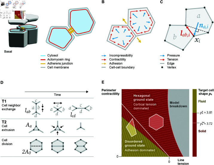
Vertex models provide a robust theoretical framework for studying epithelial tissues as a network of cell boundaries. They have been pivotal in exploring properties such as cell packing geometry and rigidity transitions. Recently, extended vertex models have become instrumental in bridging the subcellular scales to the tissue scale. Here, we review extensions of the model aiming to capture experimentally observed subcellular features of epithelial tissues including heterogeneity in myosin activity across the tissue, non-uniform contractility structures, and mechanosensitive feedback loops. We discuss how these extensions change and challenge current perspectives on observables of macroscopic tissue properties. First, we find that extensions to the vertex model can change model properties significantly, impacting the critical threshold and in some cases even the existence of a rigidity transition. Second, we find that packing disorder can be explained by models employing different subcellular mechanisms, indicating a source of stochasticity and gradual local size changes as common mesoscopic motifs in the mechanics of tissue organization. We address complementary models and statistical inference, putting vertex models in a broader methodological context and we give a brief overview of software packages utilized in increasingly complex vertex model studies. Our review emphasizes the need for more comparative, systematic studies that identify specific classes of vertex models which share a set of well-defined properties, as well as a more in-depth discussion of modeling choices and their biological motivations.


Conflict of interest statement

The authors have declared that no competing interests exist.

Figures

Fig 1
Fig 1. Forces, topological transitions, and phase transitions in a vertex model for confluent epithelial tissues.
(A) The vertex model incorporates what are considered the main mechanical components in polarized epithelial cells: cytosol filling the volume of a cell; in the apical region: an actomyosin ring along the cell perimeter and adherens junctions connecting two cells, and a cell membrane enclosing the volume. The two-dimensional model is a cross-section close to the apical surface of the tissue. (B) Cells in the tissue are approximated by polygons that form a confluent tiling. In the vertex model, we attribute specific forces to the mechanical components: The cytosol resists compression and expansion forces; the actomyosin ring exerts contractile forces on a cell and the adherens junctions result in attractive forces along the cell-cell boundary. The adhesion forces compete with the cortical tension from the cell membrane and actomyosin ring. (C) Vertices in the network satisfy force balance based on the forces from the mechanical components. Effective forces on a vertex i are along the edge length vector 𝐥ab,i pointing away from reference vertex i (tension) and the normal vector 𝐧a,i defined as perpendicular to the line connecting the two neighbors of vertex i and pointing towards the outside of the cell (pressure). (D) A common set of update rules accounts for topological transitions in the temporal evolution of a vertex network. T1 transitions are cell neighbor exchanges where an edge between cells a and b falls below a certain threshold and continues to shrink until it vanishes; then, a new edge between cells c and d is inserted perpendicular to the old edge. T2 transitions are cell extrusions where a cell with an area below a threshold value vanishes and leaves a single vertex. Cell division, as part of proliferation, occurs when a cell splits into two. (E) Phase diagram of tissue mechanical states as a function of parameters kP (perimeter contractility) and kl (line tension). When the control parameter p0=P0/A0 is below p0hex3.72 (white line), the ground state is a regular hexagonal lattice, which becomes unstable once p0 increases past p0hex [23,39]. For a disordered, metastable tissue, a solid-to-fluid transition occurs as p0 increases past p0*3.81, characterized by vanishing energy barriers for T1 transitions [29]. The exact threshold of the solid-to-fluid transition can vary depending on cell packing disorder [10] and implementation details of the dynamics [46,47]. The gray area marks where the model breaks down due to vanishing areas.
Fig 2
Fig 2. Spatiotemporal dependence of model parameters and alternative cell contractility models.
(A) Different models with spatiotemporally dependent coefficients aim to represent observed effects of (B) the complex recruitment dynamics of myosin to actin fibers in the cytoskeleton. (A) Stochastic models can describe spatial static inhomogeneity of myosin distribution. This can be expanded towards temporal stochastic processes (e.g., Ornstein-Uhlenbeck) where myosin abundance on an actin fiber varies over time. Deterministic models impose a specific condition for myosin distribution, e.g., with respect to an angle or a clock. These models are directly motivated to capture observations like convergent extension or active contractility. Some observed patterns are implemented model-free into vertex model parameters. A representative example is the contractile cable along the perimeter of the amnioserosa during dorsal closure of Drosophila embryo development. (C) The actomyosin contractility mode expressed in a vertex model with a perimeter contractility term [23] is sometimes called a purse-string mechanism; it represents a continuous actomyosin belt along the cell perimeter and generates uniform contractility. Apart from that, a series of non-uniform contractility models exist. The edge model assumes a discontinuous actomyosin belt and thus reduces coupling of the contractility structures [80]. The medial cytoskeleton observed in premature tissues has been modeled as isotropic radial fibers and anisotropic parallel fibers [35,81,82]. Parallel fibers carry a load and resist isotropic extension without introducing additional degrees of freedom [82].
Fig 3
Fig 3. The effective relation between forces and cell shapes in the vertex model can be changed by introducing submodules featuring active regulation and mechanosensitive feedback describing activities at the subcellular scale.
(A) The mechanics in the reference vertex model [23] are governed by an energy function (a phenomenological Hamiltonian) that dictates a linear relation between forces and cell shape (observed e.g., in the ground states or a proliferation-free simulation). To be able to capture realistic tissue morphologies, vertex models are commonly extended to a full simulation framework with components refining the subcellular scales, resulting in effectively nonlinear relations between forces and cell geometry [65]. The subcellular activities include the activation and turnover of cytoskeleton machinery and adhesion proteins, the relaying of mechanical signals through mechanotransduction molecules, the modulation of mechanical forces by the spatiotemporal dynamics of signaling pathways, and the activation and deactivation of genetic programs instructing protein synthesis. (B) Examples of submodules that actively regulate quantities are strain-dependent remodeling of the target edge length, edge length-dependent active tension, or strain-dependent remodeling of the density of a biomolecule, e.g., myosin. In systems with mechanosensitive feedback, these remodeled quantities are usually interdependent and formulated as coupled differential equations.
Fig 4
Fig 4. Dynamical phase diagrams describing a solid to fluid transition in various extended vertex models.
(A) Average vertex coordination number (from z¯=3 to z¯=6) against target cell shape p0 in a model with active T1 transitions [120]; (B) alignment of cell shape against average measured cell shape p¯ in an anisotropic tension model [10]; (C) Gaussian curvature against target cell shape in a vertex model restricted to a sphere (positive curvature) or hyperbolic disk (negative curvature) [121,122]; (D) alignment rate of cell polarity against traction forces in a traction force model on a spherical surface [118]. Asterisks indicate the critical target cell shape p0*=3.81 or average measured cell shape p¯=p0*) in the reference model [29,39].
Fig 5
Fig 5. Vertex-like models by resolution of cell-cell junction dynamics.
Increasing resolution of cell-cell junctions from left to right: The Voronoi model is a coarse-grained version of the vertex model with cell centers as degrees of freedom and implicit treatment of T1 transitions [125,142,143]; the vertex model as reference [28]; the deformable polygon or N-vertices model resolves the cell cortex by expanding the definition of vertices to a subjunctional flexible chain with explicit adhesion between cell membranes [,,–151]; the apposed cortex adhesion model focuses on adhesion dynamics of cell-cell junctions during topological transitions with viscoelastic continuum cortices adhering to each other by an explicit continuous adhesion energy [152].

Similar articles

References

    1. Bryant DM, Mostov KE. From cells to organs: building polarized tissue. Nat Rev Molecul Cell Biol. 2008;9(11):887–901. doi: 10.1038/nrm2523 - DOI - PMC - PubMed
    1. Mateos C, Valencia-Expósito A, Palacios IM, Martín-Bermudo MD. Integrins regulate epithelial cell shape by controlling the architecture and mechanical properties of basal actomyosin networks. PLOS Genet. 2020;16:e1008717. doi: 10.1371/journal.pgen.1008717 - DOI - PMC - PubMed
    1. Kim EJY, Sorokin L, Hiiragi T. ECM-integrin signalling instructs cellular position sensing to pattern the early mouse embryo. Development. 2022;149(1):200140. doi: 10.1242/dev.200140 - DOI - PMC - PubMed
    1. Li ZS, Adams JG, Chisholm AD. Form and function of the apical extracellular matrix: new insights from Caenorhabditis elegans, Drosophila melanogaster, and the vertebrate inner ear. Faculty Rev. 2020;9. doi: 10.12703/r/9-27 - DOI - PMC - PubMed
    1. Weaire D, Hutzler S. The physics of foams. Oxford: Oxford University Press; 2000

LinkOut - more resources