. 2025 May 20;6(5):102140.
doi: 10.1016/j.xcrm.2025.102140.

Targeting mitochondrial complex I of CD177+ neutrophils alleviates lung ischemia-reperfusion injury

Affiliations

Targeting mitochondrial complex I of CD177+ neutrophils alleviates lung ischemia-reperfusion injury

Junqi Wu et al. Cell Rep Med. .

Abstract

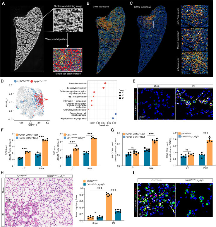
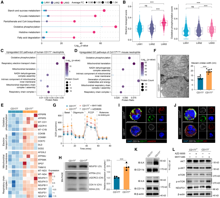
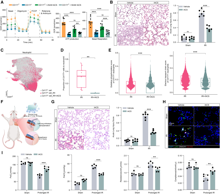
Primary graft dysfunction (PGD) is the leading cause of early morbidity and mortality following lung transplantation, with neutrophils playing a central role in its inflammatory pathology. Here, we employ single-cell RNA sequencing and spatial transcriptomics to investigate neutrophil subtypes in the lung ischemia-reperfusion injury (IRI) model. We identify CD177+ neutrophils as an activated subpopulation that significantly contributes to lung injury and serves as an early biomarker for predicting severe PGD in human lung transplant recipients (area under the curve [AUC] = 0.871). CD177+ neutrophils exhibit elevated oxidative phosphorylation and increased mitochondrial complex I activity, driving inflammation and the formation of neutrophil extracellular traps. Targeting mitochondrial function with the complex I inhibitor IACS-010759 reduces CD177+ neutrophil activation and alleviates lung injury in both mouse IRI and rat left lung transplant models. These findings provide a comprehensive landscape of CD177+ neutrophil-driven inflammation in lung IRI and highlight its potential value for future early diagnosis and therapeutic interventions.

Keywords: CD177; lung ischemia-reperfusion injury; mitochondrial complex I; neutrophil; oxidative phosphorylation.


Conflict of interest statement

Declaration of interests The authors declare no competing interests.

Figures

None
Graphical abstract
Figure 1
Figure 1
Single-cell transcriptional profiling of lung ischemia-reperfusion injury (A) Schematic diagram illustrating the experimental design. Left lung tissues and blood samples of mice subjected to lung ischemia (1 h) followed by reperfusion (3 h) were collected. Subsequent single-cell RNA sequencing (scRNA-seq) and spatial transcriptomics analyses were performed. Clinical validation involved examining CD177+ neutrophils collected pre- and post-transplantation in human lung recipients. Functional proteomics identified increased mitochondrial complex I activity in CD177+ neutrophils. Treatment with IACS-010759 (1 mg/kg), a complex I inhibitor, alleviated lung IRI. (B) Uniform manifold approximation and projection (UMAP) plot displaying 43,852 immune cells from lung tissues under sham (n = 4 mice) and IRI (n = 6 mice) conditions. These immune cells were categorized into 5 major cell types. (C) The differentially expressed genes (DEGs) of the 5 major cell clusters are listed. Myeloid cells show the highest number of DEGs. (D) Bubble heatmap highlighting genes significantly increased across 10 major subclusters of immune cells in lung IRI. Average fold change (FC) and significance (−Log10-adjusted p value) are indicated by dot size and color, respectively. (E) Relative gene expression scores of post- vs. pre-transplantation samples in human lung transplant recipients. (F) UMAP plot displaying 2,973 lung-associated neutrophils (LANs) subdivided into 5 cell types (LAN1–LAN5). (G) Heatmap of the top 6 differential genes for LAN1–LAN5. (H) Feature plots of selected marker genes (Cebpb, Ccrl2, Cd177, Camp, and Itgal) showing high expression of Cd177 in specific neutrophil sub-clusters. (I) Cell proportions of LAN1–LAN5 in IRI versus sham control groups showing LAN3 was significantly increased after lung IRI. (J) Gene Ontology enrichment analysis of LAN1–LAN3 neutrophil clusters, indicating that LAN3 is related to neutrophil activation. NK, natural killer; Neut, neutrophil; AMϕ, alveolar macrophage; Mo, monocyte. Statistical significance was assessed by two-sided Wilcoxon test adjusted using Bonferroni method in (C), (D), and (G), two-sided Wilcoxon test in (H), and Fisher’s exact test in (J). ns, not significant; ∗∗∗p < 0.001.
Figure 2
Figure 2
Enriched Cd177+ neutrophils are key contributors to lung ischemia-reperfusion injury in the mouse model (A) Nucleic acid staining and watershed-based segmentation for single-cell analysis. A representative image shows nuclei segmented using the watershed algorithm for spatial distribution analysis (n = 2). (B) Spatial expression of the pan-immune marker Cd45 in lung tissue sections reveals regions of local immune cell infiltration. (C) Spatial mapping of Cd177 expression in lung tissues after IRI. Higher-magnification images (right) show colocalization of Cd177 with the inflammatory genes Pglyrp1 and Ltf. (D) Uniform manifold approximation and projection (UMAP) visualization showing Ly6g+Cd177+ neutrophil populations (red) compared with Ly6g+Cd177 neutrophils (blue). The bubble heatmap on the right demonstrates enrichment of inflammatory Gene Ontology terms in Ly6g+Cd177+ cells. (E) Representative immunofluorescence images from the mouse left lung for Ly6G (green), CD177 (red), and citrullinated histone H3 (Cit-H3, white) showing NET formation in CD177+ neutrophils. Scale bar: 20 μm. (F) Reactive oxygen species (ROS) production was higher in CD177+ compared to CD177 neutrophils from human samples (left) and in Cd177flox/flox neutrophils compared to Cd177flox/flox; Ly6gCre neutrophils from mice (right), with or without PMA stimulation. (n = 5 per group). Neut, neutrophils; UT, untreated. (G) Quantification of MPO-DNA complexes showing increased NET formation in CD177+ compared to CD177 human neutrophils (left) and in Cd177flox/flox compared to Cd177flox/flox; Ly6gCre mouse neutrophils (right) under PMA stimulation. Neut, neutrophils; UT, untreated. (n = 5 per group). (H) Representative H&E staining of lung sections from Cd177flox/flox and Cd177flox/flox; Ly6gCre mice under sham and IRI conditions. Quantification of acute lung injury scores (right) demonstrates significant injury in Cd177flox/flox mice and minimal injury in Cd177flox/flox; Ly6gCre mice after lung IRI. Scale bar: 50 μm. (n = 6 per group). (I) Immunofluorescence staining revealing reduced NET infiltration (Ly6G, Cit-H3, and DNA/H1) in lung tissues of Cd177flox/flox; Ly6gCre mice post-lung IRI. Scale bar: 20 μm. Data are shown as mean ± SD. Statistical significance was assessed by a two-sided Wilcoxon test adjusted with the Bonferroni method in (F), (G), and (H) and Fisher’s exact test in (D). ns, not significant; ∗∗∗p < 0.001.
Figure 3
Figure 3
CD177+ neutrophils predict grade 3 primary graft dysfunction (A) UMAP plot displaying four distinct mouse peripheral blood neutrophil subsets (PBN1–PBN4) under sham and ischemia-reperfusion injury (IRI) conditions (n = 3 per group). (B) Boxplot quantifying PBN3 proportions in IRI vs. sham groups, showing a significant increase in PBN3 cells in the IRI group. (C) Bubble heatmap showing the expression of marker genes in mouse PBN1–4 and mouse lung-associated neutrophils 1–3 (LAN1–3). Both PBN3 and LAN3 exhibit high Cd177, Ngp, Camp, and Ltf expressions, indicating an activated phenotype contributing to IRI pathology. (D) Violin plots illustrating the bone marrow proximity scores of mice PBN1–4 and LAN1–3. PBN3 and LAN3 have notably high scores, indicating recent mobilization from the bone marrow. (E and F) Pseudotime analyses of murine peripheral blood neutrophil subsets (PBN1–4). (E) CytoTrace and (F) Monocle 3 highlight a neutrophil differentiation trajectory, with PBN3 cells occupying an earlier differentiation state. (G and H) Boxplots showing (G) the neutrophil-to-lymphocyte ratio across PGD 0–2 and PGD 3 groups (n = 79 vs. 25), demonstrating no significant difference, and (H) a significant increase in CD177+ neutrophil proportions in PGD 3 vs. PGD 0–2. (I) Receiver operating characteristic (ROC) curve displaying the diagnostic performance of the change in the proportion of CD177+ neutrophils (between post-4 h and pre-transplantation) in predicting grade 3 PGD within 72 h. The ROC analysis demonstrates high diagnostic accuracy (AUC = 0.871; 95% CI: 0.764–0.951). Statistical significance was assessed by a two-sided Wilcoxon test adjusted with the Bonferroni method in (B), (D), (G), and (H). ns, not significant; ∗∗p < 0.01; ∗∗∗p < 0.001.
Figure 4
Figure 4
CD177+ neutrophils exhibit enhanced mitochondrial oxidative phosphorylation and electron transport activity (A) scMetabolism analysis for lung-associated neutrophils 1–3 (LAN1–3) showing oxidative phosphorylation (OXPHOS) pathway was significantly enriched in the LAN3 subset. (B) Quantification of electron transport score (left) and OXPHOS score (right) in LAN1–3 demonstrates that the LAN3 subset has the highest values for both. (C and D) Gene Ontology (GO) enrichment analysis of upregulated pathways in human CD177+ neutrophils (C, n = 4 per group) and Cd177flox/flox mouse neutrophils (D, n = 3 per group), based on proteomic data. OXPHOS and respiratory complex I pathways were enriched. (E) Heatmap showing increased expression of mitochondrial proteins associated with OXPHOS, the electron transport chain, and the respiratory chain complexes in human CD177+ neutrophils compared to CD177 neutrophils (n = 4 per group). (F) Representative transmission electron microscopy (TEM) images depicting the mitochondrial ultrastructure of human CD177+ and CD177 neutrophils. The quantitative analysis of mitochondrial cristae width (right) reveals a significant increase in cristae width in CD177+ neutrophils (n = 8 per group). (G) Oxygen consumption rate (OCR) analysis of human CD177+ and CD177 neutrophils under basal and pharmacologically modulated conditions demonstrates enhanced mitochondrial oxidative phosphorylation in CD177+ cells. Treatment with the mTOR agonist MHY1485 markedly enhances OXPHOS activity in CD177 neutrophils, while inhibition of mTOR signaling via AZD8055 suppresses OXPHOS in CD177+ cells, indicating mTOR-dependent regulation of mitochondrial respiration. Data represent n = 9 replicates per group. (H) Blue native PAGE and immunoblot assay of mitochondrial complex subunits (CI–V) in human CD177+ and CD177 neutrophils. Quantification (right) shows elevated expression of NDUFS1 (a marker for mitochondrial complex I) in CD177+ neutrophils. Data are shown as mean ± SD (n = 4 per group). (I) Immunofluorescence staining shows a marked upregulation of MitoSOX (red) in human CD177+ neutrophils compared to CD177 neutrophils. Scale bar: 10 μm. (J) Immunofluorescence staining of human neutrophils reveals robust colocalization of ILK (red), CD177 (green), and CD11b (white) in CD177+ neutrophils. Scale bar: 10 μm. (K) Immunoblot assay of ILK protein expression in human CD177+ and CD177 neutrophils after CD11b immunoprecipitation (IP). β-actin serves as a loading control for the input. (L) Immunoblot assay of AKT, phosphorylated AKT (p-AKT), mTOR, phosphorylated mTOR (p-mTOR), and NDUFS1 expression in CD177+ and CD177 neutrophils with or without AZD8055 and MHY1485 treatment. CD177+ cells show higher p-AKT and p-mTOR levels vs. CD177 cells. AZD8055 (mTOR inhibitor) reduces p-mTOR and NDUFS1 in CD177+ cells, while MHY1485 (mTOR agonist) increases these proteins in CD177 cells. Data are shown as mean ± SD. Statistical significance assessed by a two-sided Wilcoxon test adjusted with the Bonferroni method (B and F). ns, not significant; ∗∗∗p < 0.001.
Figure 5
Figure 5
Mitochondrial complex I inhibitor IACS-010759 alleviates lung ischemia-reperfusion injury (A) OCR analysis showing mitochondrial respiration in CD177+ and CD177 neutrophils and CD177+ neutrophils treated with 25 or 50 nM IACS-010759. Quantification of ATP production and basal respiration indicates a significant reduction of oxidative phosphorylation (OXPHOS) after IACS-010759 treatment (n = 9 per group). (B) Representative images of left lung H&E staining (left) and lung injury scores (right) showing IACS-010759 (1 mg/kg) treatment reduced lung injury in the left hilar ligation/reperfusion mouse model. Scale bar: 50 μm (n = 5 per group). (C) UMAP visualization of scRNA-seq data from sorted neutrophils in lungs of IACS-treated (n = 4) and untreated mice (n = 3) under left hilar ligation/reperfusion. (D) Quantification of Cd177+ neutrophils in the IACS-treated versus untreated groups following lung IRI. IACS treatment significantly reduces the proportion of Cd177+ neutrophils. (E) Violin plots illustrating the OXPHOS (left) and electron transport chain (right) scores of Cd177+ neutrophils in IRI lungs, showing a significant decrease after IACS treatment. (F) Schematic of the orthotopic left lung transplantation rat model with prolonged cold ischemia. (G) IACS treatment reduced lung injury, as evidenced by H&E staining (left) and lung injury scores (right) from sham rats and allografts treated with vehicle or IACS-010759 (1 mg/kg) following prolonged ischemia reperfusion (n = 6 per group). Scale bar: 50 μm. (H) Immunofluorescence staining for neutrophil extracellular traps (Ly6G, DNA/H1, and Cit-H3) in lung tissues of sham and IRI rats with or without IACS treatment. Scale bar: 20 μm. (I) Lung injury indicators and pulmonary function (airway compliance, airway resistance, PaO2, and PaCO2) measured in each group showing that IACS treatment reduced lung injury in the orthotopic left lung transplant rat model (n = 5 per group). Data are shown as mean ± SD. Statistical significance was assessed by a two-sided Wilcoxon test adjusted with the Bonferroni method (A, B, D, E, G, and I). ns, not significant; ∗∗p < 0.01; ∗∗∗p < 0.001.

References

    1. Cantu E., Diamond J.M., Cevasco M., Suzuki Y., Crespo M., Clausen E., Dallara L., Ramon C.V., Harmon M.T., Bermudez C., et al. Contemporary trends in PGD incidence, outcomes, and therapies. J. Heart Lung Transplant. 2022;41:1839–1849. doi: 10.1016/j.healun.2022.08.013. - DOI - PMC - PubMed
    1. Diamond J.M., Arcasoy S., Kennedy C.C., Eberlein M., Singer J.P., Patterson G.M., Edelman J.D., Dhillon G., Pena T., Kawut S.M., et al. Report of the International Society for Heart and Lung Transplantation Working Group on Primary Lung Graft Dysfunction, part II: Epidemiology, risk factors, and outcomes-A 2016 Consensus Group statement of the International Society for Heart and Lung Transplantation. J. Heart Lung Transplant. 2017;36:1104–1113. doi: 10.1016/j.healun.2017.07.020. - DOI - PubMed
    1. Diamond J.M., Lee J.C., Kawut S.M., Shah R.J., Localio A.R., Bellamy S.L., Lederer D.J., Cantu E., Kohl B.A., Lama V.N., et al. Clinical risk factors for primary graft dysfunction after lung transplantation. Am. J. Respir. Crit. Care Med. 2013;187:527–534. doi: 10.1164/rccm.201210-1865OC. - DOI - PMC - PubMed
    1. Matthay M.A., Zemans R.L., Zimmerman G.A., Arabi Y.M., Beitler J.R., Mercat A., Herridge M., Randolph A.G., Calfee C.S. Acute respiratory distress syndrome. Nat. Rev. Dis. Primers. 2019;5:18. doi: 10.1038/s41572-019-0069-0. - DOI - PMC - PubMed
    1. Santos J., Wang P., Shemesh A., Liu F., Tsao T., Aguilar O.A., Cleary S.J., Singer J.P., Gao Y., Hays S.R., et al. CCR5 drives NK cell–associated airway damage in pulmonary ischemia-reperfusion injury. JCI Insight. 2023;8 doi: 10.1172/jci.insight.173716. - DOI - PMC - PubMed

MeSH terms

LinkOut - more resources