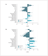
Hippocampal and amygdala subfield volumes in obsessive-compulsive disorder by medication status
- PMID: 40398928
- PMCID: PMC12114122
- DOI: 10.1503/jpn.230119
Hippocampal and amygdala subfield volumes in obsessive-compulsive disorder by medication status
Abstract
Background: Although it has been suggested that the hippocampus and amygdala (HA) are involved in the neurobiology of obsessive-compulsive disorder (OCD), volumetric findings have been inconsistent, and little work has been undertaken on the volumetry of the heterogeneous anatomic units of HA, with their specific functions and cytoarchitecture, in OCD. We sought to explore potential sources of heterogeneity in brain volumes by performing a separate analysis for people with and without psychotropic medication use, as well as the association of subfield volumes with OCD symptom severity.
Methods: We segmented T 1-weighted images from people with OCD and healthy controls in the OCD Brain Imaging Consortium to produce 12 hippocampal subfields and 9 amygdala subfields using Free-Surfer 6.0. We assessed between-group differences in subfield volume using a mixed-effects model adjusted for age and quadratic effects of age, sex, site, and whole HA volume. We also performed subgroup analyses to examine subfield volume in relation to comorbid anxiety and depression, medication status, and symptom severity. We corrected all analyses for multiple comparisons using the false discovery rate (FDR).
Results: We included images from 381 people with OCD and 338 healthy controls. These groups did not significantly differ in HA subfield volumes. However, medicated people with OCD had significantly smaller volumes in the hippocampal dentate gyrus (p FDR = 0.04, d = -0.26) and molecular layer (p FDR = 0.04, d = -0.29), and larger volumes in the lateral (p FDR = 0.049, d = 0.23) and basal (p FDR = 0.049, d = 0.25) amygdala subfields, than healthy controls. Unmedicated people with OCD had significantly smaller volumes in the hippocampal cornu ammonis sector 1 (p FDR = 0.02, d = -0.28) than controls. We did not detect associations between any subfield volume and OCD severity.
Limitations: We used cross-sectional data, which limits the interpretation of our analysis.
Conclusion: Differences in HA subfields between people with OCD and healthy controls are dependent on medication status, in line with previous work on other brain volumetric alterations in OCD. This emphasizes the importance of considering psychotropic medication in neuroimaging studies of OCD.
© 2025 CMA Impact Inc. or its licensors.
Conflict of interest statement
Competing interests:: David Mataix-Cols receives royalties from UpToDate, outside the submitted work, and is part owner of Scandinavian E-Health AB. Nynke Groenewold reports funding from the National Institutes of Health and travel support from the Society of Biological Psychiatry. Dr. Groenewold is co-chair of the Enhancing Neuroimaging Genetics through Meta-Analysis (ENIGMA) Anxiety Consortium. Dan Stein reports royalties or licences from Academic Press, American Psychiatric Publishing, Cambridge University Press, Elsevier, John Wiley, Oxford University Press, Sage, and Springer-Verlag; consulting fees from Kanna, Lundbeck, Orion, Sanofi, and Vistagen; honoraria from Discovery, Vitality, Johnson & Johnson, L’Oreal, Servier, and Takeda. Dr. Stein participates with the Canadian Institute for Obsessive Compulsive Disorders, the International College of Obsessive Compulsive Spectrum Disorders, the World Health Organization, and the World Psychiatric Association. No other competing interests were declared.
Figures


References
-
- Diagnostic and statistical manual of mental disorders. Fifth edition. Washington (DC): American Psychiatric Association; 2022.
-
- Goodman WK, Grice DE, Lapidus KAB, et al. Obsessive-compulsive disorder. Psychiatr Clin North Am 2014;37:257–67. - PubMed
-
- Mataix-Cols D, do Rosario-Campos MC, Leckman JF. A multidimensional model of obsessive-compulsive disorder. Am J Psychiatry 2005;162:228–38. - PubMed
MeSH terms
Substances
Grants and funding
LinkOut - more resources
Full Text Sources
Medical

