. 2025 May 21;16(1):408.
doi: 10.1038/s41419-025-07737-3.

CDX1 and CDX2 suppress colon cancer stemness by inhibiting β-catenin-facilitated formation of Pol II-DSIF-PAF1C complex

Affiliations

CDX1 and CDX2 suppress colon cancer stemness by inhibiting β-catenin-facilitated formation of Pol II-DSIF-PAF1C complex

Koji Aoki et al. Cell Death Dis. .

Abstract

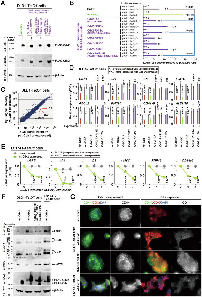
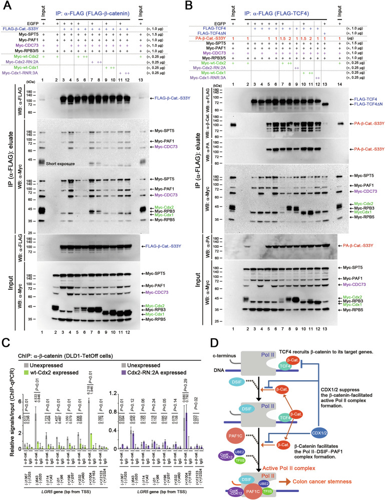
Homeobox transcription factors CDX1 and CDX2 (hereafter, CDX1/2) play key roles in determining the identity of intestinal epithelial cells and regulating their stem cell functions. However, the role of CDX1/2 in regulating colon cancer stemness and the underlying mechanisms are unclear. Here, we show that complete loss of Cdx1 or concurrent loss of Cdx1/2 increased the stemness and malignancy of intestinal tumors. Consistently, CDX1/2 reduced the expression of cancer stemness-related genes, including LGR5. CDX1/2 bound to the downstream region of the LGR5 transcription start site (TSS), a region where β-catenin also binds. Despite increased H3 acetylation and an open chromatin structure, CDX1/2 reduced the occupancy of DRB sensitivity-inducing factor (DSIF), RNA polymerase II-associated factor 1 (PAF1), and RNA polymerase II (Pol II) complexes around the LGR5 TSS. Through their homeodomains, CDX1/2 inhibited the β-catenin-facilitated formation of active Pol II complexes containing DSIF and PAF1 complexes by preventing the interaction between β-catenin and these complexes, in an additive manner. Our findings suggest that CDX1/2 cooperatively suppressed colonic tumorigenesis and cancer stemness by antagonizing β-catenin via the DSIF and PAF1 complexes. Additionally, DSIF and PAF1 complexes acted as transcriptional platforms that integrated and funneled both tumor-suppressive and oncogenic signals into the expression of genes that control colon cancer stemness.


Conflict of interest statement

Competing interests: The authors declare no competing interests. Ethical approval: All methods in this study were carried out in accordance with relevant guidelines and regulations. All animal experiments in this study were approved by the Animal Care and Use Committee of FUKUI University (approval number R04023). This study did not involve human participants or patient-derived materials; therefore, ethical approvals and informed consents were not applicable.

Figures

Fig. 1
Fig. 1. Suppression of malignant progression in colonic tumorigenesis by Cdx1 and Cdx2.
A qPCR data showing the relative expression (mean ± SD) of Cdx1, Cdx2, and Lgr5 in colonic tumor organoids derived from Apc+/− mice (compared with those in normal epithelium). P-values were calculated using a Student′s t-test (AC). Number (B) and size (C) of intestinal tumors in Apc+/−, cis-Apc+/−Cdx1+/−, and Cdx2+/−-cis-Apc+/−Cdx1+/− mice at 10–12 weeks of age (mean ± SD; n = 4–5, excluding dysplastic crypts). Hematoxylin and eosin (H&E)-stained small intestinal tumors in Apc+/− (D) and cis-Apc+/−Cdx1+/− (E) mice. Green arrows indicate invasive tumor cells (E, G). Abbreviations in DG: mm muscularis mucosa, mp muscularis propria, se serosa. Scale bars, 200 µm (DG). H&E-stained colonic tumors from cis-Apc+/−Cdx1+/− (F) and Cdx2+/−-cis-Apc+/−Cdx1+/− (G) mice.
Fig. 2
Fig. 2. Suppression of colon cancer cell stemness by CDX1 and CDX2.
A Immunoblots showing doxycycline-controlled inducible expression of FLAG-wt-Cdx2 or its homeodomain (HD) mutants in DLD1-TetOff cells. β-Actin was a loading control (A, F). B CDH17 luc reporter activities (mean ± SD) relative to those of the pGL4.10-luc2 control upon expressing wt-Cdx2 or its HD mutants. P-values were calculated using a Student′s t-test (B, D, E). C Microarray data showing the gene expression profiles of DLD1-TetOff cells after expression of wt-Cdx1 (Cy3) for 12 h (compared with those of cells not expressing wt-Cdx1; Cy5). Red arrows denote the relationship between cells with Cdx1 expression and without Cdx1 expression in terms of ID1, ID3, and c-MYC expression. qPCR data showing the relative expression (mean ± SD) of colon cancer stemness-related genes upon expressing wt-Cdx1, wt-Cdx2, or Cdx2 HD mutants for 2 days in DLD1-TetOff cells (D) and for 4 and 7 days in LS174T-TetOff cells (E), when compared with cells without their expression. F Immunoblots showing the expression of LGR5, CD44, and c-MYC upon expressing wt-Cdx1, wt-Cdx2, or Cdx2-RNR:3E in DLD1-TetOff and LS174T-TetOff cells. Note that the basal expression levels of LGR5 were different among the TetOff clones. G Immunocytochemistry showing the expression of CD44 (green) and Cdx1/2 (red) upon expression of wt-Cdx1, wt-Cdx2, or Cdx2-RNR:3E in DLD1-TetOff and LS174T-TetOff cells. The nuclei were stained with DAPI (blue). Scale bars, 20 µm.
Fig. 3
Fig. 3. Increased intestinal tumor stemness by Cdx1 and Cdx2 mutations.
A qPCR data showing the relative expression (mean ± SD) of Cdx1, Cdx2, and Lgr5 in colonic tumor organoids derived from cis-Apc+/−Cdx1+/− and Cdx2+/−-cis-Apc+/−Cdx1+/− mice (compared with those from Apc+/− mice). P-values were calculated using a Student′s t-test (AC). Growth rate (mean ± SD) of organoid cells in normal and tumor epithelial tissues derived from the small intestine (B) and colon (C) of Apc+/−, cis-Apc+/−Cdx1+/−, and Cdx2+/−-cis-Apc+/−Cdx1+/− mice. D Tumor organoids derived from Apc+/−, cis-Apc+/−Cdx1+/−, and Cdx2+/−-cis-Apc+/−Cdx1+/− colonic tumors. Scale bars, 100 µm. E, F Immunohistochemistry of Cd44 expression (brown) in a small intestinal tumor of a cis-Apc+/−Cdx1+/− mouse. F shows the magnified image of the boxed region in (E). The arrows in F indicate cells with elevated Cd44 expression, while arrowheads indicate cells with lower Cd44 expression. The tissue was also counterstained with hematoxylin (blue). Scale bars, 100 µm (E) and 20 µm (F).
Fig. 4
Fig. 4. Decreased occupancy levels of Pol II, SPT5, and PAF1 on LGR5 upon CDX2 expression.
A Integrative genome-viewer window showing the occupancy of FLAG-tagged wt-Cdx1 and wt-Cdx2 on the LGR5 gene in DLD1-TetOff cells. Genomic regions analyzed by ChIP-qPCR in B are indicated above, whereas those cloned in the LGR5 luc reporter C, D are indicated below. B ChIP-qPCR data showing the relative occupancy (mean ± SD) of endogenous CDX2 at the indicated positions of LGR5 and CDX2-target gene CDH17 (used as a positive control) in T84 cells. P-values were calculated using a Student′s t-test (BG). C LGR5 luc reporter activities (mean ± SD) relative to those of the pGL4.10-luc2 control upon expressing wt-Cdx1, wt-Cdx2, or both wt-Cdx1 and wt-Cdx2. The amounts of transfected plasmid DNA used to overexpress wt-Cdx1 and wt-Cdx2 are indicated below the bar graph. D LGR5 luc reporter activities (mean ± SD) relative to those of the pGL4.10-luc2 control upon expressing wt-Cdx2, or its homeodomain (HD) mutants, or β-catenin-S33Y. E ChIP-qPCR data showing the relative occupancy (mean ± SD) of H3K27ac and H3K4me3 at the indicated positions of LGR5 and CDH17 after expressing wt-Cdx2 in DLD1-TetOff cells for 1 day. F qPCR data quantifying MNase protection assays reflecting the chromatin architecture at the indicated positions in LGR5 after expressing wt-Cdx2 in DLD1-TetOff cells for 1 day. G ChIP-qPCR data showing the relative occupancy (mean ± SD) of Pol II, SPT5, and PAF1 at the indicated positions in LGR5 after expressing wt-Cdx2 in DLD1-TetOff cells for 1 day.
Fig. 5
Fig. 5. Suppression of Pol II–DSIF–PAF1C complex formation by CDX1/2.
A Mechanism of β-catenin-facilitated formation of the active Pol II complex through PAF1C. Step 1: TCF4 recruits β-catenin to its target genes; Step 2: β-catenin recruits the DSIF complex to Pol II; Step 3: β-catenin facilitates the formation of the Pol II–DSIF–NELF complex; Step 4: β-catenin facilitates the formation of the Pol II–DSIF–PAF1C complex; Step 5: The Pol II–DSIF–PAF1C complex forms a complex with TFIIS, UBE2, and cyclin K (CycK)-CDK12 and initiates mRNA transcription. B LGR5 luc reporter activities (mean ± SD) relative to those of the control pGL4.10-luc2 upon expressing PAF1C components, β-catenin-S33Y, and wt-Cdx2. P-values were calculated using a Student′s t-test. Immunoprecipitation (IP) assays showing the effect of wt-Cdx2 on β-catenin-S33Y (β-Cat.)-facilitated complex formation of C SPT5 (containing PAF1, TFIIS, and RPB5) and D PAF1 (containing SPT5, TFIIS, and RPB5). In the IP assays, the indicated proteins were co-expressed with either FLAG-SPT5 (C) or FLAG-PAF1 (D). The co-immunoprecipitated Myc-tagged proteins were analyzed using immunoblotting. The amounts of plasmid DNA transfected are indicated on the right. E IP assays showing the effects of wt-Cdx1, wt-Cdx2, or both wt-Cdx1 and Cdx2 on β-catenin-S33Y (β-Cat.)-facilitated formation of a complex involving TFIIS, SPT5, PAF1, and RPBs. The indicated proteins were expressed in the IP assays along with FLAG-TFIIS. The co-immunoprecipitated Myc-tagged proteins were analyzed via immunoblotting. The amounts of transfected plasmid DNA used for expressing wt-Cdx1 and wt-Cdx2 are indicated above the gel images.
Fig. 6
Fig. 6. CDX2-mediated suppression of Pol II–DSIF–PAF1C complex formation blocked by mutations of its homeodomain.
A LGR5 luc reporter activities (mean ± SD) relative to those of the pGL4.10-luc2 control upon expressing β-catenin-S33Y, PAF1C components, wt-Cdx2, and its homeodomain (HD) mutants. P-values were calculated using a Student′s t-test. IP assays showing the effects of wt-Cdx2 and its HD mutants on β-catenin-S33Y (β-Cat.)-facilitated complex formation: B SPT5-containing complexes with PAF1, TFIIS, and RPB5 or C TFIIS-containing complexes with SPT5, PAF1, and RPBs. In the IP assays, the indicated proteins were expressed along with FLAG-SPT5 (B) or FLAG-TFIIS (C).
Fig. 7
Fig. 7. Contribution of the CDX2 homeodomain to the suppression of Pol II–DSIF–PAF1C complex formation.
A LGR5 luc reporter activities (mean ± SD) relative to those of the pGL4.10-luc2 control upon expressing β-catenin-S33Y, CDC73, and wt-Cdx2 and its deletion mutants. P-values were calculated using a Student′s t-test. B IP assays showing the effects of wt-Cdx2 and its deletion mutants on the β-catenin-S33Y (β-Cat.)-facilitated formation of a complex involving TFIIS, SPT5, PAF1, and RPBs. C IP assays showing the interaction between SPT5 and Cdx2 (or its mutants) and between PAF1 and Cdx2 (or its mutants).
Fig. 8
Fig. 8. Suppression of the interaction between β-catenin and PAF1C by CDX1 and CDX2.
A IP assays showing the effects of wt-Cdx1/2 and their homeodomain (HD) mutants on the interaction of β-catenin-S33Y (β-Cat.) with SPT5, PAF1 components, and RPBs. The indicated proteins were expressed in the IP assays along with FLAG-β-catenin-S33Y. B IP assays showing the effects of wt-Cdx1/2 and their HD mutants on the interaction of FLAG-TCF4 with PA-β-catenin-S33Y (β-Cat.), Myc-tagged SPT5, PAF1 components, and RPBs. In the IP assays, the indicated proteins were expressed along with FLAG-TCF4. C ChIP-qPCR data showing the relative occupancy (mean ± SD) of β-catenin at the indicated positions in LGR5 after expressing wt-Cdx2 or Cdx2-RN:2A for 1 day in DLD1-TetOff cells, when compared with that in cells with Cdx unexpressed. P-values were calculated using a Student′s t-test. D Transcriptional mechanism underlying the inhibition of stable β-catenin by CDX1/2 via DSIF and PAF1C complexes, resulting in the suppression of colon cancer stemness. First, TCF4 recruits β-catenin to its target gene. β-Catenin then recruits DSIF and PAF1 complexes to the Pol II complex to facilitate the formation of the active Pol II complex, which promotes colon cancer stemness. CDX1/2 suppress these processes. DSIF and PAF1 complexes act as platforms that integrate and funnel oncogenic and tumor-suppressive signals into gene expression, thereby controlling cancer stemness.

References

    1. Mlodzik M, Gehring WJ. Expression of the caudal gene in the germ line of Drosophila: formation of an RNA and protein gradient during early embryogenesis. Cell. 1987;48:465–78. - PubMed
    1. Silberg DG, Swain GP, Suh ER, Traber PG. Cdx1 and cdx2 expression during intestinal development. Gastroenterology. 2000;119:961–71. - PubMed
    1. Kakizaki F, Aoki K, Miyoshi H, Carrasco N, Aoki M, Taketo MM. CDX transcription factors positively regulate expression of solute carrier family 5, member 8 in the colonic epithelium. Gastroenterology. 2010;138:627–35. - PubMed
    1. Chawengsaksophak K, James R, Hammond VE, Köntgen F, Beck F. Homeosis and intestinal tumours in Cdx2 mutant mice. Nature. 1997;386:84–87. - PubMed
    1. Stringer EJ, Duluc I, Saandi T, Davidson I, Bialecka M, Sato T, et al. Cdx2 determines the fate of postnatal intestinal endoderm. Development. 2012;139:465–74. - PMC - PubMed

MeSH terms

LinkOut - more resources