. 2025 May 21;3(1):41.
doi: 10.1038/s44259-025-00116-0.

Small-molecule screen in C. elegans identifies benzenesulfonamides as inhibitors of microsporidia spores

Affiliations

Small-molecule screen in C. elegans identifies benzenesulfonamides as inhibitors of microsporidia spores

Qingyuan Huang et al. NPJ Antimicrob Resist. .

Abstract

Microsporidia, a large group of fungal-related intracellular parasites, infect several economically significant animals, leading to substantial economic losses. As currently available anti-microsporidia therapies are either ineffective or come with numerous adverse effects, there is a need for alternative microsporidia inhibitors. Here we screen a subset of the ChemBridge DIVERset library, comprising 2500 diverse compounds, using Caenorhabditis elegans infected with its natural microsporidian parasite, Nematocida parisii. By testing these compounds at 60 μM in 96-well assay plates, we identified 26 hits that restored the ability of C. elegans to produce progeny in the presence of N. parisii. We confirmed that out of 20 tested compounds, 18 ChemBridge compounds effectively inhibit N. parisii infection in C. elegans. Of these 18, 10 were benzenesulfonamide derivatives which inhibit microsporidia infection by inactivating spores. We screened an additional 475-compound benzenesulfonamide library, successfully identifying three compounds that are effective at a lower concentration than the initial hits. We further show that one benzenesulfonamide compound displays inhibitory activity against several species of microsporidia, inhibiting infection of species belonging to the Nematocida, Enterocytozoon, and Encephalitozoon genera. Together our results suggest that benzenesulfonamides are a potential scaffold for the development of microsporidia antiseptics.


Conflict of interest statement

Competing interests: The authors declare no competing interests.

Figures
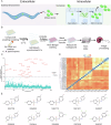
Fig. 1
Fig. 1. High-throughput screen of 2500 ChemBridge compounds identifies benzenesulfonamides which inhibit N. parisii infection of C. elegans.
A Schematic of N. parisii infection of C. elegans. The process of infection begins when the worms ingest microsporidia spores. These spores then germinate inside of the intestinal lumen of the worm, depositing the sporoplasm inside an intestinal cell. The sporoplasm then differentiates into a meront and differentiates into spores which then exit the worm. B Schematic of assay to identify microsporidia inhibitors. Compounds, microsporidia spores, and E. coli (food source of C. elegans) are incubated together and the C. elegans animals at the earliest larval stage are added. After 5 days of incubation, the worms are stained with rose bengal, imaged using a flatbed scanner, and quantified using image analysis. C Compounds at a final concentration of 60 µM were incubated with N. parisii. L1 stage C. elegans were added one hour later, cultured for five days, and progeny numbers quantified. Points represent mean progeny production of a compound as a percentage of DMSO uninfected controls. Compounds with an activity less than 40% are colored blue, compounds with an activity of at least 40% are colored red, and compound-IDs are shown. Three independent biological replicates were performed for each compound. D Clustered heat map of structural similarity of the 26 compounds with at least 40% activity. The scale indicates compound similarity with 0 (blue) being most similar and 1 (red) being least similar. Compounds tested in subsequent experiments are indicated by the dark blue dots. E Structures of benzenesulfonamides with inhibitory activity against N. parisii.
Fig. 2
Fig. 2. Validation that the identified ChemBridge compounds inhibit N. parisii.
AC N. parisii spores were incubated with compounds at a final concentration of 100 μM. One hour later, L1 stage worms were added and cultured for 4 days. Worms were then fixed and stained with DY96 to detect microsporidian spores and nematode embryos. A Representative images of continuous infection assay with the conditions DMSO uninfected (negative control), DMSO infection (positive control for infection), dexrazoxane (positive control for inhibition), and 5357859 (benzenesulfonamide compound). DAPI (top row) stains worm nuclei and DY96 (middle row) stains microsporidia spores and C. elegans embryos. Merged images (bottom row) show worms that contain microsporidia spores and/or embryos. Scale bars are 500 μm. B The percentage of animals which contain embryos. C The percentage of animals with newly formed spores. Benzenesulfonamide compounds are colored red. n = 3 biological replicates, N = ≥ 100 worms counted per biological replicate. The P values were determined by one-way ANOVA with post hoc test, with all comparisons to DMSO infected. Means ± SD (horizontal bars) are shown. (*p  < 0.05, **p  < 0.01, ****p  < 0.0001, ns means not significant).
Fig. 3
Fig. 3. Tested ChemBridge compounds do not inhibit microsporidia proliferation except for 5357859.
AF L1 stage worms were incubated for 3 h in the presence of N. parisii spores and then washed to remove undigested spores. Infected worms were incubated with compounds for 2 (D) or 4 (A–C, E) days, fixed, and stained with DY96 and a FISH probe specific to the N. parisii 18S rRNA. A Representative images of pulse infection assay with the conditions DMSO infection (positive control for infection), 5357859 (benzenesulfonamide compound), and dexrazoxane (positive control for inhibition). DAPI (top row) stains worm nuclei and FISH (middle row) stains microsporidia meronts. Merged images (bottom row) show worms that contain microsporidia meronts. B The percentage of animals which contain embryos. C The percentage of animals which contain newly formed spores. D, E Quantification of the pathogen load at 2 (D) or 4 (E) days following infection. F The percentage of animals with FISH signal. Benzenesulfonamide compounds are colored red. n = 3 biological replicates, N = ≥ 100 worms (B, C, F) or N = 10 (D, E) counted per biological replicate. The P values were determined by one-way ANOVA with post hoc test, with all comparisons to DMSO infected, except for those indicated by brackets. Means ± SD (horizontal bars) are shown. (**p < 0.01, ****p < 0.0001, ns means not significant).
Fig. 4
Fig. 4. Identified ChemBridge inhibitors prevent N. parisii invasion.
A, B After incubating N. parisii spores with compounds for 24 h, spores were washed and added to L1 stage worms. After incubation for 3 h, worms were fixed and the spores (DY96) and sporoplasms (FISH) were stained. A Percentage of empty spores in the intestinal lumen. B The mean number of sporoplasms per worm. Benzenesulfonamide compounds are colored red. (n = 3, N = ≥ 50 spores counted per biological replicate). The P values were determined by one-way ANOVA with post hoc test. Means ± SD (horizontal bars) are shown. (**p < 0.01, ***p < 0.001, ****p < 0.0001, ns means not significant).
Fig. 5
Fig. 5. Benzenesulfonamides inactivate N. parisii spores.
A, B N. parisii spores were treated with the indicated compounds for 24 h, followed by Sytox Green and Calcofluor White M2R staining. A Representative images of spore mortality assay with the conditions spore prep. (negative control), heat-treated (positive control) and 5357859 (benzenesulfonamide compound). Calcofluor White M2R (top row) stains spores and Sytox Green (middle row) stains the DNA of spores that are no longer viable. Spores that are dual stained (marked by arrowheads) in the merged image (bottom row) are counted as inviable, and those that are viable (marked by arrows) are only stained blue. Scale bars are 50 μm. B Percentage of non-viable spores. C, D N. parisii spores were incubated with the indicated compounds, acetone, or both for 24 h. Spores were then washed and stained with Sytox Green and Calcofluor White M2R (C) or mixed with L1 stage worms incubated for 3 h, fixed, and stained with DY96 and a FISH probe (D). C Percentage of non-viable spores. D Percentage of empty spores in the intestinal lumen. N = ≥ 100 spores (B, C) or N = ≥ 50 worms (D) counted per biological replicate. The P values were determined by one-way ANOVA with post hoc test. Means ± SD (horizontal bars) are shown. (*p  < 0.05, **p  < 0.01, ***p  < 0.001, ****p < 0.0001, ns means not significant).
Fig. 6
Fig. 6. Benzenesulfonamide compounds limits P. epiphaga and E. cuniculi infection.
A P. epiphaga spores were incubated with compounds at a concentration of 200 μM for 1 h and then L1 stage C. elegans were added and cultured for 4 days. Animals were then fixed and stained with DY96 and a P. epiphaga 18S rRNA FISH probe. Quantification of pathogen load. B E. cuniculi spores were incubated with 5337895, acetone, or 1% DMSO for 24 h, followed by staining with Fluorescent Brightener 28, Sytox Green, and propidium iodine. Quantification of spore mortality. C, D E. cuniculi spores were incubated with 5337895, acetone, or 1% DMSO for 24 h. Spores were then added to RK-13 cells and incubated for 3 days. C Cells were fixed, stained with Fluorescent Brightener 28 and parasitophorous vacuole number counted. D Quantitation of E. cuniculi SSU concentration. E, F RK-13 cells were incubated with E. cuniculi spores and either 5 μM dexrazoxane, 25 μM 5357859, or 200 nM albendazole for either 3 or 5 days. E Cells were fixed, stained with Fluorescent Brightener 28, and parasitophorous vacuole number counted. F Quantitation of E. cuniculi SSU concentration. Benzenesulfonamide compounds are colored red. n = 3 biological replicates, N = 10 animals (A), N = ≥ 100 spores (B), N > = 10 figure (C, E), or N = three well samples (D, F) qquantified per biological replicate. The P values were determined by one-way ANOVA (A, B) or two-way ANOVA(CF) with post hoc test. Means ± SD (horizontal bars) are shown (*p < 0.05, **p < 0.01, ****p < 0.0001, ns means not significant).
Fig. 7
Fig. 7. Screen of 475 ChemBridge benzenesulfonamide analogs identifies higher affinity N. parisii inhibitors.
A Compounds at a final concentration of 15 µM were incubated with N. parisii. L1 stage C. elegans were added one hour later and then cultured for five days. The number of worms in each well were quantified and the progeny production of each compound as a percentage of the uninfected controls is shown. Three independent biological replicates were performed for each compound. The green sulfonamide-ID represents sulfonamides that exhibited an activity of at least 40%. The red sulfonamide-ID represents the activity of the previously identified inhibitor 5357859. B The chemical structures of 5357859 and the 3 ChemBridge benzenesulfonamides with higher affinity.
Fig. 8
Fig. 8. Higher affinity benzenesulfonamides inhibit N. parisii infection.
AC, EG, IK N. parisii spores were incubated with the indicated benzenesulfonamides at different concentrations for 1 h. L1 stage C. elegans either without spore or mixed with the treated spores were cultured for four days. Worms were then fixed and stained with DY96. Effect of the indicated benzenesulfonamides at 15 µM (AC), 60 µM (EG) or 100 µM (IK) on the percentage of worms with embryos in uninfected worms (A, E, I), infected worms (B, F, J) and the percentage of worms with newly formed spores (C, F, K). N. parisii spores were incubated with 15 µM (D) 60 µM (H) and 100 µM (L) compound for 24 h and stained with Sytox Green and Calcofluor White M2R. The percentage of spores that showed Sytox Green staining. n = 3, N = ≥ 100 worms (AC, EG, IK) or N = ≥ 100 spores (D, H, L) counted per biological replicate). The P values were determined by one-way ANOVA with post hoc test. Means ± SD (horizontal bars) are shown. (*p < 0.05, **p < 0.01, ***p < 0.001, ****p < 0.0001, ns means not significant).

Similar articles

Cited by

References

    1. Han, B. & Weiss, L. M. Microsporidia: obligate intracellular pathogens within the fungal kingdom. Microbiol. Spectr.5, 5.2.03 (2017). - PMC - PubMed
    1. Keeling, P. J. & Fast, N. M. Microsporidia: biology and evolution of highly reduced intracellular parasites. Annu. Rev. Microbiol.56, 93–116 (2002). - PubMed
    1. Willis, A. R. & Reinke, A. W. Factors that determine microsporidia infection and host specificity. Exp. Suppl. 2012114, 91–114 (2022). - PubMed
    1. Vossbrinck, C. R., Andreadis, T. G. & Weiss, L. M. Phylogenetics: taxonomy and the microsporidia as derived fungi. in Opportunistic Infections: Toxoplasma, Sarcocystis, and Microsporidia (eds. Lindsay, D. S. & Weiss, L. M.) vol. 9 189–213 (Springer, 2004).
    1. Han, B., Takvorian, P. M. & Weiss, L. M. Invasion of host cells by microsporidia. Front. Microbiol. 11, 172 (2020). - PMC - PubMed

Grants and funding

LinkOut - more resources