. 2025 May 21;20(1):61.
doi: 10.1186/s13062-025-00651-w.

Mechanism of LINC01018/miR-182-5p/Rab27B in the immune escape through PD-L1-mediated CD8+ T cell suppression in glioma

Affiliations

Mechanism of LINC01018/miR-182-5p/Rab27B in the immune escape through PD-L1-mediated CD8+ T cell suppression in glioma

Su Hu et al. Biol Direct. .

Abstract

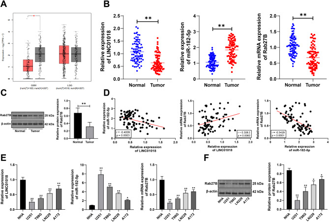
Background: Glioma is a malignant tumor associated with poorer prognosis. This study aims to elucidate the mechanism of LINC01018/miR-182-5p/Rab27B axis in PD-L1-mediated CD8+ T cell suppression in the progression of gliomas.

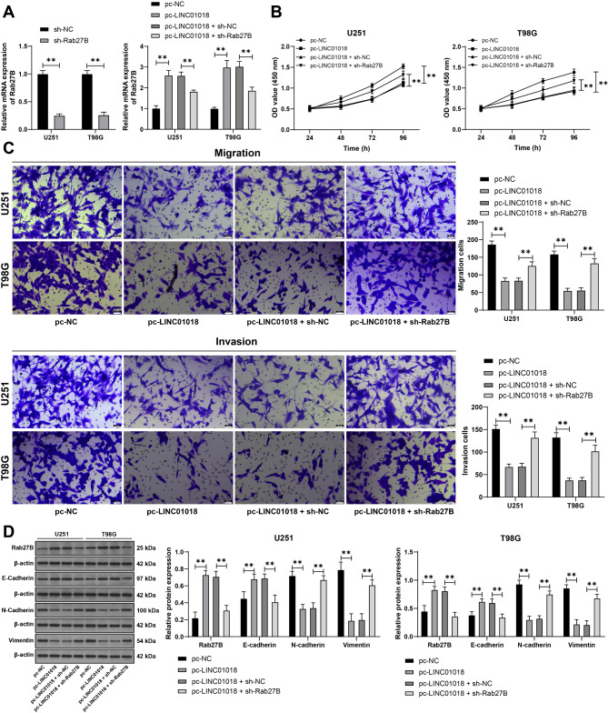
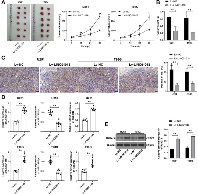
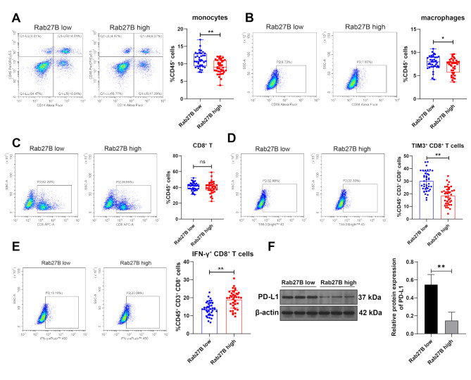
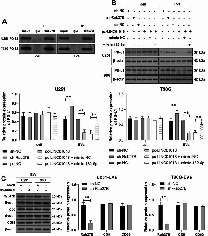
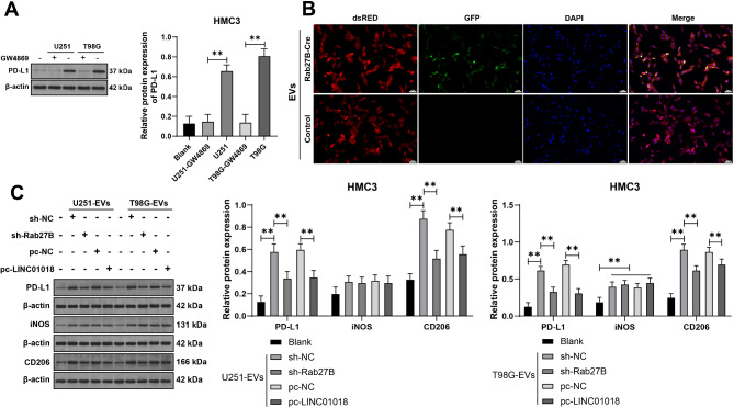
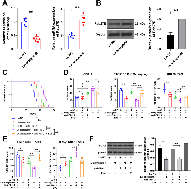
Methods: LINC01018, miR-182-5p, and Rab27B expression levels in glioblastoma tissues were measured. The proportion of infiltrating macrophages and monocytes and CD8+ T cell function were assessed. The relationship between miR-182-5p and Rab27B was analyzed. Glioma cell activity, invasion, and migration were measured. The expression of E-cadherin, N-cadherin, Vimentin, PD-L1, iNOS, and CD206 was determined. Glioma cell-derived EVs were isolated, and the co-localization of Rab27B and PD-L1 and the binding of Rab27B to PD-L1 were analyzed. The endocytosis of EVs by microglia was assayed. The impact of LINC01018/miR-182-5p/Rab27B on glioma growth was observed. The function of macrophages and CD8+ T cells in tumors was analyzed.

Results: Rab27B was downregulated, and infiltrating macrophages and monocytes were increased in glioblastoma. miR-182-5p inhibited Rab27B expression. Rab27B knockdown reverses the inhibitory effect of LINC01018 overexpression on glioma cell growth. Glioma cells-derived EVs with low Rab27B expression carried more PD-L1 to increase PD-L1 expression and M2 polarization in microglia. LINC01018 overexpression reduced macrophages in orthotopic tumors. CD8+ T cell numbers showed no significant difference, but TIM-3 increased and IFN-γ decreased. miR-182-5p inhibition enhanced the therapeutic effect of anti-PD-L1, which was reversed after glioma cell-derived EVs.

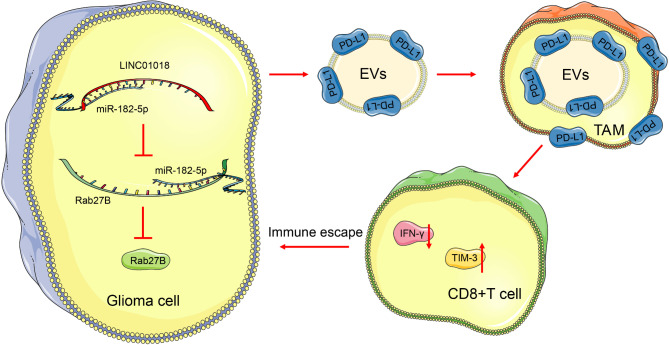
Conclusion: LINC01018 promotes PD-L1-mediated CD8+ T cell suppression via the miR-182-5p/Rab27B axis in glioma cell-derived EVs, thereby contributing to immune escape in gliomas.

Keywords: EVs; Glioma; Immune escape; LINC01018; Rab27B; miR-182-5p.


Conflict of interest statement

Declaration. Ethics approval and consent to participate: The study protocol adhered to the ethical guidelines of ethics committee of Guangzhou Institute of Cancer Research, the Affiliated Cancer Hospital, Guangzhou Medical University and the Declaration of Helsinki. All animal experiments were approved by the ethics committee of Guangzhou Institute of Cancer Research, the Affiliated Cancer Hospital, Guangzhou Medical University and conducted in accordance with the Guide for the Care and Use of Laboratory Animals [16] Consent for publication: Not applicable. Competing interests: The authors declare no competing interests.

Figures

Fig. 1
Fig. 1
Rab27B expression is reduced in glioma tissues. A: Rab27B expression in GBM (Glioblastoma multiforme) and LGG (Brain Lower Grade Glioma) was predicted online using the GEPIA2 database (http://gepia2.cancer-pku.cn/#index) [34]. B: qRT-PCR was performed to detect the expression of LINC01018, miR-182-5p, and Rab27B in tumor tissues and adjacent normal tissues from 78 glioblastoma patients. C: Western blot analysis was conducted to assess the expression of Rab27B in tumor tissues and adjacent normal tissues from 78 glioblastoma patients, with representative band images shown. D: Pearson correlation analysis was performed to evaluate the correlation between the expressions of LINC01018, miR-182-5p, and Rab27B. E: qRT-PCR was used to measure the expression of LINC01018, miR-182-5p, and Rab27B in NHA and human glioma cell lines (U251, T98G, LN229, A172). with cell experiment repeated three times. F: Western blot analysis was employed to determine the expression of Rab27B in NHA and human glioma cell lines (U251, T98G, LN229, A172), with the experiment repeated three times. In panels B and C, ** p < 0.01; In panels E and F, * versus NHA, p < 0.05, ** versus NHA, p < 0.01. Data comparison between two groups in panels B and C was analyzed using t-test, and multiple group comparison in panels E and F was analyzed using one-way ANOVA, followed by Tukey’s multiple comparisons test
Fig. 2
Fig. 2
LINC01018/miR-182-5p targets Rab27B expression. A: Dual-luciferase reporter assays were conducted to investigate the binding relationship between miR-182-5p and Rab27B. B-C: The expression of Rab27B in U251 and T98G cells under different treatments was assessed using qRT-PCR and Western blot. The cell experiments were repeated three times. ** p < 0.01. Comparison of data was analyzed using two-way ANOVA, followed by Tukey’s multiple comparisons test
Fig. 3
Fig. 3
Rab27B inhibition reverses the inhibitory effects of overexpressed LINC01018 on proliferation and metastasis of glioma cells. sh-Rab27B was transfected into U251 and T98G cells, with sh-NC as a control, followed by combined experiments with LINC01018 overexpression. A: qRT-PCR was performed to detect the expression of Rab27B in the cells. B: Cell viability was assessed using CCK-8 assay. C: Transwell assay was used to evaluate cell invasion and migration. D: Western blot analysis was conducted to measure the expression of Rab27B, E-cadherin, N-cadherin, and Vimentin in the cells. The cell experiments were repeated three times. ** p < 0.01. Comparison of data was analyzed using two-way ANOVA, followed by Tukey’s multiple comparisons test
Fig. 4
Fig. 4
Overexpression of LINC01018 suppresses glioma growth via the miR-182-5p/Rab27B axis. Glioma cells (U251, T98G) with stable overexpressed LINC01018 (Lv-LINC01018) was generated by lentiviral infection, with Lv-NC as the control. These stably expressing cells were subcutaneously injected into nude mice. A: Tumor volume was measured every 7 days, and the tumors were harvested after 28 days when the mice were euthanized. N = 12. B: Tumor weight was recorded. N = 12. C: IHC was performed to assess the expression of ki67 in the tumors. N = 6. D: qRT-PCR was used to detect the expression of LINC01018, miR-182-5p, and Rab27B in the tumors. N = 6. E: Western blot analysis was conducted to measure the expression of Rab27B in the tumors. N = 6. ** p < 0.01. Comparison of data in panels A, B, C, and E was analyzed using two-way ANOVA, followed by Tukey’s multiple comparisons test; data in panel D were analyzed using t-test
Fig. 5
Fig. 5
Rab27B is involved in immune evasion of glioma cells. Glioblastoma patients were divided into high expression group (n = 39) and low expression group (n = 39) based on the expression of Rab27B. A-C: The proportions of monocytes (CD45+CD14+), macrophages (CD45+CD14+CD68+), and CD8+ T cells (CD45+CD3+CD8+) in each glioma tissue were analyzed by flow cytometry. D-E: The functional status of CD8+ T cells was analyzed by flow cytometry. F: Western blot was performed to detect the expression of PD-L1 in the tissues. ns p > 0.05, * p < 0.05, ** p < 0.01. Data were analyzed using t-test, with representative band images shown
Fig. 6
Fig. 6
The LINC01018/miR-182-5p/Rab27B axis is involved in the transport of PD-L1 in glioma cell-derived EVs. A: Immunoprecipitation was performed to detect the binding of Rab27B to PD-L1 in cells and EVs. B: Western blot was conducted to measure the expression of PD-L1 in cells and EVs. C: Western blot was used to detect the expression of Rab27B, CD9, and CD63 in EVs. The cell experiments were repeated three times. **p < 0.01. Data in panels B and C were analyzed using two-way ANOVA, followed by Tukey’s multiple comparisons test
Fig. 7
Fig. 7
Glioma cell-derived EVs delivery PD-L1 and increase PD-L1 expression in microglial cells. A: Glioma cells (U251, T98G) with or without GW4869 were co-cultured with human microglial cells HMC3, and the expression of PD-L1 in HMC3 cells after co-culture was detected by Western blot. B: The CreLoxP system was used to evaluate the delivery of EV cargo from glioma cells to microglial cells. C: Western blot was used to detect the expression of PD-L1, iNOS and CD206 in HMC3 cells treated with different EVs. The cell experiments were repeated three times. ** p < 0.01. Data were analyzed by two-way ANOVA, followed by Tukey's multiple comparisons test.
Fig. 8
Fig. 8
Rab27B mediates glioma cell-derived EVs to regulate PD-L1 transfer and promotes immune escape in vivo. The GL261 cells with stable low expression of miR-182-5p (Lv-antagomiR) were injected into mice, with Lv-NC serving as the control. Anti-PD-L1 treatment was administered every 3 days along with intravenous injection of glioma cell-derived EVs. A: qRT-PCR was conducted to detect the expression of miR-182-5p and Rab27B in the tumor, n = 6; B: Western blot was performed to assess Rab27B expression in the tumor, n = 6; C: Survival status of mice in different treatment groups was recorded, n = 12; D: Flow cytometry was used to determine the proportions of CD8+ T cells (CD45+CD8+), macrophages (CD45+F4/80+CD11b+), and TAM (F4/80+ CD11b+ CD206+) in the tissues, n = 6; E: The functional status of CD8+ T cells was analyzed by flow cytometry, n = 6; F: Western blot was utilized to measure PD-L1 expression in the tumor, n = 6; ns p > 0.05, * p < 0.05, ** p < 0.01. Data in panels A and B were analyzed by t-test; data in panels D, E, and F were analyzed by one-way ANOVA analysis, followed by Tukey’s multiple comparisons test
Fig. 9
Fig. 9
LINC01018 is downregulated in glioma cells. LINC01018 promotes the transport of PD-L1-carrying EVs into microglia and increases PD-L1 expression through the miR-182-5p/Rab27B axis, inhibiting CD8+ T cell function and promoting immune evasion in glioma cells

Similar articles

References

    1. Weller M, Wen PY, Chang SM, Dirven L, Lim M, Monje M, Reifenberger G, Glioma. Nat Rev Dis Primers. 2024;10(1):33. - PubMed
    1. Xu S, Tang L, Li X, Fan F, Liu Z. Immunotherapy for glioma: current management and future application. Cancer Lett. 2020;476:1–12. - PubMed
    1. Yasinjan F, Xing Y, Geng H, Guo R, Yang L, Liu Z, Wang H. Immunotherapy: a promising approach for glioma treatment. Front Immunol. 2023;14:1255611. - PMC - PubMed
    1. Wang C, Chen Q, Chen M, Guo S, Hou P, Zou Y, Wang J, He B, Zhang Q, Chen L, et al. Interaction of glioma-associated microglia/macrophages and anti-PD1 immunotherapy. Cancer Immunol Immunother. 2023;72(6):1685–98. - PMC - PubMed
    1. Bridges MC, Daulagala AC, Kourtidis A. LNCcation: LncRNA localization and function. J Cell Biol. 2021;220(2). - PMC - PubMed

MeSH terms