Serum IgA levels and survival in patients with idiopathic pulmonary fibrosis: association with serum cytokine levels and peripheral monocyte counts
- PMID: 40400959
- PMCID: PMC12090133
- DOI: 10.21037/jtd-2024-2142
Serum IgA levels and survival in patients with idiopathic pulmonary fibrosis: association with serum cytokine levels and peripheral monocyte counts
Abstract
Background: Idiopathic pulmonary fibrosis (IPF) is an idiopathic fibrotic interstitial lung disease with poor prognosis. Recently, the prognostic value of serum platelet-derived growth factor (PDGF) levels in patients with IPF has been clarified. Monocyte counts in the peripheral blood have also been reported to be an important predictor of survival in IPF. This study aimed to clarify the prognostic value of serum immunoglobulin (Ig) A levels in patients with IPF to predict survival and occurrence of acute exacerbations (AE).
Methods: This retrospective study included 71 patients diagnosed with IPF based on the 2022 guidelines. Serum PDGF and interleukin (IL)-10 levels were measured using the Bio-Plex method. IgA levels were measured by a clinical testing company.
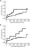
Results: Of the enrolled patients, 59 were male, and the median age of the sample was 67 [interquartile range (IQR): 61-72] years. The median serum IgA level was 307 (IQR: 232-408) mg/dL and 18 patients had serum IgA levels of >400 mg/dL. Univariate Cox proportional hazard regression analysis revealed that high IgA levels (>400 mg/dL) were a significant predictor of poor prognosis; however, monocyte counts were not. A high IgA level was a significant prognostic factor after adjusting for the percent predicted value of forced vital capacity, age, gender, and body mass index. Serum PDGF levels tended to be higher in patients with high IgA levels than in those with low IgA levels. IL-10 was not significantly correlated with IgA levels; however, IgA levels tended to be negatively correlated with monocyte counts. High IgA levels did not significantly predict AE. High monocyte counts (>600/µL) significantly predicted the early incidence of AE by univariate Cox analysis but was not confirmed by multivariate analysis. However, monocyte counts, and a monocyte count of >600/µL were significant predictors of AE occurrence for patients with low IgA ≤400 mg/dL.
Conclusions: The serum IgA level is an independent prognostic predictor of survival in patients with IPF. Serum IgA levels might suggest serum fibrogenic cytokine levels. Serum IgA levels might be associated with prognosis differently from peripheral monocyte counts. The pathophysiological role of IgA needs to be elucidated in future studies.
Keywords: Idiopathic pulmonary fibrosis (IPF); acute exacerbation (AE); interleukin-10 (IL-10); platelet-derived growth factor (PDGF); prognosis.
Copyright © 2025 AME Publishing Company. All rights reserved.
Conflict of interest statement
Conflicts of Interest: All authors have completed the ICMJE uniform disclosure form (available at https://jtd.amegroups.com/article/view/10.21037/jtd-2024-2142/coif). T.A. received lecture fees from Boehringer Ingelheim, Shionogi, AstraZeneca, and Sekisui Medical and funds from Sekisui Medical and Sysmex for activities not connected to the submitted work. The other authors have no conflicts of interest to declare.
Figures


Similar articles
-
Interleukin-11 in idiopathic pulmonary fibrosis: predictive value of prognosis and acute exacerbation.J Thorac Dis. 2023 Feb 28;15(2):300-310. doi: 10.21037/jtd-22-876. Epub 2023 Jan 10. J Thorac Dis. 2023. PMID: 36910057 Free PMC article.
-
Platelet-derived growth factor can predict survival and acute exacerbation in patients with idiopathic pulmonary fibrosis.J Thorac Dis. 2022 Feb;14(2):278-294. doi: 10.21037/jtd-21-1418. J Thorac Dis. 2022. PMID: 35280478 Free PMC article.
-
Prognostic value of IFN-γ, sCD163, CCL2 and CXCL10 involved in acute exacerbation of idiopathic pulmonary fibrosis.Int Immunopharmacol. 2019 May;70:208-215. doi: 10.1016/j.intimp.2019.02.039. Epub 2019 Mar 6. Int Immunopharmacol. 2019. PMID: 30851700
-
Serum Soluble Toll-Like Receptor 4 is a Predictive Biomarker for Acute Exacerbation and Prognosis of Idiopathic Pulmonary Fibrosis: A Retrospective Study.Lung. 2025 Mar 12;203(1):43. doi: 10.1007/s00408-025-00800-y. Lung. 2025. PMID: 40074958 Free PMC article.
-
Acute exacerbation of idiopathic pulmonary fibrosis: lessons learned from acute respiratory distress syndrome?Crit Care. 2018 Mar 23;22(1):80. doi: 10.1186/s13054-018-2002-4. Crit Care. 2018. PMID: 29566734 Free PMC article. Review.
References
LinkOut - more resources
Full Text Sources
Miscellaneous
