. 2025 Jul;13(7):e0305724.
doi: 10.1128/spectrum.03057-24. Epub 2025 May 22.

Gut opportunistic pathogens contribute to high-altitude pulmonary edema by elevating lysophosphatidylcholines and inducing inflammation

Affiliations

Gut opportunistic pathogens contribute to high-altitude pulmonary edema by elevating lysophosphatidylcholines and inducing inflammation

Xianduo Sun et al. Microbiol Spectr. 2025 Jul.

Abstract

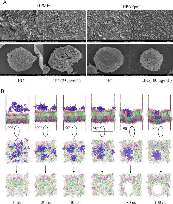
Gut microbiota have been found to promote hypoxia-induced intestinal injury. However, the role of gut microbiota in high-altitude pulmonary edema (HAPE), the preventive effect of synbiotic on HAPE, and the mechanisms by which they might work remain unknown. In this study, we aimed to investigate the role of gut microbiota in the pathogenesis of HAPE and to explore the underlying mechanisms involved. We performed a fecal microbiome analysis and found a significant decrease in intestinal Klebsiella and Escherichia-Shigella, along with a notable increase in intestinal Bifidobacterium and Lactobacillus, as volunteers recovered from acute mountain sickness (AMS). Gavage colonization with Klebsiella pneumoniae and Escherichia coli induced plasma inflammation, increased plasma lysophosphatidylcholine (LPC) levels, and contributed to HAPE in rats at a simulated altitude of 6,500 m. Conversely, a synbiotic containing Bifidobacterium, Lactiplantibacillus, fructooligosaccharides, and isomaltose-oligosaccharides significantly reduced the severity of HAPE. Cellular experiments and molecular dynamics simulations revealed that LPCs can cause damage and permeability to human pulmonary microvascular endothelial cell (HPMEC) and human pulmonary alveolar epithelial cell (HPAEpiC) monolayers under hypoxic conditions by disrupting cell membrane integrity. In addition, tail vein injection of LPCs promoted HAPE in mice at a simulated altitude of 6,500 m. In conclusion, this study describes a gut microbiota-LPCs/inflammation-HAPE axis, an important new insight into HAPE that will help open avenues for prevention and treatment approaches.

Importance: The role of the gut microbiota in high-altitude pulmonary edema (HAPE) is currently unknown. This study found that intestinal Klebsiella pneumoniae and Escherichia coli contribute to HAPE by inducing inflammation and increasing lysophosphatidylcholine (LPC) levels under hypoxic conditions. Conversely, a synbiotic containing Bifidobacterium, Lactiplantibacillus, fructooligosaccharides, and isomaltose-oligosaccharides significantly reduced the severity of HAPE. Further investigation revealed that LPCs can cause damage and permeability to human pulmonary microvascular endothelial cell (HPMEC) and human pulmonary alveolar epithelial cell (HPAEpiC) monolayers under hypoxic conditions by disrupting cell membrane integrity. These findings contribute to the understanding of the pathogenesis of HAPE and will benefit populations living at high altitude or traveling from low to high altitude.

Keywords: gut microbiota; high-altitude pulmonary edema; inflammation; lysophosphatidylcholines.


Conflict of interest statement

The authors declare no conflict of interest.

Figures

Fig 1
Fig 1
Hypoxia alters the human gut microbiota. (A, B) Pan/Core analysis. The Pan species curve (A) and Core species curve (B) illustrate the changes in total and core species as the sample size increases. The flatness of the pan/core species curve indicates the adequacy of the sequencing sample size. (C, D) Rarefaction curves based on the Shannon index (C) and the Chao1 index (D) offer valuable insights into the sufficiency of the sequencing data. A leveling off of the curve suggests that the sequencing data are sufficient. (E, F) Comparison of gut microbiota alpha diversity among all groups using the Shannon index (diversity) (E) and Chao1 index (richness) (F). (G) PLS-DA of all groups. Each point represents one sample, and points with the same color and shape belong to the same group. (H, I) Comparison of the MDI between the LG and HG groups (H), as well as between the HA and HR groups (I). The MDI serves as a metric to assess the degree of microbial imbalance, with higher values indicating a greater extent of bacterial disturbance. (J) The species composition at the genus level is illustrated in the bar chart, which displays the top 25 genera based on their relative abundance. (K, L) Results of LEfSe analysis showing significantly differentiated bacterial taxa between different groups. The color red indicates a significant increase in the HA (K) and LG (L) groups, while blue indicates a significant increase in the HR (K) and HG (L) groups. Statistical analysis was performed using one-way ANOVA (E), Kruskal-Wallis test (F), or Student’s t-test (H, I). **P < 0.01, ***P < 0.001; “ns” indicates no significance. Data are means ± SD.
Fig 2
Fig 2
E. coli and K. pneumoniae contribute to HAPE under hypoxic conditions. (A, B) Taxonomic cladogram obtained from LEfSe analysis displaying significantly differentiated bacterial taxa. Red indicates a significant increase in the NC group (A, B); blue indicates a significant increase in the HC group (A, B); green indicates a significant increase in the NKE group (A) or the HKE group (B). Nodes labeled S1 and S2 represent species K. pneumoniae and E. coli (A, B), respectively (n = 6). (C) Results of LEfSe analysis showing significantly differential bacterial taxa between the HKE and HKE+S groups (n = 6). Red indicates a significant increase in the HKE group, while blue indicates a significant increase in the HKE+S group. (D–G) Comparisons of body weight (D), food intake (E), water intake (F), and lung water content (G) between different treatment groups (n = 8). (H) H&E staining of lung tissues. Bar = 100 µm, magnification 200×. Red arrows indicate inflammatory exudation; black arrows indicate hemorrhage. (I) Semi-quantitative histopathological score of lung injury (n = 4). Statistical analysis was performed by one-way ANOVA. *P < 0.05, **P < 0.01, ***P < 0.001 (D–G and I). Data are means ± SD.
Fig 3
Fig 3
E. coli and K. pneumoniae induced inflammation during HAPE under hypoxic conditions. (A–E) Comparisons of TNF-α (A), IL-1β (B), IL-6 (C), IL-17 (D), and MCP-1 (E) levels in rat plasma between different groups (n = 7). (F) KEGG pathway enrichment analyses using the differentially expressed genes between the HKE and HC groups, and heat maps showing the differentially expressed genes in the chemokine signaling pathway, TNF signaling pathway, and lipid and atherosclerosis pathway. Genes labeled a, b, and c represent differential expression in the TNF signaling pathway, chemokine signaling pathway, and lipid and atherosclerosis pathway, respectively. (G) KEGG pathway enrichment analyses using the differentially expressed genes between the HKE and NC groups, and heat maps showing the differentially expressed genes in the lipid and atherosclerosis pathway. Statistical analysis was performed by one-way ANOVA (A, B, and E) or Kruskal-Wallis test (C, D). *P < 0.05, **P < 0.01, ***P < 0.001. Data are means ± SD.
Fig 4
Fig 4
E. coli and K. pneumoniae disrupt lipid metabolism under hypoxic conditions. (A, C) PLS-DA between the HC and HKE groups (A), as well as between the HC and HKE+S groups (C). Each point represents one sample, and points of the same color belong to the same group. (B, D) Volcano plot of differential metabolites between HKE and HC (B), as well as between HKE+S and HC (D). (E) The heat map displays the pairwise Spearman’s correlation matrix of the 12 differential LPCs between the HKE and HC groups. The color gradient represents Spearman’s correlation coefficients, while *, **, and *** indicate significant correlations at P < 0.05, P < 0.01, and P < 0.001, respectively. The relationships of the differential LPCs with E. coli and K. pneumoniae were assessed using Spearman correlation analysis. The width of the borders reflects the Spearman’s r statistic, and the color of the borders indicates statistical significance. (F) KEGG pathway-based differential abundance (DA) scoring between the HC and HKE groups. The horizontal axis represents the DA score, while the vertical axis indicates the names of the KEGG pathways. The size of each dot reflects the number of differential metabolites associated with that pathway; larger dots signify a greater quantity of differential metabolites. When the dots are placed to the right of the central axis, the longer the line segment, the more the overall expression of the pathway tends to be upregulated. Conversely, when the dots are placed to the left of the central axis, the longer the line segment, the more the overall expression of the pathway tends to be downregulated.
Fig 5
Fig 5
LPCs cause damage and permeability to HPMEC and HPAEpiC monolayer. (A) HPMEC and HPAEpiC monolayers were treated with different concentrations of LPCs, and cell viability was assessed (n = 4). (B) HPMEC and HPAEpiC monolayers were treated with different concentrations of PEs, and cell viability was determined (n = 4). (C) HPMEC and HPAEpiC monolayers were treated with different concentrations of LPCs, and permeability was determined (n = 3). (D) HPAEpiCs were treated with 100 µg/mL LPCs, either with or without various inhibitors, and cell viability was assessed (n = 4). (E) HPMECs were treated with 100 µg/mL LPCs, with or without different inhibitors, and cell viability was evaluated (n = 4). (F) HPMEC and HPAEpiC monolayers were treated with different concentrations of LPCs, and LDH release assay was performed (n = 4). Statistical analysis was performed by Student’s t-test (A, C through F). Kruskal-Wallis test was applied to compare the HC group with the 1.56 µg/mL LPC group (HPAEpiCs), while the Student’s t-test was utilized for the remaining comparisons (B). *P < 0.05, **P < 0.01, ***P < 0.001; “ns” indicates no significance. Data are means ± SD.
Fig 6
Fig 6
LPCs disrupt the cell membranes. (A) SEM images of HPMECs and HPAEpiCs after different treatments. (B) Molecular dynamics simulation of the interaction process between LPCs and cell membranes. The cell membrane is composed of a variety of lipids represented by different colors: green for CHL1, white for PSM, pink for POPC, cyan for POPS, and purple for POPE. LPC molecules are shown in blue and are randomly distributed in the upper part of the membrane. Water molecules are shown in a cotton-like transparent shape that surrounds the outside of the entire system.
Fig 7
Fig 7
LPCs promote HAPE in mice under hypoxic conditions. (A) Lung water content across different treatment groups was analyzed (n = 8). (B) H&E staining of lung tissues. Bar = 200 µm, magnification 100×. Red arrows indicate inflammatory exudation; green arrows denote the accumulation of white blood cells against the walls of blood vessels; and black arrows indicate interstitial pulmonary edema. (C) Semi-quantitative histopathological score of lung injury (n = 3). Statistical analysis was conducted using either the Student’s t-test (A) or the Mann-Whitney U test (C). *P < 0.05, **P < 0.01; “ns” indicates no significance. Data are presented as means ± SD.

Similar articles

References

    1. Liu Y, Feng X, Tang Y, Sun Y, Pu X, Feng X. 2023. Clinical characteristics of venous thromboembolism onset from severe high-altitude pulmonary edema in plateau regions. Thromb J 21:22. doi: 10.1186/s12959-023-00469-4 - DOI - PMC - PubMed
    1. Netzer N, Strohl K, Faulhaber M, Gatterer H, Burtscher M. 2013. Hypoxia-related altitude illnesses. J Travel Med 20:247–255. doi: 10.1111/jtm.12017 - DOI - PubMed
    1. Hultgren HN, Wilson R, Kosek JC. 1997. Lung pathology in high-altitude pulmonary edema. Wilderness Environ Med 8:218–220. doi: 10.1580/1080-6032(1997)008[0218:lpihap]2.3.co;2 - DOI - PubMed
    1. Luks AM, Beidleman BA, Freer L, Grissom CK, Keyes LE, McIntosh SE, Rodway GW, Schoene RB, Zafren K, Hackett PH. 2024. Wilderness medical society clinical practice guidelines for the prevention, diagnosis, and treatment of acute altitude illness: 2024 update. Wilderness Environ Med 35:2S–19S. doi: 10.1016/j.wem.2023.05.013 - DOI - PubMed
    1. Bärtsch P, Swenson ER. 2013. Acute high-altitude illnesses. N Engl J Med 369:1666–1667. doi: 10.1056/NEJMc1309747 - DOI - PubMed

MeSH terms

Substances

Supplementary concepts

LinkOut - more resources