. 2025 May 21;113(10):1507-1524.e17.
doi: 10.1016/j.neuron.2025.04.035.

Enhancer AAV toolbox for accessing and perturbing striatal cell types and circuits

Avery C Hunker  1 Morgan E Wirthlin  1 Gursajan Gill  2 Nelson J Johansen  1 Marcus Hooper  1 Victoria Omstead  1 Sara Vargas  1 M Nathaly Lerma  1 Naz Taskin  1 Natalie Weed  1 William D Laird  1 Yemeserach M Bishaw  1 Jacqueline L Bendrick  2 Bryan B Gore  1 Yoav Ben-Simon  1 Ximena Opitz-Araya  1 Refugio A Martinez  1 Sharon W Way  1 Bargavi Thyagarajan  1 Sven Otto  1 Raymond E A Sanchez  1 Jason R Alexander  1 Avalon Amaya  3 Adam Amster  1 Joel Arbuckle  1 Angela Ayala  1 Pam M Baker  1 Tyler Barcelli  1 Stuard Barta  1 Darren Bertagnolli  1 Cameron Bielstein  1 Prajal Bishwakarma  1 Jessica Bowlus  1 Gabriella Boyer  1 Krissy Brouner  1 Brittny Casian  1 Tamara Casper  1 Anish Bhaswanth Chakka  1 Rushil Chakrabarty  1 Peter Chong  1 Michael Clark  1 Kaity Colbert  1 Scott Daniel  1 Tim Dawe  1 Maxwell Departee  1 Peter DiValentin  1 Nicholas P Donadio  1 Nadezhda I Dotson  1 Deepanjali Dwivedi  1 Tom Egdorf  1 Tim Fliss  1 Amanda Gary  1 Jeff Goldy  1 Conor Grasso  3 Erin L Groce  1 Kathryn Gudsnuk  1 Warren Han  3 Zeb Haradon  1 Sam Hastings  1 Olivia Helback  1 Windy V Ho  1 Cindy Huang  1 Tye Johnson  3 Danielle L Jones  1 Zoe Juneau  1 Jaimie Kenney  3 Madison Leibly  1 Su Li  1 Elizabeth Liang  1 Henry Loeffler  3 Nicholas A Lusk  1 Zachary Madigan  1 Jessica Malloy  1 Jocelin Malone  1 Rachel McCue  1 Jose Melchor  1 John K Mich  1 Skyler Moosman  1 Elyse Morin  1 Robyn Naidoo  3 Dakota Newman  1 Kiet Ngo  1 Katrina Nguyen  3 Aaron L Oster  1 Ben Ouellette  3 Alana A Oyama  1 Nick Pena  1 Trangthanh Pham  1 Elliot Phillips  1 Christina Pom  1 Lydia Potekhina  1 Shea Ransford  1 Patrick L Ray  1 Melissa Reding  1 Dean F Rette  1 Cade Reynoldson  1 Christine Rimorin  1 Ana Rios Sigler  1 Dana B Rocha  1 Kara Ronellenfitch  1 Augustin Ruiz  1 Lane Sawyer  1 Josh P Sevigny  1 Nadiya V Shapovalova  1 Noah Shepard  1 Lyudmila Shulga  1 Sherif Soliman  1 Brian Staats  1 Michael J Taormina  1 Michael Tieu  1 Yimin Wang  1 Josh Wilkes  3 Toren Wood  1 Thomas Zhou  1 Ali Williford  3 Nick Dee  1 Tyler Mollenkopf  1 Lydia Ng  1 Luke Esposito  1 Brian E Kalmbach  4 Shenqin Yao  1 Jeanelle Ariza  1 Forrest Collman  1 Shoaib Mufti  1 Kimberly Smith  1 Jack Waters  1 Ina Ersing  5 Marcella Patrick  5 Hongkui Zeng  1 Ed S Lein  6 Yoshiko Kojima  7 Greg Horwitz  8 Scott F Owen  2 Boaz P Levi  1 Tanya L Daigle  4 Bosiljka Tasic  1 Trygve E Bakken  1 Jonathan T Ting  9
Affiliations

Enhancer AAV toolbox for accessing and perturbing striatal cell types and circuits

Avery C Hunker et al. Neuron. .

Abstract

We present an enhancer-AAV toolbox for accessing and perturbing striatal cell types and circuits. Best-in-class vectors were curated for accessing major striatal neuron populations including medium spiny neurons (MSNs), direct- and indirect-pathway MSNs, Sst-Chodl, Pvalb-Pthlh, and cholinergic interneurons. Specificity was evaluated by multiple modes of molecular validation, by three different routes of virus delivery, and with diverse transgene cargos. Importantly, we provide detailed information necessary to achieve reliable cell-type-specific labeling under different experimental contexts. We demonstrate direct pathway circuit-selective optogenetic perturbation of behavior and multiplex labeling of striatal interneuron types for targeted analysis of cellular features. Lastly, we show conserved in vivo activity for exemplary MSN enhancers in rats and macaques. This collection of striatal enhancer AAVs offers greater versatility compared to available transgenic lines and can readily be applied for cell type and circuit studies in diverse mammalian species beyond the mouse model.

Keywords: Pvalb; Sst-Chodl; basal ganglia; caudoputamen; cholinergic; macaque; medium spiny neuron; spiny projection neuron; striatonigral; striatopallidal.


Conflict of interest statement

Declaration of interests B.P.L., B.B.G., J.K.M., and E.S.L. are founders and board members of EpiCure Therapeutics. J.T.T., B.P.L., E.S.L., T.L.D., B. Tasic, H.Z., and J.K.M. are co-inventors on patent application PCT/US2021/45995 Artificial expression constructs for selectively modulating gene expression in striatal neurons. J.T.T., B.P.L., T.L.D., B. Tasic, and T.E.B. are co-inventors on provisional patent application US 63/582,759 Artificial expression constructs for modulating gene expression in the basal ganglia. H.Z. is on the scientific advisory board of MapLight Therapeutics, Palo Alto, CA.

Figures

Figure 1.
Figure 1.. Pipeline for the discovery, validation, and distribution of striatal enhancer AAVs
(A) Enhancer selection process. (B) Putative enhancer sequences were screened for cell-type specificity following RO injection using SYFP2 reporter. (C) “Best-in-class” enhancers were validated across multiple data modalities. (D) Characterization data are available through the Genetic Tools Atlas (https://portal.brain-map.org/genetic-tools/genetic-tools-atlas). Plasmid DNA and select virus aliquots are available at Addgene. See also Figures S1 and S2.
Figure 2.
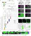
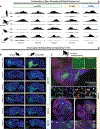
Figure 2.. Putative enhancers drive cell-type-specific expression in striatum
(A) Heatmap of median transcript detection for striatal cell type marker genes. (B) Sankey diagram indicating the sources for all putative enhancer peaks for each cell target, their predicted expression specificities, and their validated expression. Values indicate the number of enhancers at each node. (C and D) Putative enhancer peaks selected for striatal cell types from BOCA (C) and CATlas (D) datasets. Tracks are grouped by brain region for BOCA (PMC, primary motor cortex; PVC, primary visual cortex; AMY, amygdala; HIPP, hippocampus; MDT, medio-dorsal thalamus; NAC, nucleus accumbens; PUT, putamen) and by whole-mouse brain taxonomy cell subclass for CATlas. The proximal marker gene is shown above each peak. (E) Top: diagram of expression patterns indicative of striatal MSN cell populations. Middle: sagittal sections of native SYFP2 expression from enhancer AAVs for each MSN population. Bottom: zoomed-in views of axonal projection patterns (GPi, globus pallidus internal segment; GPe, globus pallidus external segment; SNr, substantia nigra pars reticulata). (F) Left: diagram of expression patterns indicative of striatal interneuron populations. Right: sagittal section of native SYFP2 from enhancer-AAV-targeting cholinergic interneurons. (G) Native SYFP2 illustrating morphological differences for distinguishing striatal interneuron populations. See Figure S3.
Figure 3.
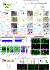
Figure 3.. Striatal enhancer AAVs demonstrate significant improvement in cell-type specificity over existing tools
(A) Diagram of enhancer-AAV validation approaches. (B) Enhancer specificities determined by SSv4 scRNA-seq (n = 1–2 mice and, on average, 50 cells per enhancer). Cell types in dendrogram encompass all neuronal basal ganglia subclasses from the whole-mouse brain taxonomy, with the six cell types targeted in this study highlighted in color. (C) Quantification of on-target enhancer-AAV specificities across all three validation methods. Black boxes indicate no data and stars indicate three of the best enhancers. (D and E) Top: example sagittal sections of (D) hChATp- or (E) AiE0743m_3xC2-driven native SYFP2 fluorescence. Bottom: identification of striatal cholinergic interneurons by IHC using ChAT marker antibody. Each panel is a representative ROI from three different brain regions. (F) RNAscope using probes against SYFP2 for three versions of the AiE0779m D1 MSN enhancer (FL, full-length; 3xC2, 3x core region; 6xC2, 6x core region). (G) Quantification of completeness of labeling from RNAscope images. Fisher’s exact test revealed differences in the proportions of SYFP2-positive cells between AiE0779m enhancer versions (p < 0.0001). Post hoc pairwise comparisons with Bonferroni correction showed significant differences between all enhancers (***p < 0.001). (H) SYFP2 signal intensity per cell from RNAscope images. The Kruskal-Wallis test showed a significant difference in SYFP2 intensity across enhancers (χ = 424.39, df = 2, p < 0.0001). Post hoc Dunn’s test with Bonferroni correction showed that all pairwise comparisons were significant: 3xC2 versus 6xC2 (p < 0.0001), 3xC2 versus FL (p < 0.0001), and 6xC2 versus FL (p < 0.0001). n = 1–2 mice and 1 ROI per enhancer version for RNAscope quantification. See also Figures S4–S6.
Figure 4.
Figure 4.. Specificity of striatal enhancer AAVs across different delivery routes and cargos
(A) AAV vector components and recommended dosages for different routes of administration in mice. (B–E) Representative sagittal sections of native SYFP2 across all three delivery routes for enhancers targeting (B and C) D1 MSNs, (D) D2 MSNs, and (E) cholinergic interneurons. (F) Summary of D1 and D2 MSN enhancer and interneuron enhancer specificities by route of administration. Each point represents a single enhancer. A Kruskal-Wallis test revealed no significant difference in specificity across routes for MSN enhancers (p = 0.7963), while a significant difference was observed for interneuron enhancers (p = 0.04383). Post hoc Dunn’s test with Bonferroni correction indicated that the RO route differed significantly from the STX route (p < 0.05), but no significant difference was found between ICV and other routes. (G) Heatmap of on-target specificities for striatal enhancers for the three routes of administration determined by scRNA-seq and mapping (D1 and D2 MSN enhancers) or IHC with cell type marker antibodies (interneuron enhancers). (H) Images of IHC comparing two cholinergic enhancers by STX injection. White arrows, SYFP2+/ChAT+ cells; white circles, off-target SYFP2+ MSNs. (I) Diagram of enhancer AAV for delivery of functional transgenes by STX injection into dorsal striatum. (J) Representative sagittal images of native SYFP2 for enhancer AAVs driving functional transgenes. AAVs used in images from left to right: AiP13278, AiP14035, and AiP14134.
Figure 5.
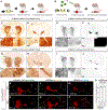
Figure 5.. Comparison of D1 MSN and D2 MSN enhancer AAVs to GENSAT Cre-driver lines
(A and B) Diagrams depicting generation of Cre-dependent reporter expression by crossing Cre-driver and reporter mice (A) or enhancer AAVs injected RO into Cre reporter mice (B). (C and D) ISH from https://www.gensat.org of GENSAT EY217 Drd1a-Cre-driver line crossed with Rosa26-EGFP reporter line. (E and F) STPT images of D1 MSN enhancer AiE0779m driving iCre(R297T) (AiP15260) injected into Ai14 reporter mouse line. (G and H) ISH from https://www.gensat.org of GENSAT ER44 Drd2-Cre-driver line crossed to Rosa26-EGFP reporter line. (I and J) STPT images of D2 MSN enhancer AiE0452h driving iCre (R297T) (AiP13781) injected into Ai14 reporter mouse line. (K–P) Example sagittal sections of enhancer AAVs driving iCre(R297T) (AiP14825, AiP15578, and AiP13781) in the Ai14 reporter mouse line through either STX (K–M) or RO (N–P) injection. Images are of native tdTomato fluorescence. (Q) Comparison of on-target cell-type specificity using scRNA-seq between RO and STX injection methods. Bars represent mean ± standard error (SE). Individual data points indicate values for each enhancer. A Wilcoxon rank-sum test was performed to assess statistical differences between groups (W = 5, p = 1), indicating no significant difference between injection methods. STR, striatum; OT, olfactory tubercle; CTX, cortex; SNr, substantia nigra pars reticulata; SNc, substantia nigra pars compacta; VTA, ventral tegmental area. See also Figure S7.
Figure 6.
Figure 6.. Optogenetic stimulation with D1 MSN enhancer AAV is sufficient to induce locomotion and contralateral rotations
(A and B) Experimental design with ICV injection of AAV vectors (AiP13278 for D1-ChR2 and AiP13044 for matched D1-SYFP2 control) into postnatal day 2 (P2) mouse pups. (C) Exemplar tracks of mouse locomotion before (light gray; 10 s), during (blue, orange; 10 s), and after (gray; 10 s) optogenetic stimulation in a circular arena. (D and E) Unilateral light delivery through the optical fiber (450 nm, 0.3 mW, 10 s) increased locomotion speed in D1-ChR2 mice (***p ≤ 0.001 by paired t test; n = 8 mice) but not D1-SYFP2 controls (ns, not significant; n = 9 mice). (F and G) Unilateral light delivery increased rotation bias toward contralateral rotations in D1-ChR2 mice (***p ≤ 0.001 by paired t test; n = 8 mice) but not D1-SYFP2 control mice (ns; n = 9 mice). (H and I) Optogenetic stimulation induced contralateral rotations in D1-ChR2 mice but not D1-SYFP2-only controls. Rotations persisted throughout the stimulation. See also Figure S8.
Figure 7.
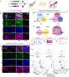
Figure 7.. Concurrent striatal cell type identification and targeting by multiplexed enhancer-AAV delivery in C57 mice
(A) Enhancer AAVs were individually packaged, pooled, and injected RO into adult mice. (B and C) Example images of two different triple-labeling combinations. AAVs used in (B): AiP12609 (D1 MSN-SYFP2), AiP12700 (D2 MSN-mTFP1), and AiP13738 (cholinergic-tdTomato). AAVs used in (C): AiP12689 (Sst-Chodl-SYFP2), AiP13808 (Pvalb-Pthlh-mTFP1), and AiP13738 (cholinergic-tdTomato). (D and E) Quantification of cell counts in dorsal striatum (D) and cortex (E). Barnard’s test was used to evaluate enhancer expression overlap in cortex versus STRd (n = 1 mouse and ROI per region). For D1 MSN+D2 MSN+cholinergic, no significant difference was found (two-sided p = 0.8882), while for Sst-Chodl+Pvalb-Pthlh+cholinergic enhancers, a significant difference in enhancer expression overlap between cortex and STRd was observed (two-sided p < 0.0001). (F–H) Single-cell patch-clamp recordings from coronal brain slices derived from a mouse with multiplex injection of AiP15050 (Sst-Chodl-SYFP2), AiP13808 (Pvalb-Pthlh-mTFP1), and AiP13738 (cholinergic-tdTomato). Representative responses to hyperpolarizing and depolarizing current steps recorded from the same mouse (top) with example single action potential traces (middle) and current injection steps (bottom). (I and J) 3D plots comparing electrophysiological features that distinguish the three striatal interneuron types. Diamonds represent the mean values for each interneuron type. Circles with black outline correspond to neuron traces in (F)–(H). PERMANOVA using all five electrophysiological features revealed significant differences across the three cell type populations (F = 51.071, p = 0.001). A pairwise PERMANOVA post hoc analysis indicated significant differences between all cell type pairwise comparisons (p < 0.01). Number of cells recorded: Pvalb-Pthlh, n = 13; Sst-Chodl, n = 16; cholinergic, n = 8. A total of 7 mice were used for triple-RO injection patch-clamp recording experiments. See also Figure S9.
Figure 8.
Figure 8.. Cross-species conservation of striatal enhancer open chromatin and enhancer-AAV activity in vivo
(A) Bulk ATAC-seq open chromatin regions in cortex and striatum of humans, macaques, rats, and mice at conserved orthologs of five enhancer loci. (B) Conservation of striatal enhancer specificity in vivo in mouse and rat brain. Sagittal brain sections with native SYFP2 (green) and DAPI (blue). (C–H) Stereotaxic injection of AiE0780m driving SYFP2 expression (AiP12610) into macaque caudate and putamen. (C) Coronal section of striatum injection site with propidium iodide (red), DAPI (blue), and native SYFP2 (green). (D) Example RNAscope images from putamen injection site. Yellow circles, SYFP+DRD1+PPP1R1B+ cells. (E–H) Coronal sections posterior to the injection site showing axon terminal labeling in GPi and SNr. See also Figure S10.

Update of

  • Enhancer AAV toolbox for accessing and perturbing striatal cell types and circuits.
    Hunker AC, Wirthlin ME, Gill G, Johansen NJ, Hooper M, Omstead V, Vargas S, Lerma MN, Taskin N, Weed N, Laird WD, Bishaw YM, Bendrick JL, Gore BB, Ben-Simon Y, Opitz-Araya X, Martinez RA, Way SW, Thyagarajan B, Otto S, Sanchez REA, Alexander JR, Amaya A, Amster A, Arbuckle J, Ayala A, Baker PM, Barcelli T, Barta S, Bertagnolli D, Bielstein C, Bishwakarma P, Bowlus J, Boyer G, Brouner K, Casian B, Casper T, Chakka AB, Chakrabarty R, Clark M, Colbert K, Daniel S, Dawe T, Departee M, DiValentin P, Donadio NP, Dotson NI, Dwivedi D, Egdorf T, Fliss T, Gary A, Goldy J, Grasso C, Groce EL, Gudsnuk K, Han W, Haradon Z, Hastings S, Helback O, Ho WV, Huang C, Johnson T, Jones DL, Juneau Z, Kenney J, Leibly M, Li S, Liang E, Loeffler H, Lusk NA, Madigan Z, Malloy J, Malone J, McCue R, Melchor J, Mich JK, Moosman S, Morin E, Naidoo R, Newman D, Ngo K, Nguyen K, Oster AL, Ouellette B, Oyama AA, Pena N, Pham T, Phillips E, Pom C, Potekhina L, Ransford S, Reding M, Rette DF, Reynoldson C, Rimorin C, Sigler AR, Rocha DB, Ronellenfitch K, Ruiz A, Sawyer L, Sevigny J, Shapovalova NV, Shepard N, Shulga L, Soliman S, Staats B, Taormina MJ, Tieu M, Wang Y, Wilkes J, Wood T, Zhou T, Williford A, Dee N,… See abstract for full author list ➔ Hunker AC, et al. bioRxiv [Preprint]. 2025 Mar 20:2024.09.27.615553. doi: 10.1101/2024.09.27.615553. bioRxiv. 2025. Update in: Neuron. 2025 May 21;113(10):1507-1524.e17. doi: 10.1016/j.neuron.2025.04.035. PMID: 39386678 Free PMC article. Updated. Preprint.

References

    1. DeLong MR, and Wichmann T (2007). Circuits and circuit disorders of the basal ganglia. Arch. Neurol. 64, 20–24. - PubMed
    1. Crittenden JR, and Graybiel AM (2011). Basal Ganglia disorders associated with imbalances in the striatal striosome and matrix compartments. Front. Neuroanat. 5, 59. - PMC - PubMed
    1. Lobo MK, and Nestler EJ (2011). The striatal balancing act in drug addiction: distinct roles of direct and indirect pathway medium spiny neurons. Front. Neuroanat. 5, 41. - PMC - PubMed
    1. Lipton DM, Gonzales BJ, and Citri A (2019). Dorsal striatal circuits for habits, compulsions and addictions. Front. Syst. Neurosci. 13, 28. - PMC - PubMed
    1. Kawaguchi Y (1993). Physiological, morphological, and histochemical characterization of three classes of interneurons in rat neostriatum. J. Neurosci. 13, 4908–4923. - PMC - PubMed