. 2025 Sep 17;66(3):2401610.
doi: 10.1183/13993003.01610-2024. Print 2025 Sep.

Particulate matter-related ITIH4 deficiency is associated with an emphysema phenotype of COPD through JNK-dependent and JNK-independent signalling

Affiliations

Particulate matter-related ITIH4 deficiency is associated with an emphysema phenotype of COPD through JNK-dependent and JNK-independent signalling

Sheng-Ming Wu et al. Eur Respir J. .

Abstract

Background: Prolonged exposure to airborne particulate matter (PM) is associated with emphysema and COPD; however, the precise underlying mechanism remains unclear. In a previous high-throughput screen, we identified inter-α-trypsin inhibitor heavy chain 4 (ITIH4) as a biomarker of long-term PM exposure. We hypothesised that ITIH4 is implicated in PM-related emphysema.

Methods: We investigated the association between ITIH4 expression and ambient PM exposure through a clinical cohort analysis (220 patients with COPD and 61 healthy participants) and in vitro studies.

Results: The COPD cohort studies revealed significant correlations between emphysema severity, ambient PM exposure and serum ITIH4 levels. In primary small airway epithelial cells from COPD patients with low basal levels of ITIH4, exposure to PM and oxidative stress led to increased apoptosis. However, ITIH4 overexpression significantly inhibited oxidative-stress-induced apoptosis in normal and COPD airway epithelial cells. Acute exposure to hydrogen peroxide resulted in the rapid degradation of ITIH4 protein with no effect on transcriptional level, although ITIH4 gene expression is downregulated in the lung tissue of patients with COPD. A human apoptosis antibody array revealed that ITIH4 overexpression attenuated hydrogen-peroxide-induced apoptotic signalling. Furthermore, extracellular ITIH4 protein confers cytoprotective functions in cells exposed to PM or oxidative stress. Mechanistically, ITIH4 attenuated oxidative-stress-induced c-Jun N-terminal kinase (JNK) activation and β-catenin decrease. A deficiency of ITIH4 exacerbated the effects of oxidative stress.

Conclusions: We identified a novel pathogenetic mechanism involving ITIH4, where chronic exposure to air pollution induces or promotes emphysema.


Conflict of interest statement

Conflict of interest: The authors have no potential conflicts of interest to disclose.

Figures

None
Ambient particulate matter exposure is implicated in decreased inter-α-trypsin inhibitor heavy chain 4 (ITIH4) levels and the development of emphysema in individuals with COPD. Acute exposure to particulate matter with aerodynamic diameter <2.5 µm (PM2.5) leads to a reduction in ITIH4 expression through oxidative-stress-mediated protein depletion, an effect that is more pronounced in individuals with COPD. Patients with COPD also have reduced ITIH4 expression at the mRNA level, which might be related to chronic exposure to PM2.5 and is not oxidative-stress-mediated (not shown in this model). Significantly, ITIH4 demonstrates protective roles against PM2.5 and oxidative-stress-induced apoptosis in lung epithelial cells via the activation of caspase-3. Additionally, the extracellular ITIH4 protein effectively mitigates PM2.5- or oxidative-stress-induced apoptosis. Mechanistically, ITIH4 alleviates the activation of c-Jun N-terminal kinase (JNK) induced by oxidative stress and decreases the levels of β-catenin. In contrast, an ITIH4 deficiency intensified the impact of oxidative stress. The effects of increased reactive oxygen species (ROS) may inactivate β-catenin survival signalling in a JNK-independent pathway.
FIGURE 1
FIGURE 1
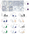
Correlation between particulate-matter-related inter-α-trypsin inhibitor heavy chain 4 (ITIH4) reduction and emphysema. a) ITIH4 levels were negatively correlated with the mean daily concentrations of exposure to particulate matter with aerodynamic diameter <10 µm (PM10) (rs=−0.366, p<0.001) and <2.5 µm (PM2.5) (rs=−0.414, p<0.001) in 170 COPD patients over the past 10 years. b) Box-and-whisker plots illustrating the differential ITIH4 levels in nonsmoker normal controls (NS, n=31), smoker normal controls (S, n=30) and patients with Global Initiative for Chronic Obstructive Lung Disease (GOLD) COPD stages I (n=27), II (n=77), III (n=50) and IV (n=16). c) The correlation between forced expiratory volume in 1 s (FEV1) percentage and serum ITIH4 levels was analysed in the total cohort, which included 61 normal controls and 170 participants with COPD. d) Total lung emphysema (low attenuation area (LAA%)) levels were correlated with the mean daily concentrations of PM10 and PM2.5 exposure in 220 COPD patients over the past 10 years. e) Analysis of the pack-years of smoking in 170 COPD patients correlated with total lung LAA% and serum ITIH4 levels. f) The correlation between total lung LAA% and ITIH4 level was analysed in 170 COPD patients. Representative computed tomography images depicting total LAA% and the corresponding ITIH4 expression in patients with COPD. g) ITIH4 protein expression levels in lung tissue sections from patients with COPD (n=16) and normal controls (n=20) were analysed through immunohistochemical (IHC) staining. An enlarged view of two rectangular regions in the airway and alveolar epithelium is shown in the right panel. Airway epithelial cells are represented by pink dashed lines. Staining signals were observed under a conventional light microscope. Scale bar=200 μm. Original magnification ×200. The IHC scores of ITIH4 protein levels in the airway or alveolar areas in COPD patients were compared with those in normal controls. Data are presented mean±sd of each group. ***: p<0.001; ****: p<0.0001; #: p<0.01 versus NS; : p<0.01 versus S; +: p<0.01 versus GOLD stage I; §: p<0.05 versus GOLD stage II.
FIGURE 2
FIGURE 2
Role of reactive oxygen species (ROS) in mediating low-level inter-α-trypsin inhibitor heavy chain 4 (ITIH4) protein expression. a) Secretory ITIH4 levels in bronchial epithelial (BEAS-2B), normal human small airway epithelial cells (HSAEpCs) or COPD HSAEpC cells exposed to irritants, including vehicle control, lipopolysaccharide (LPS) (20 ng·mL−1), interleukin (IL)-1β (10 ng·mL−1) and hydrogen peroxide (H2O2) (100 μM). ITIH4 levels in cells treated with the antioxidant N-acetyl-l-cysteine (NAC; 5 mM). b) Secretory ITIH4 levels in cells subject to 50 μg·mL−1 carbon black (CB), diesel exhaust particles (DEP), urban dust (UD) or vehicle control exposure. c) Levels of normalised ITIH4 gene expression (log2FPKM) in lung tissues from 98 patients with COPD (GSE57148 dataset cohort), compared with the normal group (n=91). d) Histograms comparing ITIH4 expression levels in BEAS-2B cells exposed to irritants, including LPS (20 ng·mL−1), IL-1β (10 ng·mL−1), 100 μM H2O2 and vehicle control (n=5−8 per group). The gene expression levels were measured using a quantitative real-time reverse transcription (qRT)-PCR assay. The levels of ITIH4 expression were examined in cells subject to 50 μg·mL−1 CB, DEP, UD or vehicle control exposure. e) ITIH4 gene expression levels in normal and COPD HSAEpCs exposed to irritants including LPS, IL-1β, H2O2 and vehicle control. ITIH4 expression levels were measured in normal control and COPD HSAEpCs subject to CB, DEP, UD or vehicle control exposure (n=5−8 per group). f) Effect of 100 μM H2O2 or vehicle with or without 50 μg·mL−1 cycloheximide on ITIH4 in BEAS-2B cells over a duration of 2 h. The half-life (t1/2) of ITIH4 protein in vehicle and H2O2 was analysed (n=5 per group). Relative values are expressed as the fold change calculated by comparison with vehicle control cells. Data are presented as mean±sd of at least five for each independent group. *: p<0.05; **: p<0.01.
FIGURE 3
FIGURE 3
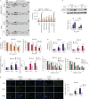
Inter-α-trypsin inhibitor heavy chain 4 (ITIH4) protects epithelial cells from particulate matter with aerodynamic diameter <2.5 µm (PM2.5)-induced and reactive oxygen species (ROS)-induced apoptosis. a) Cleaved caspase-3 levels in the airway or alveolar epithelium of normal control participants or patients with COPD. Cleaved caspase-3 levels in lung tissue sections from patients with COPD (n=16) and normal controls (n=20) were analysed through immunohistochemical (IHC) staining. An enlarged view of two rectangular regions in the airway and alveolar epithelium is shown in the right panel. Airway epithelial cells are represented by pink dashed lines. Staining signals were observed under a conventional light microscope. Scale bar=200 μm. Original magnification ×200. The IHC scores of cleaved caspase-3 levels in the airway or alveolar areas in COPD patients were compared with that in normal controls. b) ROS accumulation levels in control and N-acetyl-l-cysteine (NAC)-treated bronchial epithelial cells (BEAS-2B) exposed to 0–100 μg·mL−1 PM2.5 (n=5−8 per group). Moreover, ROS levels were measured in normal human small airway epithelial cells (HSAEpCs) and COPD HSAEpCs exposed to 0–50 μg·mL−1 PM2.5 (n=5–9 per group). c) Secreted ITIH4 levels in control and NAC-treated BEAS-2B, normal and COPD HSAEpCs exposed to PM2.5 (n=5–8 per group). d) Apoptotic cell population (%) in si-ITIH4-knockdown and ITIH4-overexpressing BEAS-2B cells in comparison with scramble si-RNA and pcDNA vector control cells exposed to 20 μg·mL−1 PM2.5, respectively. Apoptosis was measured by knockdown and overexpression of ITIH4 in HSAEpCs and COPD HSAEpCs exposed to 20 μg·mL−1 PM2.5, respectively (n=5–9 per group). e) Apoptosis was analysed in ITIH4-knockdown and ITIH4-overexpressing cells exposed to hydrogen peroxide (H2O2) as indicated (n=5–8 per group). Relative values are expressed as the fold change calculated through comparison with vehicle-treated si-scramble or pcDNA control cells. Data are presented as mean±sd of at least five for each independent group. *: p<0.05; **: p<0.01.
FIGURE 4
FIGURE 4
Inter-α-trypsin inhibitor heavy chain 4 (ITIH4) deficiency triggers oxidative-stress-mediated apoptosis signalling in lung epithelial cells. a) Human apoptosis antibody array image and histogram depicting the differential expression of proteins, including p21, FAS, HSP60, TRAIL, BAD and cleaved caspase-3 in bronchial epithelial (BEAS-2B) cells transfected with empty vector control (EV ctrl) or ITIH4 overexpression plasmid, treated with or without hydrogen peroxide (H2O2). b) Expression levels of cleaved caspase-3 and actin proteins in ITIH4-overexpression or control cells exposed to 0–200 μM H2O2 for 24 h (n=5 per group). c) Cell viability was examined in BEAS-2B cells treated with 20−100 μg·mL−1 particulate matter with aerodynamic diameter <2.5 µm (PM2.5) and 50−200 μM H2O2 for 24 h. Additionally, the effect of administration of 0.1 μg·mL−1 human recombinant full-length ITIH4 protein (rITIH4) on PM2.5 or H2O2-mediated cell death was evaluated. Cell viability was determined in cells treated with the indicated concentrations of PM2.5 and H2O2 in combination with rITIH4 (n=6 per group). d) Apoptotic cell population (%) was determined in BEAS-2B cells treated with the indicated 50 μg·mL−1 PM2.5 and 100 μM H2O2 in combination with rITIH4 using propidium iodide and annexin V-FITC staining. Furthermore, apoptosis analysis was performed in BEAS-2B cells overexpressing Flag-ITIH4 after PM2.5 or H2O2 treatment compared with empty vector cells treated with vehicle control (n=5 per group). e) Moreover, reactive oxygen species (ROS) production levels were also measured in cells administered rITIH4 or overexpressing Flag-ITIH4 after PM2.5 or H2O2 treatment compared with control cells (n=6–7 per group). f) Cell viability was examined in BEAS-2B cells treated with 1–10 μM protein secretion inhibitors (brefeldin A) for 24 h. Moreover, cell viability was determined in cells treated with 50 μg·mL−1 PM2.5 in combination with 0.1 μg·mL−1 rITIH4. Cell viability was also measured in cells overexpressing Flag-ITIH4 after brefeldin A and/or PM2.5 treatment compared with vector control cells (EV ctrl) (n=5–6 per group). g) DNA fragmentation in apoptotic cells in BEAS-2B cells was determined using the terminal deoxynucleotidyl transferase-mediated dUTP nick-end labelling (TUNEL) assay. The proportion of TUNEL-positive cells after treatment with 50 µg·mL−1 PM2.5 and 100 µM H2O2 was compared with cells given 0.1 µg·mL−1 rITIH4 and control (n=6 per group). Scale bar=200 μm. Original magnification ×200. Relative values are expressed as the fold change calculated through comparison with vehicle-treated control cells. Data are presented as mean±sd of at least five for each independent group. DAPI: 4′,6-diamidino-2-phenylindole. *: p<0.05; **: p<0.01.
FIGURE 5
FIGURE 5
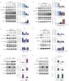
Overexpression of inter-α-trypsin inhibitor heavy chain 4 (ITIH4) inhibits oxidative-stress-induced activation of c-Jun N-terminal kinase (JNK) signalling and reduction of β-catenin. a) Levels of ITIH4, β-catenin, p-JNK and JNK in bronchial epithelial (BEAS-2B) cells exposed to lipopolysaccharide (LPS) (20 ng·mL−1), interleukin (IL)-1β (10 ng·mL−1) or hydrogen peroxide (H2O2) (100–300 μM), examined through an immunoblot analysis (n=6 per group). b) Levels of ITIH4, β-catenin, p-JNK and JNK in BEAS-2B cells exposed to 50–100 μg·mL−1 carbon black (CB), diesel exhaust particles (DEP), urban dust (UD) and vehicle control (ctrl) were measured. Moreover, levels of ITIH4, β-catenin, p-JNK and JNK were determined in c) bronchial epithelial (BEAS-2B) cells and d) human small airway epithelial cells (HSAEpCs) exposed to 50–100 μg·mL−1 DEP and DEP-derived particulate matter with aerodynamic diameter <2.5 µm (PM2.5) (n=6 per group). e) The effects of exposure to 100 μM H2O2-mediated expression of ITIH4, β-catenin, p-JNK and JNK proteins were assessed in BEAS-2B cells transfected with ITIH4-overexpression vector or empty vector control (EV) (n=6–7 per group). f) By contrast, the effects of exposure to H2O2-mediated β-catenin expression and JNK activation were evaluated in BEAS-2B cells transfected with siRNA control or si-ITIH4 (n=6 per group). Levels of p-JNK and JNK were measured in cells exposed to stimuli for 2 h. Levels of ITIH4, β-catenin and actin were measured in cells treated with irritants for 24 h. Relative values are expressed as the fold change calculated through comparison with vehicle-treated cells. Data are presented as mean±sd of at least five for each independent group. *: p<0.05; **: p<0.01.
FIGURE 6
FIGURE 6
Inter-α-trypsin inhibitor heavy chain 4 (ITIH4) protects epithelial cells from reactive oxygen species (ROS)-induced injury through c-Jun N-terminal kinase (JNK)-dependent and JNK-independent mechanisms. a) Apoptotic effects of mitogen-activated protein kinases (MAPKs) including 10 µM JNK inhibitor (SP600125), p38 MAPK inhibitor (SB203580) and extracellular signal-regulated kinase inhibitor (PD98059) in ITIH4-knockdown or control cells exposed to 100 µM hydrogen peroxide (H2O2) (n=5–7 per group). b) Apoptotic effects in ITIH4-knockdown or control cells treated with 5 mM N-acetyl-l-cysteine (NAC) (n=5–7 per group). c) Apoptosis was also detected in cells exposed to pan caspase inhibitor Z-VAD-FMK (50 μM) (n=5–6 per group). d) Effects of JNK inhibitor treatment on ITIH4, p-JNK/JNK, cleaved caspase-3 and β-catenin protein expression in bronchial epithelial (BEAS-2B) cells transfected with si-control or si-ITIH4 in the presence of 100 μM H2O2 (n=5 per group). e) Levels of ITIH4, p-JNK/JNK, cleaved caspase-3 and β-catenin proteins measured in cells exposed to H2O2 following NAC treatment (n=5 per group). The levels of p-JNK and JNK were measured in cells exposed to H2O2 for 2 h. Levels of ITIH4, β-catenin, cleaved caspase-3 and actin measured in cells treated with H2O2 for 24 h. Relative values are expressed as the fold change calculated through comparison with vehicle-treated knockdown control cells. All data are presented as the mean±sd of at least five for each independent group. *: p<0.05, **: p<0.01.

Comment in

References

    1. Christenson SA, Smith BM, Bafadhel M, et al. Chronic obstructive pulmonary disease. Lancet 2022; 399: 2227–2242. doi: 10.1016/S0140-6736(22)00470-6 - DOI - PubMed
    1. Lu Z, Coll P, Maitre B, et al. Air pollution as an early determinant of COPD. Eur Respir Rev 2022; 31: 220059. doi: 10.1183/16000617.0059-2022 - DOI - PMC - PubMed
    1. Rice MB, Ljungman PL, Wilker EH, et al. Long-term exposure to traffic emissions and fine particulate matter and lung function decline in the Framingham Heart Study. Am J Respir Crit Care Med 2015; 191: 656–664. doi: 10.1164/rccm.201410-1875OC - DOI - PMC - PubMed
    1. Guo C, Zhang Z, Lau AKH, et al. Effect of long-term exposure to fine particulate matter on lung function decline and risk of chronic obstructive pulmonary disease in Taiwan: a longitudinal, cohort study. Lancet Planet Health 2018; 2: e114–e125. doi: 10.1016/S2542-5196(18)30028-7 - DOI - PubMed
    1. Tung NT, Ho SC, Lu YH, et al. Higher alveolar deposition of particulate matter in emphysematous lobes of COPD. ERJ Open Res 2021; 7: 00324-2021. doi: 10.1183/23120541.00324-2021 - DOI - PMC - PubMed

MeSH terms