. 2025 May 22;16(1):4752.
doi: 10.1038/s41467-025-59835-7.

Bioresorbable, wireless dual stimulator for peripheral nerve regeneration

Affiliations

Bioresorbable, wireless dual stimulator for peripheral nerve regeneration

Hak-Young Ahn et al. Nat Commun. .

Abstract

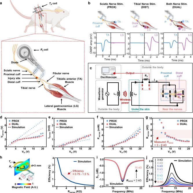
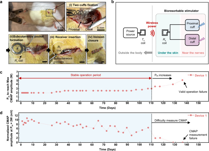
Wireless bioresorbable electrical stimulators have broad potential as therapeutic implants. Such devices operate for a clinically relevant duration and then harmlessly dissolve, eliminating the need for surgical removal. A representative application is in treating peripheral nerve injuries through targeted stimulation at either proximal or distal sites, with operation for up to one week. This report introduces enhanced devices with additional capabilities: (1) simultaneous stimulation of both proximal and distal sites, and (2) robust operation for as long as several months, all achieved with materials that naturally resorb by hydrolysis in surrounding biofluids. Systematic investigations of the materials and design aspects highlight the key features that enable dual stimulation and with enhanced stability. Animal model studies illustrate beneficial effects in promoting peripheral nerve regeneration, as quantified by increased total muscle and muscle fiber cross-sectional area and compound muscle action potentials. These findings expand the clinical applications of bioresorbable stimulators, particularly for long-term nerve regeneration and continuous neuromodulation-based monitoring.


Conflict of interest statement

Competing interests: The authors declare no competing interests.

Figures

Fig. 1
Fig. 1. Bioresorbable wireless dual-stimulator for nerve regeneration.
a Illustrations of the overall envisioned use case for a device to accelerate recovery from a sciatic nerve injury. b Illustration of the site of implantation for this clinical use case. c Exploded-view schematic illustration of the device. d Photographs of the device integrated with a phantom nerve, with red indicator LEDs, resting on part of a Tx coil (top), and in a bent state on the finger (bottom). e Micro-CT image of a device implanted in a rat. f Images of the accelerated dissolution of a device captured at various times after immersion in PBS (pH 7.4) solution at 95 °C. Scale bar, 5 mm.
Fig. 2
Fig. 2. Designs for improved lifetime for stable operation.
a Top-view schematic illustration of the device. Schematic illustrations of key engineering features, including (i) an interlocking folded via structure for joining the Rx coils, (ii) a diode transferred onto a PA substrate, (iii) a pocket for diode insertion, (iv) a diode connection pad with a hole and sawtooth structure and (v) an enlarged view of the connection with W/wax paste, (vi) a connected coil and extension electrode structure fabricated from a single sheet of Mo by a laser-cutting process. Scale bar, 1 mm. b Measured RF behavior (S11) and resonance frequency as a function of time of immersion in 1 × PBS (pH 7.4) at 37 °C. c I-V characteristics before connection (black) and after connection (red) of the bioresorbable PIN diode. d Changes in resistance of serpentine traces for the optimized joined design during cyclic loading (40% compression, 5 mm/s velocity, 70,000 cycles). e Comparison of the degradation period (y-axis), operating lifetime (x-axis), and the change in device stability (color change by stability index) over time for the bioresorbable devices reported here and those in previous reports. The stability index on the right displays the rate of change for each of the following parameters: (i) Maximum EMG amplitude induced by the bioresorbable stimulator over 7 days, (ii) changes in intracranial pressure (ICP), (iii) resistance changes in the electrode, and (iv) minimum operating voltage required to induce muscle contractions. The yellow solid line indicates the case where the degradation period and operational lifetime are equal.
Fig. 3
Fig. 3. In vivo acute tests for characterization of wireless bioresorbable dual stimulators in a rodent model.
a Schematic illustration of the implantation location. Top: Placement of the device in the animal and wireless coupling to an external transmission (Tx) coil. Bottom: Device with cuffs wrapped around the sciatic nerve (proximal cuff) and tibial nerve branch (distal cuff). Due to a transaction between the cuffs on the tibial nerve, the proximal cuff only stimulates the TA through the fibular nerve branch. The distal cuff uniquely stimulates the LG. Rx= receiver. Tx= transmission. [Created with Biorender.com]. b Schematic illustration of the experimental design (top) and EMG signals from the TA and LG muscles for each case. Corresponding changes in CMAP amplitude (bottom). TA tibialis anterior, LG lateral gastrocnemius, CMAP compound muscle, action potential, SHAM no stimulation using a sham device model, PROX stimulation of only the proximal sciatic nerve, DUAL stimulation of both the proximal sciatic nerve and the distal tibial nerve. [Created with Biorender.com]. c Circuit and block diagrams of the wireless stimulation system. A resistor (R3) facilitates measurements of the change in voltage between points 4 and 5 as a function of input voltage. dg Voltage (d), current (e), and power (f) applied to the nerve, and (g) resistance of the nerve as a function of input voltage after nerve damage, as determined through in vivo tests. h Schematic illustration and modeling results for the magnetic inductive coupling between a Tx coil and the Rx coils (left). Modeling results for the efficiency of power transfer as a function of the resistance of the nerve (Rnerves) (right). i, j Modeling results for the efficiency (i) and the reflection coefficient as a function of frequency (j) for various values of Rnerve.
Fig. 4
Fig. 4. In vivo tests of a wireless bioresorbable dual-stimulator in a rodent model.
a Surgical procedure for implantation. From left to right: an incision in the skin allows attachment of the stimulation cuffs to the sciatic nerve and tibial nerve branch; a subcutaneous pocket forms a point of insertion for the radio frequency (RF) harvester and its connection to the cuffs through serpentine traces; the Rx coil resides in the subcutaneous space; sutures and clips close the muscle and the skin, respectively; passing RF current through the Tx coil activates stimulation. b Circuit and block diagram of the system. c Changes in minimum input power required to achieve maximum CMAP amplitude and (d) normalized changes in CMAP amplitude under the lowest RF power to the Tx coil (2 W), for n = 3. Rx = receiver. Tx= transmission.
Fig. 5
Fig. 5. Effects of dual electrical stimulation on preventing muscle atrophy during the early stages of nerve injury.
a Schematic of ultrasound acquisition and representative ultrasound image of the LG in cross-section. TIB Tibia. [Created with Biorender.com]. b Normalized ultrasound CSA of ipsilateral LG muscle to contralateral 1 week after injury. SHAM (n = 8); PROX (n = 7); DUAL (n = 6). n= biologically independent animals. c Muscle fiber CSA of MG 2 weeks post-injury. SHAM (n = 10); PROX (n = 9); DUAL (n = 8). n biologically independent animals. A one-way ANOVA with Fisher’s LSD post hoc was used to determine the effect of treatment. Data were expressed as mean ± SEM. CSA cross-sectional area, LG lateral gastrocnemius, MG medial gastrocnemius. SHAM no stimulation using a sham device model, PROX stimulation of only the proximal sciatic nerve, DUAL stimulation of both the proximal sciatic nerve and the distal tibial nerve.
Fig. 6
Fig. 6. Effects of proximal and dual electrical stimulation on muscle reinnervation 6 weeks after injury.
a Ultrasound CSA of ipsilateral LG normalized to the contralateral at 6 weeks. SHAM (n = 12); PROX (n = 11); DUAL (n = 11). n biologically independent animals. b Muscle fiber CSA of MG 6 weeks post-injury, plus example images of MG sections stained with Hematoxylin and Eosin (H&E) for each treatment. Scale bars, 30 µm. SHAM (n = 12); PROX (n = 11); DUAL (n = 11). n biologically independent animals. c Representative 6-week CMAP traces (bottom, colored waveform) are baseline traces (top, black waveform) for each treatment. Scale bar, 2 mv (vertical), 0.2 ms (horizontal). d CMAP amplitude of LG 6 weeks after injury, normalized to baseline values. SHAM (n = 10); PROX (n = 11); DUAL (n = 11). n biologically independent animals. e Percentage of small, angulated muscle fibers (less than 250 μm2) in MG 6 weeks post-injury. SHAM (n = 12); PROX (n = 11); DUAL (n = 10). n biologically independent animals. A one-way ANOVA with Fisher’s LSD post hoc was used to determine the effect of treatment. Data were expressed as mean ± SEM. CSA cross-sectional area. LG lateral gastrocnemius, MG medial gastrocnemius, CMAP compound muscle action potential. SHAM no stimulation using a sham device model. PROX stimulation of only the proximal sciatic nerve. DUAL stimulation of both the proximal sciatic nerve and the distal tibial nerve.

References

    1. Hardy, P. B., Wang, B. Y., Chan, K. M., Webber, C. A. & Senger, J. L. B. The use of electrical stimulation to enhance recovery following peripheral nerve injury. Muscle Nerve70, 1151–1162 (2024). - PubMed
    1. Gordon, T., Amirjani, N., Edwards, D. C. & Chan, K. M. Brief post-surgical electrical stimulation accelerates axon regeneration and muscle reinnervation without affecting the functional measures in carpal tunnel syndrome patients. Exp. Neurol.223, 192–202 (2010). - PubMed
    1. Wong, J. N., Olson, J. L., Morhart, M. J. & Chan, K. M. Electrical stimulation enhances sensory recovery: a randomized controlled trial. Ann. Neurol.77, 996–1006 (2015). - PubMed
    1. Power, H. A., Morhart, M. J., Olson, J. L. & Chan, K. M. Postsurgical electrical stimulation enhances recovery following surgery for severe cubital tunnel syndrome: a double-blind randomized controlled trial. Neurosurgery86, 769–777 (2020). - PubMed
    1. Barber, B. et al. Intraoperative brief electrical stimulation of the spinal accessory nerve (BEST SPIN) for prevention of shoulder dysfunction after oncologic neck dissection: a double-blinded, randomized controlled trial. J. Otolaryngol. Head. Neck Surg.47, 7 (2018). - PMC - PubMed

LinkOut - more resources