. 2025 May 22;15(1):17846.
doi: 10.1038/s41598-025-03017-4.

Elucidating the tumor microenvironment interactions in breast, cervical, and ovarian cancer through single-cell RNA sequencing

Affiliations

Elucidating the tumor microenvironment interactions in breast, cervical, and ovarian cancer through single-cell RNA sequencing

Xiaoyue Zhu et al. Sci Rep. .

Abstract

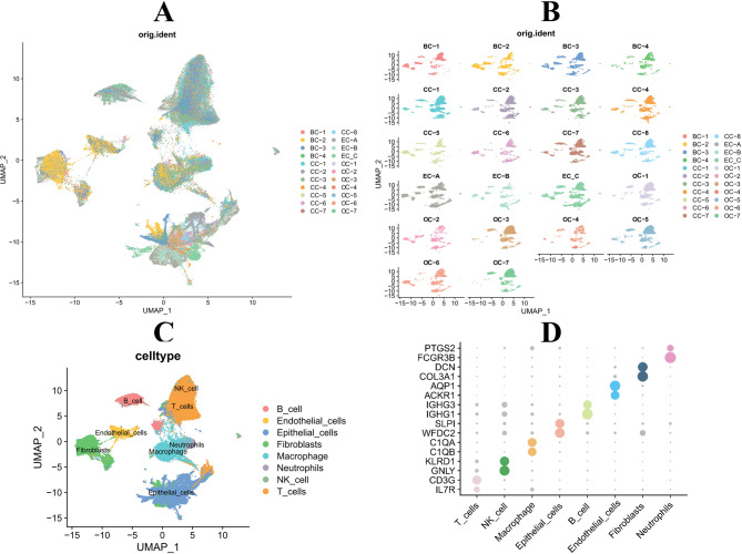
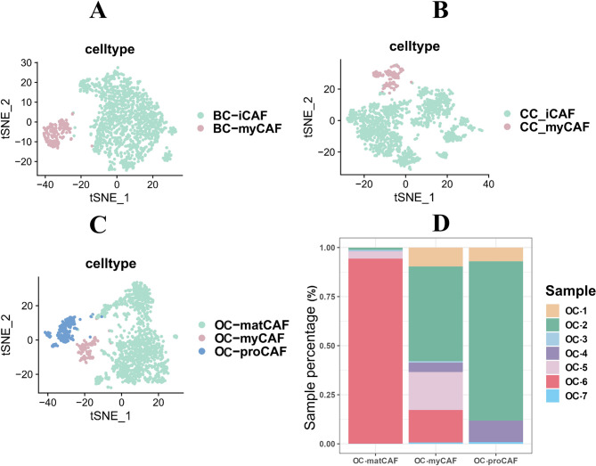
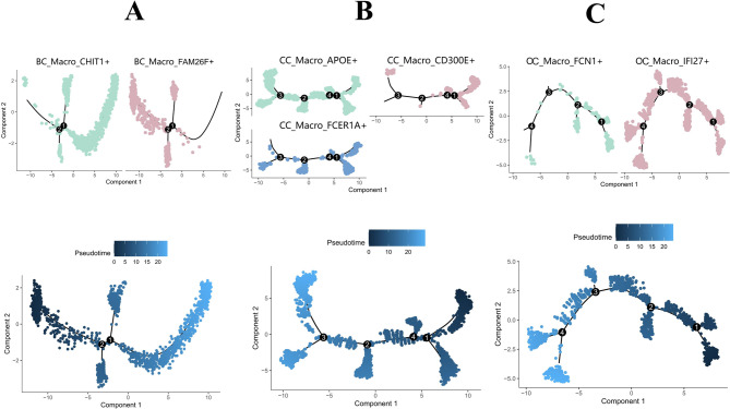
This study aimed to identify the key cell types and their interactions in gynecological oncology of breast cancer, cervical cancer, and ovarian cancer. Single-cell RNA sequencing was performed on tumor samples of gynecological oncology from the GEO database. Cell types were identified using SingleR and cell composition was analyzed to understand the tumor microenvironment (TME). CellChat was used to analyze cell interactions, and pseudotemporal analysis was conducted on cancer-associated fibroblasts (CAFs) and tumor-associated macrophages (TAMs) to understand their differentiation status. Four CAF subtypes were identified: iCAF, myCAF, proCAF, and matCAF. The iCAF subpopulation secreted COL1A1 and promoted tumor cell migration, while myCAF was involved in angiogenesis. The matCAF subpopulation was present throughout tumor development. TAMs were found to promote angiogenesis through the VEGFA_VEGFR2 signaling pathway. CAFs and TAMs play pivotal roles in tumor progression through their interactions and signaling pathways.

Keywords: Angiogenesis; Cancer-associated fibroblasts; Single-cell RNA sequencing; Tumor microenvironment; Tumor-associated macrophages.


Conflict of interest statement

Declarations. Competing interests: The authors declare no competing interests.

Figures

Fig. 1
Fig. 1
scRNA-seq reveals diverse cell types and tumor heterogeneity in different types of diseases. (A) UMAP plots displays the effects of different samples after batch processing. (B) UMAP plots illustrates the cell distribution across different samples. (C) UMAP plots showing the different samples into eight main cell types: B cells, Endothelial cells, Epithelial cells, Fibroblast cells, Macrophages, Neutrophils, NK_cell, and T_cells. (D) Bubble plots depicting the expression levels of marker genes for each cell types.
Fig. 2
Fig. 2
Cell type content maps for different diseases and interaction communication UMAP plots. (A,B) Cell content bar charts for different disease types and different samples. (C) The interaction communication intensity of major cell types in BC, CC, and OC.
Fig. 3
Fig. 3
Signaling patterns in BC, CC, and OC. (A) Outgoing and incoming signaling patterns in BC. (B) Outgoing and incoming signaling patterns in CC. (C) Outgoing and incoming signaling patterns in OC.
Fig. 4
Fig. 4
Identification and classification of malignant fibroblast subtypes in different cancer types. (A). The tSNE plots shows two distinct subtypes of fibroblasts in BC, labeled as BC-iCAF (CXCL14) and BC-myCAF (MYH11). (B) The tSNE plots shows two subtypes of fibroblasts are identified in CC, namely CC-iCAF (CXCL14) and CC-myCAF (RGS5). (C) In OC, three fibroblast subtypes are identified, which are OC-proCAF (C7), OC-myCAF (MYH11), and OC-matCAF (POSTN). (D) The cell composition plot demonstrates a high abundance of matCAF in late-stage OC samples, indicating its association with advanced tumor development and progression.
Fig. 5
Fig. 5
Pseudotemporal analysis of cancer-associated fibroblast (CAFs) subpopulations across different cancer types. The pseudotemporal trajectory analysis using “monocle” reveals that the iCAF subpopulation is at the initial stage of differentiation, while the myCAF subpopulation is at the late stage in both BC (A) and CC (B) samples. (C) In contrast to BC and CC, the differentiation trend in OC samples shows an opposite sequence, with the proCAF subpopulation at the early stage, followed by differentiation into myCAF and a small portion of matCAF.
Fig. 6
Fig. 6
Subtyping of macrophages based on marker gene expression in different cancer types. (A) The tSNE plots illustrates the two subtypes of macrophages identified in BC, labeled as BC_Macro_FAM26F + and BC_Macro_CHIT1+. (B) In CC, macrophages are categorized into three distinct subtypes, named CC_Macro_APOE+, CC_Macro_CD300E+, and CC_Macro_FCER1A+. (C) The typing results for OC macrophages reveal two subtypes, identified as OC_Macro_IFI27 + and OC_Macro_FCN1+.
Fig. 7
Fig. 7
Pseudotemporal analysis of macrophage subtypes differentiation within the TME using “Monocle”. (A) The pseudotemporal trajectory shows the differentiation path of macrophages in BC samples, transitioning from BC_Macro_FAM26F + to BC_Macro_CHIT1+. (B) The analysis reveals that the CC_Macro_APOE + cell cluster is present throughout the differentiation process, while CC_Macro_CD300E + and CC_Macro_FCER1A + cell clusters are active primarily in the early and middle stages of differentiation. (C) The differentiation state in OC samples resembles that, the OC_Macro_IFI27 + subpopulation is persistent throughout the differentiation process and the OC_Macro_FCN1 + subpopulation being active only in the early stages of differentiation.
Fig. 8
Fig. 8
Cell-to-cell interaction analysis between CAFs and macrophage subtypes in the tumor microenvironment using “CellChat”. (A) The CellChat analysis for BC samples shows that iCAF and Macro_CHIT1 + subtypes exhibit the highest level of communication among the cell subtypes within the tumor microenvironment. (B) In CC samples, the iCAF and Macro_FCER1A + subtypes are identified as having the most active cell-to-cell interactions. (C) In OC, matCAF and Macro_FCN1 + subtypes demonstrate the most significant cell-to-cell interactions.

References

    1. Bray, F., Laversanne, M., Weiderpass, E. & Soerjomataram, I. The ever-increasing importance of cancer as a leading cause of Prematu re death worldwide. Cancer127, 3029–3030. 10.1002/cncr.33587. - PubMed
    1. Bray, F. et al. Global cancer statistics 2022: GLOBOCAN estimates of incidence and Mor tality worldwide for 36 cancers in 185 countries. CA: Cancer J. Clin.74, 229–263. 10.3322/caac.21834. - PubMed
    1. Barzaman, K. et al. Breast cancer: biology, biomarkers, and treatments. Int. Immunopharmacol.84, 106535. 10.1016/j.intimp.2020.106535. - PubMed
    1. Abu-Rustum, N. R. et al. NCCN Guidelines® Insights: Cervical Cancer, Version 1.2024. J. Natl. Compr. Canc Netw21, 1224–1233. 10.6004/jnccn.2023.0062. - PubMed
    1. Stewart, C., Ralyea, C. & Lockwood, S. in Semin Oncol Nurs. 151–156 (Elsevier). - PubMed

MeSH terms