. 2025 May 22;11(1):132.
doi: 10.1038/s41531-024-00849-1.

Rational selection of the monoclonal α-synuclein antibody amlenetug (Lu AF82422) for the treatment of α-synucleinopathies

Affiliations

Rational selection of the monoclonal α-synuclein antibody amlenetug (Lu AF82422) for the treatment of α-synucleinopathies

Pekka Kallunki et al. NPJ Parkinsons Dis. .

Abstract

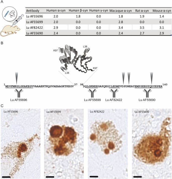
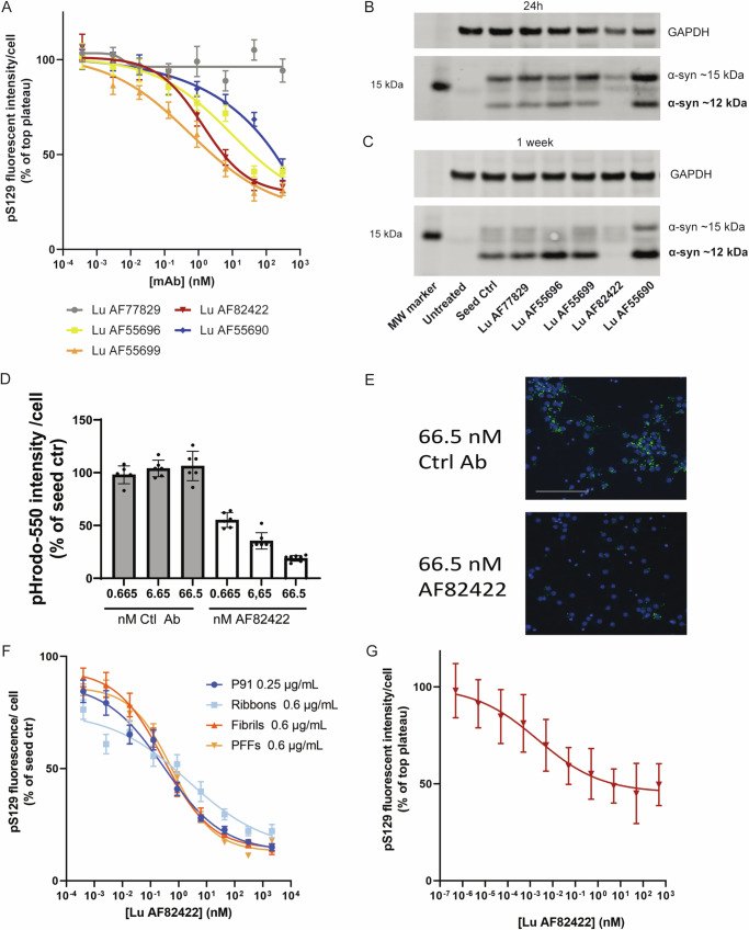
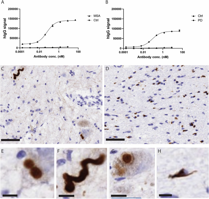
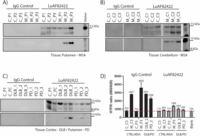
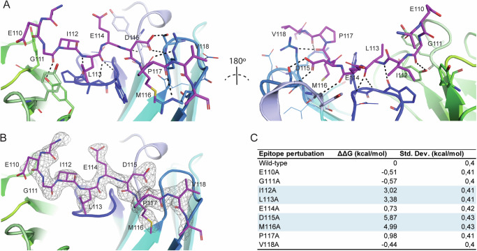
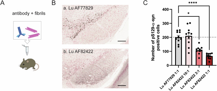
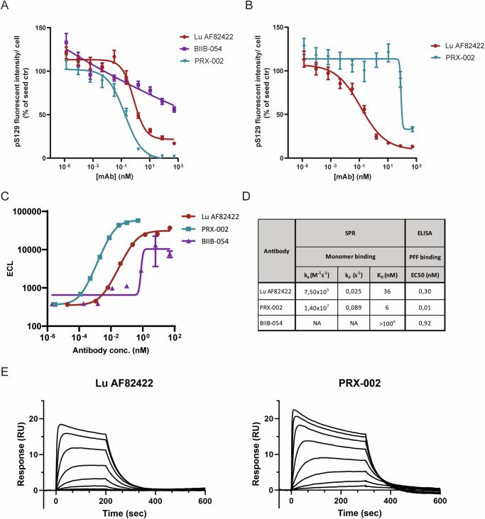
Amlenetug (Lu AF82422) is a human monoclonal antibody targeting α-synuclein in clinical development for multiple system atrophy. We describe a series of studies that characterize its functional properties and supported its selection as a viable clinical candidate. Amlenetug inhibits seeding induced in mouse primary neurons by various α-synuclein fibrillar assemblies and by aggregates isolated from MSA brain homogenate. In vivo, both co-injection of amlenetug with α-synuclein assemblies in mouse brain and peripheral administration inhibit α-synuclein seeding. Amlenetug inhibits uptake of α-synuclein seeds as well as accumulation of C-terminal truncated α-synuclein seeds and demonstrates binding to monomeric, aggregated, and truncated forms of human α-synuclein. The epitope of amlenetug was mapped to amino acids 112-117 and further characterized by crystallographic structure analysis. Based on our data, we hypothesize that targeting α-synuclein will potentially slow further disease progression by inhibiting further pathology development but be without impact on established pathology and symptoms.


Conflict of interest statement

Competing interests: P.K., F.S., K.W., M.L., L.D., M.A., L.B., B.O.K., L.R.O., S.V., A.J., M.N.H., D.S.M.D., K.B.-A., K.F. are full-time employees of H. Lundbeck A/S. A.-L.B., I.M., S.N., T.T.E., J.B.S., K.J.A., L.Ø.P., S.C., J.E., J.N.S., P.G.W.-L. were full-time employees of H. Lundbeck A/S at the time of this work. E.N.V.D.B., R.R., D.S., P.P. were full-time employees of Genmab BV at the time of this work. The research was funded by H. Lundbeck A/S, Copenhagen, Denmark.

Figures

Fig. 1
Fig. 1. Basic binding characteristics and epitope overview of four representative antibodies identified from the immunization campaign: Lu AF55696, Lu AF55699, Lu AF82422 and Lu AF55690.
A Octet binding (octet response units; nm) to human, macaque, mouse, and rat monomeric α-syn as well as human β- and γ-syn. B Overview of linear epitope (underlined) locations on α-syn N- and C-terminal sequences on either side of the NAC domain. Common truncation sites marked. C Immunohistochemical staining pattern in substantia nigra of a PD patient. Lewy Body like inclusion bodies were identified in dopaminergic neurons (neuromelanin positive brown deposits inside the cells) with all antibodies. Scalebar = 10 nm. A file containing Octet binding data and the raw data from the epitope mapping is included in Supplementary File 1.
Fig. 2
Fig. 2. Antibodies inhibit seeded α-syn aggregation in mouse primary neuronal cells and affect uptake and truncations of seeds differently.
A Antibody inhibition of seeding was examined in F28 primary mouse neurons with pS129 α-syn immunolabelling as a readout for seeding. Cells were treated at DIV5 with 0.4 µg/ml α-syn assemblies of the P91 type premixed with a dilution series of antibody and subsequently fixed and analyzed for pS129 immunolabelling and cell count after 7 days, n = 4 independent cultures. B, C Antibody effect on the uptake and proteolytic cleavage of full length human α-syn assemblies of the P91 type after 24 h and 1 week in wt mouse primary neuronal cultures. “Untreated” denotes naïve primary neurons while “Seed Ctrl” signifies primary neurons treated with full length human α-syn P91 but no antibody, n = 2 independent cultures. Full WB’s are included in Supplementary Fig. 2A, B. D Lu AF82422 dose-dependent inhibits uptake of PFFs labeled with pHrodo. 0.5 µg/ml PFF-pHrodo premixed with a dilution series of Lu AF82422 added to F28 primary mouse neurons and fluorescence intensity per cell normalized to “seed only” quantified 48 h later, n = 2–3 independent experiments with n = 2–3 technical replicates each (=6–9 datapoints) cultures ± SD. E Representative images from 2D. Cells treated with either 66.5 nM control (Lu AF77829) or AF82422. Overlay of Hoechst in blue and PFF-pHrodo in green. Scale bar = 100 µm. F Lu AF82422 dose-dependently inhibits seeding induced by various recombinant α-syn assemblies (P91 0.25 µg/ml, Ribbons 0.6 µg/ml, Fibrils 0.6 µg/ml and PFF’s 0.6 µg/ml) in F28 primary mouse neurons, n > 3 independent cultures ± SEM. G Lu AF82422 dose-dependently inhibits seeding induced by enriched MSA brain extract in F28 mouse primary neurons. Two different enriched MSA brain extracts originating from putamen were used, n = 2 independent cultures ± SD. Raw data from Cellomics quantifications reported in A, F and G are included in Supplementary File 2A–C. Full western blots of blots in B and C are included in Supplementary Fig. 2. Raw data from PFF uptake assays is included in Supplementary File 3.
Fig. 3
Fig. 3. Lu AF82422 binds to pathological forms of α-syn in patient derived homogenates and brain sections.
A, B ELISA measure of Lu AF82422 binding to α-syn aggregates derived from MSA and PD brains, respectively and compared to control brain. Lu AF82422 used for staining of C section from Locus coeruleus in PD patient showing corkscrew shaped neurite and cell with cytoplasmic Lewy bodies, D section from cerebellum in MSA patient showing extensive white matter pathology with multiple glial cytoplasmic inclusions. E, F Increased magnifications of Lewy bodies and Lewy Neurites from (C). G section from cortex in DLB patient showing a cytoplasmic Lewy body. H Glial cytoplasmic inclusion from (D). Scale bar = 50 nm in (C, D), 10 nm in (EH).
Fig. 4
Fig. 4. Immunoprecipitation and immunodepletion of α-syn from MSA, PD and DLB brain tissue material.
WB analysis of α-syn IP’ed with Lu AF82422 from MSA brain homogenates, putamen (A) and cerebellum (B), and corresponding controls. α-Syn is visualized using total or pS129-α-syn antibodies. C IP and western blotting analysis performed as in (A) but using PD (putamen) and DLB (cortex) brain homogenates, and matching controls. D HTRF-based analysis of α-syn aggregates in brain homogenates immunodepleted with Lu AF82422 or control antibody. Samples were selected to represent similar pathology load (Supplementary Fig. 5A). HTRF ratio is plotted as measure of aggregated α-syn over experimental background. See Supplementary Table 1 for additional information on tissue samples. Full western blots of AC and raw data for the HTFR assay are included in Supplementary Fig. 5B–D and Supplementary File 5, respectively.
Fig. 5
Fig. 5. Crystal structure of Lu AF82422 binding to α-syn.
A Crystal structure of the Lu AF82422:α-syn complex rotated 180°. Light chain of Lu AF82422 shown in green, heavy chain in blue and α-syn in red. Key residues are shown as sticks and hydrogen bonds as dashes (black). B Difference electron density (mFo-DFc) of the epitope peptide indicates that it is well resolved in the binding pocket. C In silico free energy perturbation by alanine scanning through the epitope based on the crystal structure. Key affinity driving residues with contributions to ΔΔG > 1 kcal/mol are highlighted (blue).
Fig. 6
Fig. 6. Lu AF82422 dose-dependently inhibits seeding in a co-injection paradigm in mice.
A Antibodies and α-syn assemblies of the Fibrillar type were pre-mixed prior to intrastriatal injection; for each injection, 2 µg of Fibrils were mixed with varying amounts of antibodies, at Fibril:antibody monomer molar ratio of 1:1, 3:1 or 10:1. B Representative images depicting pS129-α-syn positive staining in the substantia nigra of animals injected with Fibrils mixed with Lu AF77829 (control hIgG) or Lu AF82422 at 1:1 ratio; scale bar: 100 µm. C The number of pS129-α-syn positive cell bodies was quantified in animals injected with Fibrils mixed with Lu AF82422 at 1:1, 3:1 or 10:1 and compared to the numbers from animals injected with Fibrils mixed with Lu AF77829 at 1:1. Data were analyzed by a one-way ANOVA followed by Kruskal–Wallis post hoc comparison.
Fig. 7
Fig. 7. Lu AF82422 inhibit seeding following systemic dosing in mice.
A Mice were assigned to three different treatment groups: Control hIgG, Lu AF77829 administered once weekly (100 mg/kg i.p.) from 2 weeks before and up to 6 weeks after seeding initiation (−2 to 6 weeks post-injection, −2 to 6 wpi); Lu AF82422 administered once weekly (100 mg/kg i.p.) from 2 weeks before and up to 6 weeks after seeding initiation (−2 to 6 wpi); Lu AF82422 administered once weekly (100 mg/kg i.p.) from 1 weeks after and up to 6 weeks after seeding initiation (+1 to 6 wpi). All mice were injected intrastriatally with α-syn assemblies of the P91 type and sacrificed 7 days after the last administration of antibody. A blood sample was collected at the end of week 1, as well as terminally, 7 days after the last administration of antibody. B Representative images depicting pS129-α-syn positive staining in the motor cortex (MC; i, iii) and substantia nigra (SN; ii, iv) of animals injected with P91 and treated weekly with Lu AF77829 (a) or Lu AF82422 (b) from −2 to 6 wpi. Scale bar: 100 µm. C Number of pS129-α-syn positive cell bodies quantified in the substantia nigra (SN) and motor cortex (MC) indicate a reduction in pathology load in animals treated with Lu AF82422 from −2 to 6 wpi. Data were analyzed by a one-way ANOVA followed by Holm-Sidak’s post-hoc comparison. D Plasma hIgG levels measured by LC-MS at the end of week 1 and week 6.
Fig. 8
Fig. 8. Side-by-side comparison of Lu AF82422, cinpanemab and prasinezumab in primary neuron α-syn seeding assay.
A Antibody inhibition of seeding with Lu AF82422, cinpanemab and prasinezumab in F28 primary neurons. Cells were treated at DIV5 with 0.5 ug/mL α-syn assemblies of the PFF type premixed with dilution series of antibody and analyzed for pS129 immunolabelling and cell count after 7 days, n = 4 independent cultures ± SEM. B Lu AF82422 effectively inhibits seeding of truncated α-syn (1-119) in F28 primary neurons but prasinezumab does not. Cells were treated with 0.4 μg/mL 1-119 truncated α-syn assemblies of the PFF type premixed with dilution series of antibody and analyzed for pS129 immunolabelling and cell count after 7 days, n = 3 independent cultures ± SEM. C Binding of Lu AF82422, prasinezumab and cinpanemab to α-syn assemblies of the PFF type in ELISA. D ka, kd and KD for Lu AF82422, cinpanemab and prasinezumab binding to monomeric alpha-synuclein determined by SPR, as well as EC50 values for binding to PFF determined by ELISA. Poor SPR kinetics observed for cinpanemab. NA non-applicable. E Representative SPR sensorgram of Lu AF82422 and PRX-022 binding to monomeric α-syn using 0–600 nM and 0–300 nM dilution series. Raw data from Cellomics quantifications reported in A and B are reported in Supplementary File 6.

References

    1. Papp, M. I. & Lantos, P. L. The distribution of oligodendroglial inclusions in multiple system atrophy and its relevance to clinical symptomatology. Brain J. Neurol.117, 235–243 (1994). - PubMed
    1. Papp, M. I. & Lantos, P. L. Accumulation of tubular structures in oligodendroglial and neuronal cells as the basic alteration in multiple system atrophy. J. Neurol. Sci.107, 172–182 (1992). - PubMed
    1. Engelhardt, E. Lafora and Trétiakoff: the naming of the inclusion bodies discovered by Lewy. Arq. Neuropsiquiatr.75, 751–753 (2017). - PubMed
    1. Lewy, F. H. in Handbuch der Neurologie, Vol. 3 (ed. Lewandowsky, M.) Ch. I. Pathologische Anatomie, 920–958 (Springer, 1912).
    1. Papp, M. I., Kahn, J. E. & Lantos, P. L. Glial cytoplasmic inclusions in the CNS of patients with multiple system atrophy (striatonigral degeneration, olivopontocerebellar atrophy and Shy-Drager syndrome). J. Neurol. Sci.94, 79–100 (1989). - PubMed

LinkOut - more resources