. 2025 May 22;5(1):190.
doi: 10.1038/s43856-025-00920-9.

Inter-seizure variability in thalamic recruitment and its implications for precision thalamic neuromodulation

Affiliations

Inter-seizure variability in thalamic recruitment and its implications for precision thalamic neuromodulation

Pariya Salami et al. Commun Med (Lond). .

Abstract

Background: Thalamic stimulation is a promising approach to controlling seizures in patients with intractable epilepsy. It does not, however, provide good control for everyone. A big issue is that the role of the thalamus in seizure organization and propagation is unclear. When using responsive stimulation devices, they must detect seizure activity before sending stimulation. So, it's important to know which parts of the thalamus are involved in different seizures.

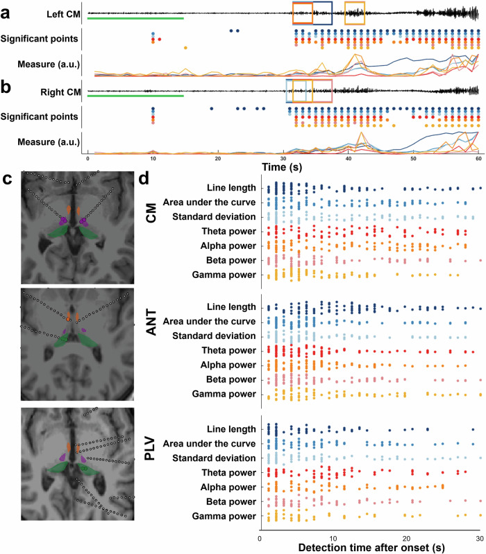
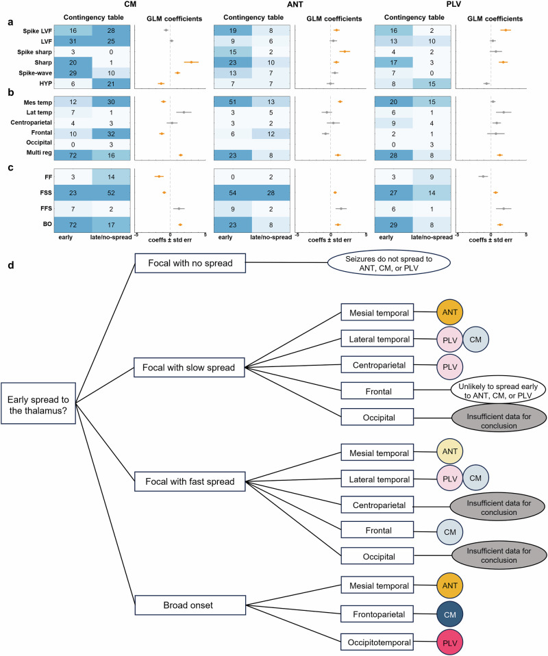
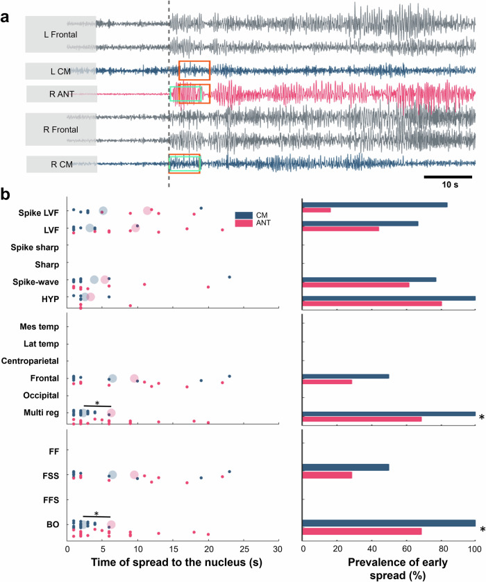
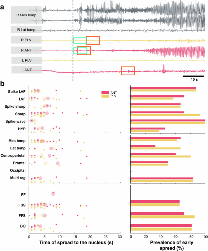
Methods: To better choose thalamic targets for stimulation, we studied how different seizures spread to each stimulation target. Expert reviews and automated tools were used to identify seizure spread recorded from invasive recordings. We categorized seizures based on how they start and spread, and determined whether seizures reached thalamic areas early or late. We used generalized linear models (GLM) to evaluate which seizure properties are predictive of time of spread to the thalamus, testing effect significance using Wald tests.

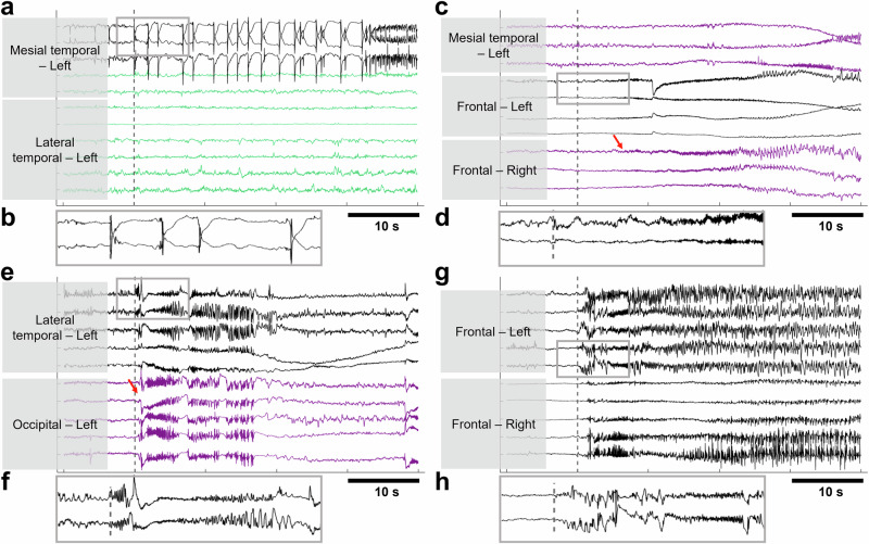
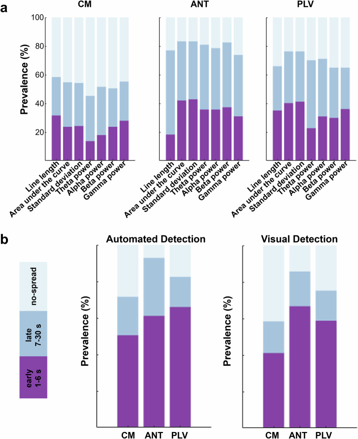
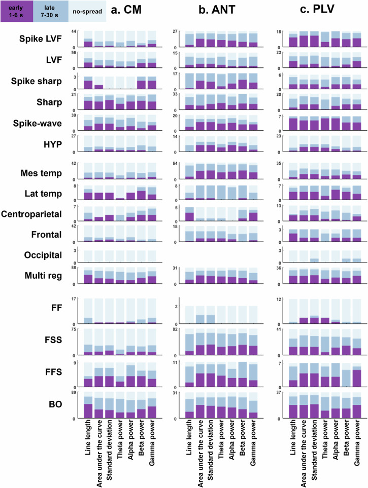
Results: We show that seizures with <2 Hz synchronized-spiking patterns do not spread early to the thalamus, while seizures starting with faster activity (<20 Hz) spread early to all thalamic areas. Most importantly, seizures that begin broadly across the brain quickly recruit the centromedian and pulvinar areas, suggesting these may be better stimulation targets in such cases. Alternatively, seizures that start deep in the temporal lobe tend to involve the anterior part of the thalamus, meaning the centromedian might not be the best choice for those seizures.

Conclusions: Our results suggest that by analyzing electrical activity during seizures, we can better predict which parts of the thalamus are involved. This could lead to more effective stimulation treatments for people with epilepsy.

Plain language summary

Some people with epilepsy don’t get relief from standard treatments and may need brain stimulation to help control their seizures. The thalamus, a deep brain structure, is a promising area for this kind of therapy, but clinicians still don’t know exactly which people with epilepsy will benefit most. In our study, we found that certain brainwave patterns during seizures can help identify which part of the thalamus is most involved. This could lead to more personalized and effective brain stimulation treatments for different types of epilepsy.


Conflict of interest statement

Competing interests: The authors declare no competing interests.

Figures

Fig. 1
Fig. 1. Examples of seizures with different onset and spread patterns.
a A seizure recorded from patient 24 with an onset in left mesial temporal region, with no spread to other regions (FF seizure). b The expanded trace of the selected gray rectangle in a shows a hypersynchronous (HYP) spike pattern at the onset of the seizure. c A low voltage fast (LVF) activity at a seizure from patient 18 with a left frontal onset that propagates (red arrow) after a few seconds to other areas such as right frontal and left mesial temporal regions. d The expanded trace shows LVF activity at the onset of the seizure. e A seizure recorded from patient 9 with a sharp (f) onset in left lateral temporal area that spreads within milliseconds (red arrow) to other areas such as left occipital. g A spike-wave (h) broad seizure started simultaneously in left and right frontal regions of patient 16. Black traces indicate the contacts that show seizure activity simultaneously at the onset. Purple traces are the contacts with seizure spread. Green traces are the contacts with no ictal activity during the seizure.
Fig. 2
Fig. 2. Detecting seizure spread in the thalamus with different measures.
a, b (bottom traces) Changes in the measures over time. a, b (middle traces) The time at which these measures were significant (i.e., exceeded twice the standard deviation of the reference period (green line)). a, b (top traces) A recorded signal from left and right CM, where colored rectangles signify detecting a spread of activity by a specific measure. The time at which the 3 s significant increase from the background has started is identified as the time of the spread for that specific measure, ranging from 1–30 s. c Example of the anatomical location of the electrodes (dotted lines) within CM (purple), ANT (orange), and PLV (green). d Swarm chart showing the time of detected spread by any of the measures to CM (n = 190 seizures), ANT (n = 126 seizures), and PLV (n = 97 seizures). Line length and gamma power measure could detect a spread of activity within the CM faster than other measures, but this was only significant in comparison to detection by theta and alpha power (Kruskal–Wallis p = 0.002). Within the ANT line length detected the spread later than all measures other than gamma power (Kruskal–Wallis p = 0.0004). Otherwise, these distributions were broadly similar across other measures and across all measures within PLV. (CM Centromedian, ANT anterior nucleus of thalamus, PLV pulvinar, a.u. arbitrary unit, s second).
Fig. 3
Fig. 3. Distribution of spread to different thalamic nuclei.
a The prevalence of early, late, and no-spread as detected by each measure. For example, line length detected more early spread in the CM cohort while missing most of the spread in ANT. b Automated and visual detection performed similarly in identifying the seizure spread, with the automated method detecting higher early spread only within the CM cohort. Around 75% of seizures spread to CM (purple and steel blue), with around 50% of seizures having an early spread (purple). Seizure spread (purple and steel blue) to ANT is prominent, however this spread is early (purple) for ~60% of seizures. Around 60% of seizures have an early spread to PLV. Comparing a and b, it is clear that while each single measure may only detect an early spread in less than 40% of seizures, using a combination of the measures can help identify spread of more seizures.
Fig. 4
Fig. 4. Distribution of different categories of spread to different thalamic nuclei.
a Most seizures with sharp and spike-wave patterns spread to CM. Seizures with a broad onset in multi regions often spread to CM. b Most seizures with an onset in mesial temporal areas, or with a spread (non-FF seizures) may have an early or late spread to the ANT. c Seizures with spike LVF and spike-wave patterns, lateral temporal seizures or seizures with an onset in multiple regions may have an early (purple) spread to PLV. (LVF low-voltage fast, HYP hypersynchronous, Mes temp mesial temporal, Lat temp lateral temporal, FF focal onset remaining focal, FSS focal onset with slow spread, FFS focal onset with fast spread, BO broad onset, s second).
Fig. 5
Fig. 5. Evaluating the effect of each seizure type on the time of spread to different thalamic nuclei.
Seizures are classified based on their onset pattern (c), onset region (b), and pattern of spread (c). ac Contingency tables showing the number of seizures with early and late/no-spread in each category. The shades of blue indicate higher (dark blue) or lower (light blue) number of occurrences. The plots show the coefficients of GLMs with an “early-or-not-early” response variable. Markers show the coefficients ± standard errors for each category predictor (seizure type). For each category, positive (negative) coefficients suggest a positive (negative) association with the occurrence of the response variable. The coefficients highlighted in yellow indicate that the specific predictor significantly affects the response variable (p < 0.05; the exact p values can be found in Supplementary Table 1). The missing coefficients correspond to variables where their response is invariant (always 0 or 1; e.g., Spike sharp in CM group). Therefore, no meaningful coefficient could explain the relationship between the predictor and response variable. d Decision tree showing possible nuclei to which seizures may have an early spread. Darker coloring indicates significance (binomial test; dark shades p values from top to bottom: 0.000108, 0.0016, 2.82 × 10⁹, 7.53 × 10⁶). We note that in groups with lighter coloring, while at least 60% of seizures show early spread to selected nuclei, there are not enough seizures (<10) to perform a meaningful statistical analysis. (CM centromedian, ANT anterior nucleus of the thalamus, PLV pulvinar, LVF low-voltage fast, HYP hypersynchronous, Mes temp mesial temporal, Lat temp lateral temporal, FF focal onset remaining focal, FSS focal onset with slow spread, FFS focal onset with fast spread, BO broad onset).
Fig. 6
Fig. 6. Seizure spread to centromedian (CM) and anterior nucleus of the thalamus (ANT).
a A seizure recorded from Pt 22 with a broad multi-regional spike-wave pattern at the onset (example recordings of right and left frontal region onsets are shown here). Automated (orange rectangle) and visual (green rectangle) methods of spread detection agree on the time of spread to the right ANT and right CM. b Time of spread to the CM and ANT in different seizure types. The small solid circles indicate the time of spread for each seizure. The larger circles with light shading indicate the average time of spread for each category. There are no significant differences between the two nuclei for the times of spread of seizures in most of the cases, except for the seizures with broad onset from multi-regions where seizures spread significantly earlier to CM than ANT (p = 0.01), and the early spread occurs in significantly larger number of seizures (p = 0.02). (LVF low-voltage fast, HYP hypersynchronous, Mes temp mesial temporal, Lat temp lateral temporal, FF focal onset remaining focal, FSS focal onset with slow spread, FFS focal onset with fast spread, BO broad onset).
Fig. 7
Fig. 7. Seizure spread to centromedian (CM) and pulvinar (PLV).
a A sharp onset seizure recorded from Pt 23 with an onset in the right centroparietal region that spreads to the rest of the brain after a few seconds. The time of spread to ipsilateral CM and PLV, identified by both automated (orange rectangle) and visual (green rectangle) methods, shows that the spread was detected slightly earlier in the PLV compared to CM. b The small solid circles indicate the time of spread for each seizure. The larger circles with light shading indicate the average time of spread for each category. Seizures recorded from patients with both CM and PLV implants tend to spread faster to PLV, especially the seizures with spike LVF patterns and onset within mesial temporal regions, but this did not reach significance, as the number of seizures in these categories is small within this cohort. (LVF low-voltage fast, HYP hypersynchronous, Mes temp mesial temporal, Lat temp lateral temporal, FF focal onset remaining focal, FSS focal onset with slow spread, FFS focal onset with fast spread, BO broad onset).
Fig. 8
Fig. 8. Seizure spread to anterior nucleus of the thalamus (ANT) and pulvinar (PLV).
a A seizure with a sharp mesial temporal onset recorded from Pt 13 with identifiable spreads to both ANT and PLV (orange: automated detection; green: visual detection). b The small solid circles indicate the time of spread for each seizure. The larger circles with light shading indicate the average time of spread for each category. No significant differences were found between the time of spread of different seizures to ANT and PLV. Note that most of the patients with electrodes within both these nuclei had seizures rising from mesial temporal areas. We showed that mesial temporal seizures have a high likelihood of spreading early to ANT, and when comparing simultaneously, these seizures may equally have an early spread to PLV. (LVF low-voltage fast, HYP hypersynchronous, Mes temp mesial temporal, Lat temp lateral temporal, FF focal onset remaining focal, FSS focal onset with slow spread, FFS focal onset with fast spread, BO broad onset).

References

    1. Kwan, P. & Brodie, M. J. Early identification of refractory epilepsy. N. Engl. J. Med.342, 314–319 (2000). - PubMed
    1. Thijs, R. D., Surges, R., O’Brien, T. J. & Sander, J. W. Epilepsy in adults. Lancet393, 689–701 (2019). - PubMed
    1. Englot, D. J. & Chang, E. F. Rates and predictors of seizure freedom in resective epilepsy surgery: an update. Neurosurg. Rev.37, 389–405 (2014). - PMC - PubMed
    1. Ma, B. B. et al. Responsive neurostimulation for regional neocortical epilepsy. Epilepsia61, 96–106 (2020). - PubMed
    1. Ryvlin, P., Rheims, S., Hirsch, L. J., Sokolov, A. & Jehi, L. Neuromodulation in epilepsy: state-of-the-art approved therapies. Lancet Neurol.20, 1038–1047 (2021). - PubMed

LinkOut - more resources