. 2025 Aug;39(8):1848-1856.
doi: 10.1038/s41375-025-02642-2. Epub 2025 May 22.

KK2845, a PBD dimer-containing antibody-drug conjugate targeting TIM-3-expressing AML

Affiliations

KK2845, a PBD dimer-containing antibody-drug conjugate targeting TIM-3-expressing AML

Jian Zou et al. Leukemia. 2025 Aug.

Abstract

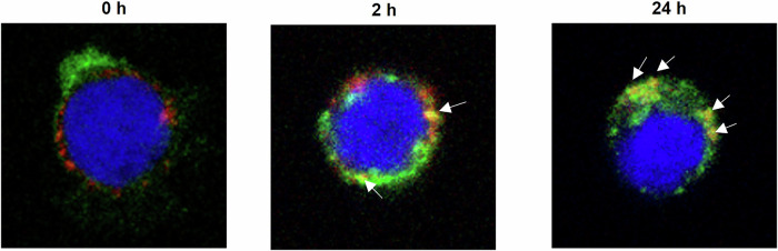
Acute myeloid leukemia (AML) is a common hematopoietic malignancy with high recurrence rates, and there is an urgent need for new therapeutic agents. T-cell immunoglobulin mucin-3 (TIM-3) is expressed on the surface of both LSCs and blasts in most AML patients, but not in normal hematopoietic stem cells (HSCs). We have developed KK2845, an antibody drug conjugate (ADC) that consists of an anti-TIM-3 fully human IgG1 antibody, a valine-alanine linker and a highly potent DNA cross-linking pyrrolobenzodiazepine (PBD) dimer SG3199. KK2845 exhibited potent cytotoxicity against AML cells both in vitro and in vivo. The cytotoxicity against AML cells was almost comparable between KK2845 and CD33-ADC, an anti-CD33 antibody conjugated with PBD dimer that has shown high remission rates in clinical studies. In addition to the cytotoxicity depending on PBD dimer, KK2845 also showed potent antibody-dependent cell cytotoxicity (ADCC) activity against AML cells. KK2845 showed less cytotoxicity against human normal bone marrow cells than CD33-ADC. The pharmacokinetics of KK2845 in cynomolgus monkey after intravenous infusion demonstrated a favorable profile. Taken together, these data suggest that KK2845 could be a novel ADC therapeutic in AML.


Conflict of interest statement

Competing interests: J.Z., H.K., S.T., T.I., T.A., K.N., S.K., M.A., H.T., and H.S. are employees of Kyowa Kirin. T.S., Y.K., and K.A. are collaborators who received financial support from Kyowa Kirin. Ethics statement: The use of human-derived materials was approved by the Research Ethics Review Committee of Kyowa Kirin Co., Ltd., Kyushu University, or the vendor’s ethical regulations. Informed consent was obtained from all human subjects. All experiments involving animals were approved by the Institutional Animal Care and Use Committee and performed in accordance with the animal welfare guidelines of Kyowa Kirin Co., Ltd., and Shin Nippon Biomedical Laboratories, Ltd., both of which are accredited by AAALAC International.

Figures

Fig. 1
Fig. 1. Structure of KK2845.
KK2845 is an ADC that consists of an anti-TIM-3 fully human IgG1 antibody, a valine-alanine linker, and a highly potent DNA cross-linking PBD dimer SG3199.
Fig. 2
Fig. 2. Localization and trafficking of KK2845.
KK2845-Alexa488 was incubated with Kasumi-3 cells on ice (0 hour), or at 37 °C for 2 hours or 24 hours. The Alexa Fluor 488 signal is displayed in green. The LysoTracker signal is displayed in red. The DAPI signal is displayed in blue. Yellow colored signals indicated by arrows show colocalization of KK2845-Alexa488 and Lysotracker. Original magnification ×100 for all the imaging.
Fig. 3
Fig. 3. Cytotoxicity of KK2845 against AML cells.
A-C Kasumi-3, EoL-1/hTIM-3, or EoL-1 cells were incubated with KK2845 (0.01-10000 ng/mL) or KM8047-SG3249 (0.01-10000 ng/mL) at 37 °C for 4 days. CCK-8 was then added to these cells, and the absorbance was measured. The mean ± SD cell viability (%) is shown (n = 3). The data presented are representative results from three independent tests.
Fig. 4
Fig. 4. Cytotoxicity of KK2845 against primary AML cells.
A-D The primary AML cells were incubated with KM8047-SG3249, KK2845, or CD33-ADC at 37 °C for 4 days. The payload of the CD33-ADC used in this study was SGD-1910. The living cells in the CD34+ fraction, CD34+CD38- fraction, or CD34- fraction were then counted by flow cytometry. Using the number of viable cells in the untreated group as 100%, the percentage of living cells in each treatment group was calculated. A, B Lot: AML818BM. The CD34- fraction was not evaluated due to a low number of cells. C Lot: AML817BM. The CD34+ fraction was not evaluated due to a low number of cells. D Lot: AML942 II BM. The CD34- fraction was not evaluated due to a low number of cells.
Fig. 5
Fig. 5. ADCC activity of KK2845.
A-C EoL-1/hTIM-3 cells were incubated with KK2845 (0.001-10 μg/mL) or KM8047-SG3249 (10 μg/mL) in the presence of human PBMCs at 37 °C for 4 hours. After incubation, the number of viable EoL-1/hTIM-3 cells and BD Trucount beads were counted by flow cytometry. The number of living EoL-1/hTIM-3 cells was normalized by the number of beads. ADCC activity was calculated as follows: ADCC activity (%) =100 - (Normalized cell number of each sample / Average (n = 4) of normalized cell number of sample A) ×100. The mean + SD is shown (n = 4). A PBMC donor 1, B PBMC donor 2, C PBMC donor 3.
Fig. 6
Fig. 6. Cytotoxicity of KK2845 against human normal bone marrow cells.
A-B CD34+ bone marrow cells, or myeloid progenitor cells were incubated with PBS or increasing concentrations of KM8047-SG3249, KK2845, or CD33-ADC (0.1-1000 ng/mL) for 4 days. The payload of the CD33-ADC used in this study was SG3249. To assess cell viability, CellTiter-Glo® reagent was then added to these cells, and the luminescence was measured. Using cell luminescence, the percentage in each treatment group was calculated by setting the PBS treatment group (untreated control) as 100%. The mean ± SD of cell viability (%) is shown (n = 3). The data presented are representative results from two independent tests.
Fig. 7
Fig. 7. Anti-tumor activity of KK2845 in murine models of AML.
SCID mice were subcutaneously inoculated with Kasumi-3 (A) or CMK11-5 cells B. Vehicle, KK2845, or KM8047-SG3249 was intravenously administered once on day 1. Each plot represents the mean ± SE of tumor volume. (A: n = 7, B: n = 5). The difference in tumor volume on day 14 (A, B) between KK2845 and KM8047-SG3249 treated groups at each dose, was analyzed by Student’s t test or the Aspin-Welch test. *: p < 0.05. C A scheme for KK2845 efficacy evaluation using a disseminated model is presented. EoL-1/hTIM-3 cells were intravenously inoculated into NOD-SCID mice. After confirming EoL-1/TIM-3 engraftment in the bone marrow of satellite mice (n = 2) by detecting human CD45+ cells using flow cytometry, vehicle, 0.3 mg/kg of KM8047-SG3249, KK2845, or CD33-ADC was intravenously administered once. The payload of the CD33-ADC used in this study was SG3249. Five days after the administration of ADC, bone marrow cells were collected from the femur of each mouse. The residual cancer cells were analyzed by flow cytometry. D Each plot represents the individual value for the percentage of residual cancer cells. Horizontal lines represent the mean of each group (n = 10). E Overall survival curves in the disseminated EoL-1/hTIM-3 xenograft model. EoL-1/hTIM-3 cells were intravenously inoculated into NOD-SCID mice. After confirming EoL-1/TIM-3 engraftment in the bone marrow of satellite mice, vehicle, 0.3 mg/kg of KM8047-SG3249 or KK2845 was intravenously administered once. The survival time of the mice was monitored. The difference in overall survival between KK2845 and KM8047-SG3249 treated groups was evaluated by Log-Rank test (n = 8/group). *: p < 0.001. F The combination of KK2845, Venetoclax and Aza using subcutaneous xenograft model. SCID mice were inoculated with Kasumi-3 cells. Mice received 0.5 mg/kg KK2845 alone (single administration, i.v.), 100 mg/kg venetoclax (qd×7, p.o.) in combination with 5 mg/kg Aza (qd×7, i.v.), or 0.5 mg/kg KK2845 in combination with 100 mg/kg venetoclax and 5 mg/kg Aza from day 1. The time course of the mean tumor volume is shown. Each plot represents the mean ± SE of tumor volume (n = 5). The difference in tumor volume on day 21 between non-treatment and the triple-combination (KK2845, venetoclax and Aza), the dual-combination (venetoclax and Aza) and the triple-combination, or KK2845 monotherapy and the triple-combination were analyzed by the Aspin-Welch test. ‡: p < 0.05, difference between non-treatment and the triple-combination. #: p < 0.05, difference between the dual-combination and the triple-combination. *: p < 0.05, difference between KK2845 monotherapy and the triple-combination.

References

    1. Bonnet D, Dick JE. Human acute myeloid leukemia is organized as a hierarchy that originates from a primitive hematopoietic cell. Nat Med. 1997;3:730. - PubMed
    1. Hope KJ, Jin L, Dick JE. Acute myeloid leukemia originates from a hierarchy of leukemic stem cell classes that differ in self-renewal capacity. Nat Immunol. 2004;5:738–43. - PubMed
    1. Lapidot T, Sirard C, Vormoor J, Murdoch B, Hoang T, Caceres-Cortes J, et al. A cell initiating human acute myeloid leukaemia after transplantation into SCID mice. Nature. 1994;367:645–8. - PubMed
    1. Shimony S, Stahl M, Stone RM. Acute myeloid leukemia: 2023 update on diagnosis, risk-stratification, and management. Am J Hematol. 2023;98:502–26. - PubMed
    1. Siegel RL, Miller KD, Fuchs HE, Jemal A. Cancer Statistics, 2021. CA Cancer J Clin. 2021;71:7–33. - PubMed

MeSH terms