. 2025 Jul;30(7):1309-1318.
doi: 10.1007/s10147-025-02775-5. Epub 2025 May 22.

Distant parenchymal recurrence during long-term use of TTFields treatment for glioblastoma

Affiliations

Distant parenchymal recurrence during long-term use of TTFields treatment for glioblastoma

Yuhei Takido et al. Int J Clin Oncol. 2025 Jul.

Abstract

Background: Tumor treating fields (TTFields) treatment has been an important option for the treatment of glioblastoma. The introduction of novel treatment options may lead to distinct recurrence patterns compared to those observed with conventional therapies; however, the specific recurrence pattern during TTFields treatment has not been elucidated.

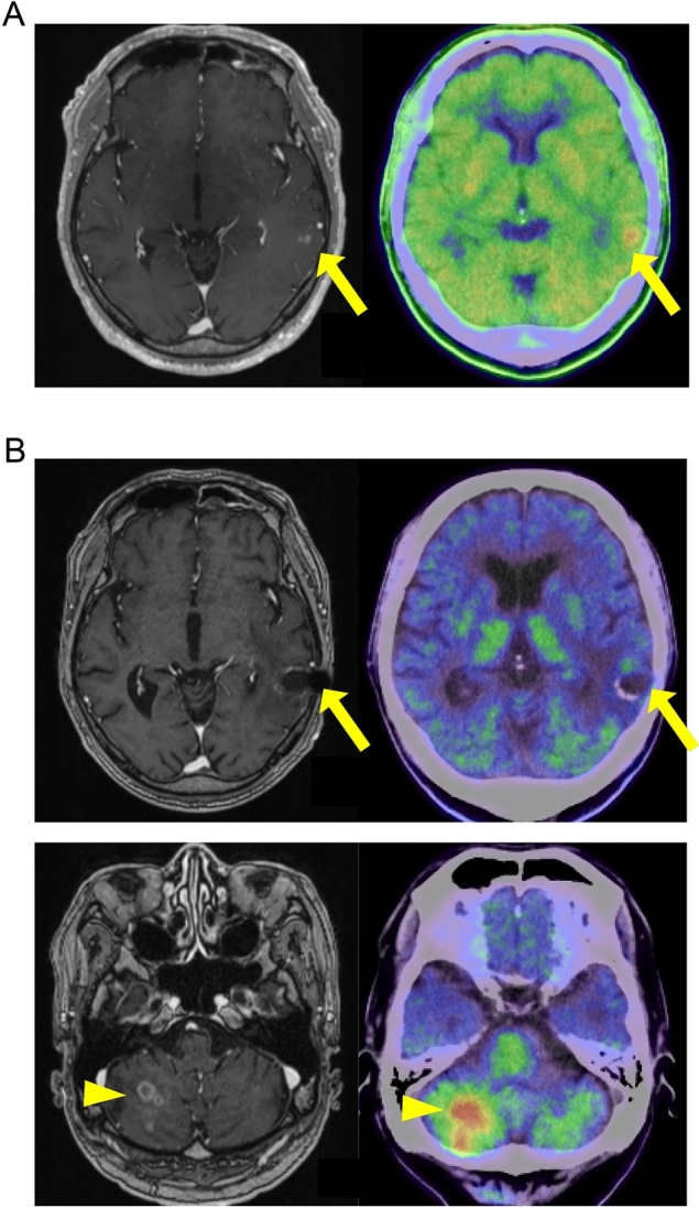
Methods and results: Here, we analyzed 39 cases of glioblastoma treated with TTFields. Although a usage rate of more than 75% is recommended, among 39 cases, 18 discontinued TTFields treatment owing to requests by patients with lower usage rates. In these discontinued cases, patients exhibiting sensory aphasia were more frequently included compared to those who continued TTFields (44.4%, p < 0.001). Among 21 cases involving patients who continued TTFields, tumor recurrence was observed in 15 of those cases. Five out of 15 cases (33.3%) exhibited recurrence in distant parenchyma from the primary lesion. A higher usage rate and relatively longer use of TTFields were observed in these five cases, along with more favorable progression-free survival than those in the other 10 cases (p = 0.019, p = 0.040, and p = 0.024, respectively). In one case, recurrent tumors with lower grade glioma histology but molecular markers characteristic for glioblastoma, IDH-wildtype were indentified. This tumor arose in an area that received a lower local minimum power density of TTFields compared to the primary lesion, following long-term TTFields therapy.

Conclusions: Long-term use of TTFields might be correlated with a high frequency of distant parenchymal recurrence in cases with favorable response.

Keywords: Distant recurrence; Glioblastoma; TTFields treatment.


Conflict of interest statement

Declarations. Conflict of interest: Fumiharu Ohka: lecture fee from Novocure. Shoichi Deguchi: lecture fee from Novocure. Kazuya Motomura: lecture fee from Novocure. Ryuta Saito: lecture fee from Novocure.

Figures

Fig. 1
Fig. 1
Kaplan–Meier curve indicating PFS. Kaplan–Meier curve indicating PFS of patients with distant parenchymal recurrence (n = 5; blue line) and patients without distant parenchymal recurrence (n = 10; orange line; p = 0.024)
Fig. 2
Fig. 2
MRI and Methionine-PET images of Case 1. (A) Pre-operative MRI contrast-enhanced T1-weighted image (left) and Methionine-PET (right). Yellow arrow indicates tumor lesion. (B) MRI contrast-enhanced T1-weighted image after radiation therapy concomitant with TMZ. Yellow arrow indicates tumor lesion. (C) MRI contrast-enhanced T1-weighted image (left) and Methionine-PET (right) of the primary lesion (upper) and cerebellum (lower) on recurrence. Yellow arrows and arrow heads indicate primary lesions and recurrent lesions, respectively
Fig. 3
Fig. 3
MRI, Methionine-PET, and Hematoxylin and Eosin (HE) images of Case 2. (A) Pre-operative (left) and post-operative (right) MRI contrast-enhanced T1-weighted image. Yellow arrows indicate tumor lesion. (B) HE image of primary tumor. A scale bar indicates 100 um. (C) MRI contrast-enhanced T1-weighted image (left), FLAIR images (middle), and Methionine-PET (right) of the recurrent lesion. Axial image (upper and middle) and coronal image (lower). Yellow arrows and arrow heads indicate recurrent tumors around primary lesions and on distant parenchyma, respectively. (D) HE image (left) and IHC using anti-Ki67 antibody (right) of recurrent tumor around the primary lesion (upper) and on distal parenchyma (lower). Scale bars indicate 100 um
Fig. 4
Fig. 4
Electric fields map for recurrent lesions of Case 2. (A) Axial (left) and sagittal (right) images of electric fields for a recurrent tumor on the primary lesion. The purple, red or green line indicates gross tumor volume (GTV; 1.8 LMiPD), clinical target volume (CTV; 1.5 LMiPD) and peritumoral boundary zone (PBZ; 1.5 LMiPD), respectively. (B) Axial (left, upper), sagittal (right, upper), and coronal (left, lower) image of electric fields for recurrent tumor on distant parenchyma. The purple line indicates GTV (1.1 LMiPD) of a recurrent lesion

References

    1. Louis DN, Perry A, Wesseling P et al (2021) The 2021 WHO classification of tumors of the central nervous system: a summary. Neuro Oncol 23(8):1231–1251 - PMC - PubMed
    1. Ceccarelli M, Barthel FP, Malta TM et al (2016) Molecular profiling reveals biologically discrete subsets and pathways of progression in diffuse glioma. Cell 164(3):550–563 - PMC - PubMed
    1. Brennan CW, Verhaak RG, McKenna A et al (2013) The somatic genomic landscape of glioblastoma. Cell 155(2):462–477 - PMC - PubMed
    1. Kirson ED, Gurvich Z, Schneiderman R et al (2004) Disruption of cancer cell replication by alternating electric fields. Can Res 64(9):3288–3295 - PubMed
    1. Kirson ED, Dbalý V, Tovarys F et al (2007) Alternating electric fields arrest cell proliferation in animal tumor models and human brain tumors. Proc Natl Acad Sci U S A 104(24):10152–10157 - PMC - PubMed

LinkOut - more resources