. 2025 May 22;25(1):184.
doi: 10.1186/s12935-025-03813-y.

N-phenylmaleimide induces bioenergetic switch and suppresses tumor growth in glioblastoma tumorspheres by inhibiting SLC25A11

Affiliations

N-phenylmaleimide induces bioenergetic switch and suppresses tumor growth in glioblastoma tumorspheres by inhibiting SLC25A11

Hye Joung Cho et al. Cancer Cell Int. .

Abstract

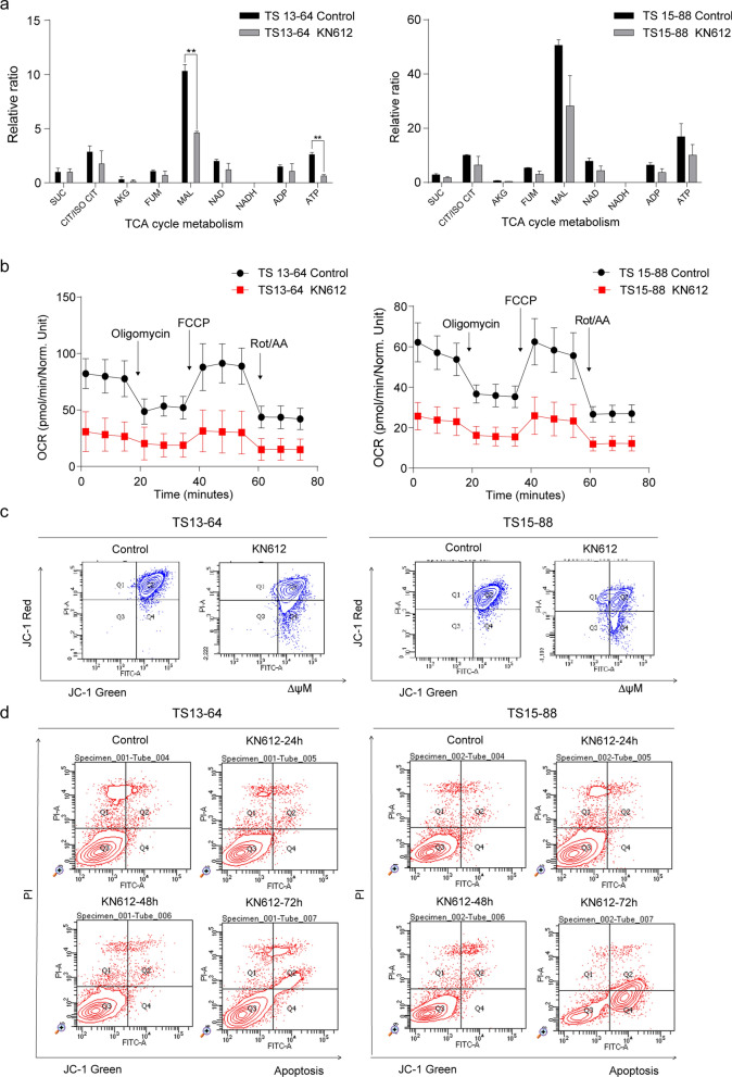
Background: Glioblastoma (GBM) is a highly resistant tumor, and targeting its bioenergetics could be a potential treatment strategy. GBM cells depend on cytosolic nicotinamide adenine dinucleotide (NADH), which is transported into the mitochondria via the malate-aspartate shuttle (MAS) for ATP production. N-phenylmaleimide (KN612) is a MAS inhibitor that targets SLC25A11, an antiporter protein of the MAS. Therefore, this study investigated the effects of KN612 in GBM treatment using in vitro and in vivo models.

Methods: We examined the biological effects of KN612 in GBM tumorspheres (TSs), including its effects on cell viability, ATP level, cell cycle, stemness, invasive properties, energy metabolic pathways, and transcriptomes. Additionally, we investigated the in vivo efficacy of KN612 in a mouse orthotopic xenograft model.

Results: Transcriptomic analysis showed that SLC25A11 mRNA expression was significantly higher in GBM TSs than in normal human astrocytes. Additionally, siRNA-mediated SLC25A11 knockdown and KN612-mediated MAS inhibition decreased the oxygen consumption rate, ATP levels, mitochondrial activity, and cell viability in GBM TSs and decreased the stemness and invasion ability of GBM cells. Moreover, gene ontology functional annotation indicated that KN612 treatment inhibited cell-cycle and mitotic processes. Furthermore, KN612 treatment reduced tumor size and prolonged survival in an orthotopic xenograft model.

Conclusions: Targeting GBM bioenergetics using KN612 may represent a novel and effective approach for GBM treatment.

Keywords: Bioenergetics; Glioblastoma; KN612; Malate-aspartate shuttle; SLC25A11.


Conflict of interest statement

Declarations. Ethics approval and consent to participate: All applicable international, national, and/or institutional guidelines for the care and use of animals were followed. All experiments involving animals followed the ethical standards of the institution or practice at which the studies were conducted. Consent for publication: Not applicable. Competing interests: The authors declare no competing interests.

Figures

Fig. 1
Fig. 1
Targeting the SLC25A11 gene in GBM TSs. Cell viability, ATP production and mitochondrial function were measured 48 h after transfection with si-SLC25A11. a SLC25A11 expression levels in normal human astrocytes (NHA; n = 5) and GBM TS (n = 35) were determined using RNA-Seq. b Western blotting to detect SLC25A11 expression in GBM TSs. c Western blotting to detect SLC25A11 expression in TS13-64 and TS15-88 following transfection with siRNA. d Cell viability and e ATP levels were measured in TS13-64 and TS15-88 after SLC25A11 knockdown. f Oxygen consumption rate (OCR) was measured in TS13-64 and TS15-88 after SLC25A11 knockdown. g Mitochondrial membrane potential was measured in TS13-64 and TS15-88 after SLC25A11 knockdown. Differences between groups were evaluated using Welch’s ANOVA and Games-Howell post hoc tests. The data are presented as the mean ± standard deviation (SD); *p < 0.05, **p < 0.01 and ***p < 0.001
Fig. 2
Fig. 2
Effects of KN612 on the expression of malate-aspartate shuttle (MAS)-related genes, cell viability, and energy levels in GBM TS. a Chemical structure of KN612. b Western blotting to detect MAS-related proteins following KN612 treatment. c Heatmaps visualizing changes in the mRNA levels of MAS-related genes after KN612 treatment. d Cell viability was determined using the MTT assay. e ATP level was measured using the luminescent ATP assay kit. f NADH/NAD+ ratio was measured in GBMTSs treated with the indicated concentration of KN612. Differences between groups were evaluated using Welch’s ANOVA and Games-Howell post hoc tests. Data are presented as the mean ± standard deviation (SD); *p < 0.05, **p < 0.01 and ***p < 0.001
Fig. 3
Fig. 3
Effects of KN612 on mitochondria function and cell death in GBM TSs. Determination of mitochondrial membrane potential and energy metabolism levels was performed 72 h after KN612 treatment. a Metabolite levels were measured using LC–MS/MS. b Oxygen consumption rate (OCR) was measured using the Seahorse XFe96 analyzer. c Mitochondrial membrane potential (∆ψM) was assessed using JC-1 staining and flow cytometry, with green fluorescence indicating increased apoptosis. d Representative dot plot data illustrating both early and late apoptosis were used to assess apoptosis via the annexin V assay. *p < 0.05, **p < 0.01, and ***p < 0.001 relative to the control group
Fig. 4
Fig. 4
Effects of KN612 on the stemness and invasiveness of GBM TSs. Evaluation of stemness and invasiveness was performed 72 h after KN612 treatment. a Stemness was assessed using neurosphere formation assay, which measured both the sphere formation capacity and the percentage of sphere radius. b Invasive potential was assessed using a 3D invasion assay. c Western botting was performed to examine the expression of proteins associated with stemness and d invasion. e Heatmaps displaying expression levels of genes associated with stemness and invasion. Significant differences were determined unpaired Student's t-test (means ± SD; *p < 0.05, **p < 0.01, and ***p < 0.001)
Fig. 5
Fig. 5
Effects of KN612 on gene expression profile. RNA-seq was performed to examine the transcriptomic profile of TS13-64 after KN612 treatment for 72 h. a Heatmap of DEGs between the control and KN612 treatment groups. DEGs were defined as genes with p < 0.05 and log2 fold change cutoff of 0.1. b Volcano plot showing DEGs based on p-value and fold change. Red, yellow, and blue dots indicate genes with p < 0.05, log2 (fold change) > 0.5, or both, respectively. c Visualization of genes related to the MAS pathway based on REACTOME pathway analysis. d Upregulated and downregulated enriched genes following KN612 treatment
Fig. 6
Fig. 6
Therapeutic effects of KN612 in an orthotopic xenograft model. TS13-64 luc cells were pretreated with 10 µM of KN612 for 72 h. Control (n = 5), KN612 (n = 5). a Tumor volume was measured using bioluminescence imaging. Signal intensity was quantified as total photon flux from tissues. One-way ANOVA with Tukey’s post hoc test (*p < 0.05). b Kaplan–Meier curves were used to estimate survival probabilities, and statistical significance was evaluated using the log-rank test. Log-rank test (p < 0.05). c Hematoxylin and eosin (H&E) staining (magnification × 2, × 20) was performed on brain sections from sacrificed mice to assess the size and extent of tumor masses. d, e Immunohistochemistry was used to examine SLC25A11 protein expression via brown staining, and to identify invading cells via Zeb1 staining (magnification × 2, × 20, × 40). Zeb1 quantification was conducted using images captured at × 40 magnification. In total, 10 randomly selected images per group were analyzed to quantify the number of infiltrating Zeb1-positive cells (means ± standard error of mean [SEM]; *p < 0.01 compared with control). Hematoxylin (blue) was used to counterstain nuclei in all images. f Schematic overview of the study. KN612 inhibits SLC25A11 expression, inducing bioenergetic reprogramming and effectively reducing mitochondrial function, stemness, and invasiveness in GBM cells, ultimately leading to suppressed tumor growth

Similar articles

References

    1. Lee JH, Lee JE, Kahng JY, Kim SH, Park JS, Yoon SJ, Um JY, Kim WK, Lee JK, Park J, et al. Human glioblastoma arises from subventricular zone cells with low-level driver mutations. Nature. 2018;560(7717):243–7. - PubMed
    1. Park J, Shim JK, Kang JH, Choi J, Chang JH, Kim SY, Kang SG. Regulation of bioenergetics through dual inhibition of aldehyde dehydrogenase and mitochondrial complex I suppresses glioblastoma tumorspheres. Neuro Oncol. 2018;20(7):954–65. - PMC - PubMed
    1. Stupp R, Hegi ME, Mason WP, van den Bent MJ, Taphoorn MJ, Janzer RC, Ludwin SK, Allgeier A, Fisher B, Belanger K, et al. Effects of radiotherapy with concomitant and adjuvant temozolomide versus radiotherapy alone on survival in glioblastoma in a randomised phase III study: 5-year analysis of the EORTC-NCIC trial. Lancet Oncol. 2009;10(5):459–66. - PubMed
    1. Yoon SJ, Park J, Jang DS, Kim HJ, Lee JH, Jo E, Choi RJ, Shim JK, Moon JH, Kim EH, et al. Glioblastoma cellular origin and the firework pattern of cancer genesis from the subventricular zone. J Korean Neurosurg Soc. 2020;63(1):26–33. - PMC - PubMed
    1. Roh TH, Park HH, Kang SG, Moon JH, Kim EH, Hong CK, Ahn SS, Choi HJ, Cho J, Kim SH, et al. Long-term outcomes of concomitant chemoradiotherapy with temozolomide for newly diagnosed glioblastoma patients: a single-center analysis. Medicine. 2017;96(27): e7422. - PMC - PubMed

LinkOut - more resources