. 2025 May 22;17(1):114.
doi: 10.1186/s13195-025-01759-x.

Novel fully human high-affinity anti-TREM2 antibody shows efficacy in clinically relevant Alzheimer´s mouse model

Affiliations

Novel fully human high-affinity anti-TREM2 antibody shows efficacy in clinically relevant Alzheimer´s mouse model

Markus Kraller et al. Alzheimers Res Ther. .

Abstract

Background: New drugs to treat Alzheimer´s disease (AD) are urgently needed. Human triggering receptor expressed on myeloid cells 2 (hTREM2) is a validated drug target which is genetically associated with AD. Existing anti-hTREM2 antibodies were raised in animal immune systems, and subsequently humanized, which may incur immunological complications upon repeated preventive or therapeutic applications in vivo in AD patients. In addition, anti-hTREM2 antibodies should be optimized for both, efficacy and safety.

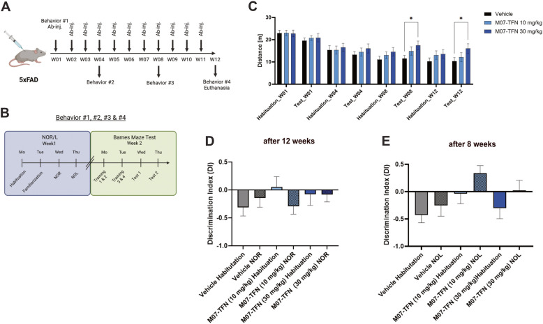
Methods: A novel fully human monoclonal brain-targeting anti-hTREM2 antibody M07-TFN was created. Binding affinities, cell viabilities, and agonist potencies were investigated on rhTREM2 and in human microglia. Transcytosis assays modeled blood-brain barrier translocation (BBB). Behavior tests were carried out in 5 × familiar AD (5xFAD) mice of both genders, to test for brain function and cognition as well as hippocampus-dependent spatial memory using the Barnes maze. In addition, amyloid plaque formation was determined on brain sections at the end of the study.

Results: M07-TFN showed higher binding affinities and stronger activation of hTREM2 signaling than all previously described anti-hTREM2 antibodies. p-Syk activation was increased 30-fold in hTREM2-overexpressing HEK293 cells and fourfold in human microglia cells compared to baseline. Human microglia viability significantly improved after stress testing. M07-TFN showed strong BBB translocation in a human BBB model, and exerted cross-reactivity to the mouse TREM2 stalk region, which allowed us to investigate M07-TFN directly in an AD mouse model. In 5xFAD mice, M07-TFN resulted in improved novel object location and better spatial orientation and memory, and significantly reduced plaque load. Additional safety investigations in mice showed no negative effects on blood cells or major organs.

Conclusion: Compared to existing humanized anti-hTREM2 antibodies that have been investigated in clinical trials, M07-TFN showed best-in-class affinities and agonist potencies. Being a fully human anti-hTREM2 antibody, M07-TFN holds the promise of reduced immunogenicity for use in human patients.

Keywords: Alzheimer’s disease; Antibody; Microglia; Neurodegeneration; TREM2.


Conflict of interest statement

Declarations. Ethics approval and consent to participate: All animal experiments were permitted by the local Ethics board in accordance with European and German animal welfare legislations (5.1–231 5682/LMU/BMC/CAM). The most recent version of the Animal Research: Reporting In Vivo Experiments (ARRIVE) guidelines was strictly followed. Consent for publication: Not applicable. Competing interests: Several authors are employees of the biotech company ISAR Bioscience which is, however, owned (100%) by a non-profit foundation.

Figures

Fig. 1
Fig. 1
M07 binds human and mouse TREM2 efficiently A Schematic representation of M07-WT and M07-TFN. Both antibodies contain the LALA modification (yellow stars). M07-TFN was modified with a blood–brain-barrier shuttle in both CH3 domains of the Fc region within the heavy chain (orange bars) B Binding of M07 to human full-length TREM2 or TREM1. Data represent the mean ± SEM (n = 3) C Binding of M07 to human or mouse TREM2 peptide fragment of the stalk region. Data represent the mean ± SEM (n = 4). D Competition ELISA with hTREM2 peptide to determine specific binding of M07 to hTREM2 peptide. Each dot represents an independent experiment. Data represent the mean ± SEM (n = 3)
Fig. 2
Fig. 2
M07 potently activates Syk signaling, decreased shedding and is shuttled across a human in vitro blood–brain barrier model A Titration curves of the anti-hTREM2 antibodies M07-WT and M07-TFN demonstrating strong activation of p-Syk signaling in HEK293 Flp-In hTREM2/hDAP12 cells. Data represent mean ± SEM (n = 3) B Titration of M07 antibodies for inhibition of TREM2 shedding in HEK293 Flp-In hTREM2/hDAP12 cells. M07-WT and M07-TFN inhibited TREM2 cleavage (one-way ANOVA; *, p < 0.05; **, p < 0.01). Each dot represents an independent experiment. The mean ± SEM is plotted (n = 3) C M07-TFN and anti-hTfR1 antibody MEM-189 were present in hCMEC/D3 lysates immediately after the pulse (0 h) with decreasing levels over time. M07-WT was not detected at significant levels. Two-way ANOVA; *, p < 0.05; **, p < 0.01; and ***, p < 0.001; and ****, p < 0.0001. Each dot represents an independent experiment. The mean ± SEM is plotted D M07-TFN and MEM-189 were detected in the chase media of both the apical and basolateral compartments at 5 and 24 h. M07-WT, at both 5 and 24 h, was present at background levels similar to the blank. Two-way ANOVA; *, p < 0.05; **, p < 0.01; and ***, p < 0.001; and ****, p < 0.0001. Each dot represents an independent experiment. The mean ± SEM is plotted
Fig. 3
Fig. 3
M07 activates hiPSC-derived microglia cells and increases their viability A Titration of p-Syk signaling in hiPSC-derived microglia upon antibody treatment: One-way ANOVA; *, p < 0.05, **, p < 0.01; ***, p < 0.001; ****, p < 0.0001; Data represent mean ± SEM (n = 5) B Titration of p-Syk signaling in hiPSC-derived microglia upon antibody treatment: One-way ANOVA; *, p < 0.05, **, p < 0.01; ***, p < 0.001; ****, p < 0.0001; Data represent mean ± SEM (n = 12) C Microglia viability assay upon stimulation with M07-WT and M07-TFN antibody. One-way ANOVA; **, p < 0.01; ***, p < 0.001; ****, p < 0.0001; Data represent mean ± SEM (n = 3)
Fig. 4
Fig. 4
Pharmacokinetic study of M07-TFN in 5xFAD mice. Serum concentrations of M07-TFN were determined in mice over time after injection of 30 mg/kg M07-TFN. 6 independent animals were used. Each dot represents one mouse. ELISA evaluation was repeated three times. Means are shown with standard deviations (SD)
Fig. 5
Fig. 5
Single application study of M07-TFN displays mild improvement of cognitive behavior in male 5xFAD mice A Timeline for Novel Object Recognition (NOR) and Novel Object Location (NOL) test setup with a 24 h delay between individual test phases B Scheme for Novel Object Recognition (NOR) and Novel Object Location (NOL) test. Mice were habituated to empty arenas and thereafter familiarized to two identical objects (L and R). Afterwards, a novel, equally attractive object was presented to the mice or the familiar object was moved to a new location (Relocated object) C Novel Object Recognition (NOR) after single antibody application followed by a 3 week chase. Animals in the different treatment groups showed similar preference for the familiar and novel object. DI = (Timenovel – Timefamiliar)/(Timenovel – Timefamiliar); One-way ANOVA; Data represent the mean ± SEM (of n = 12 independently measured animals in each group) D Novel Object Location (NOL) after single antibody application followed by a 3 week chase. DI = (Timenovel – Timefamiliar)/(Timenovel – Timefamiliar); One-way ANOVA; *, p < 0.05, Data represent the mean ± SEM (n = independently measured 12 animals in each group)
Fig. 6
Fig. 6
12-Week study – NOR/NOL in mixed gender 5xFAD mice A Design of the mouse study. Equal numbers of male and female mice were included in all study groups, respectively. Behavior was assessed prior to the first antibody application (W01). The behavior tests were performed every 4 weeks. Legend: behavior = AD-specific behavioral tests; Ab-inj. = administration of antibody; W = week; 5xFAD = AD mouse model B Timing of the behavioral study. NOR/L was performed in the first week on 4 consecutive days followed by the Barnes maze experiment in the second week C Distance the animals travel during the NOR was measured and showed reduced activities in the vehicle-treated group as compared to the animals treated with 30 mg/kg M07-TFN antibody. One-way ANOVA; *, p < 0.05, Data represent the mean ± SEM (n = independently measured 12 animals in each group) D Novel Object Recognition (NOR) after weekly antibody application followed by a 12-week chase. DI = (Timenovel – Timefamiliar)/(Timenovel – Timefamiliar); One-way ANOVA; Data represent the mean ± SEM (n = 12 independently measured animals in each group) E Novel Object Location (NOL) after weekly antibody application followed by an 8-week chase. DI = (Timenovel – Timefamiliar)/(Timenovel – Timefamiliar); One-way ANOVA; Data represent the mean ± SEM (n = 12 independently measured animals in each group)
Fig. 7
Fig. 7
12-week study—Barnes Maze in mixed gender 5xFAD mice A Scheme for Barnes Maze behavior testing. Mice were located at the middle of the platform (‘start’) and had to find a flight box located below one of the 20 holes. In the Training phase the box was located at the same location. For the Test situation the box was moved by 45°C (relocated flight box) B The time the animals needed to find the flight box for 0 (Training 1–1 to Test 1–2), 4 (Training 2–1 to Test 2–2) and 8 weeks (Training 3–1 to Test 3–2). Highlighted in the yellow box are the time points that are shown in C including statistical analysis C Statistical analysis of the 8-week time point of the Barnes Maze study. One-way ANOVA; *, p < 0.05, **, p < 0.01; Data represent the mean ± SEM (n = 12 independently measured animals in each group) D Representative images of strategies during the Barnes Maze study. Direct (mice move directly to the flight box), Wall (mice go through consecutive holes), Random (arbitrary pattern). Percentage of search strategies used to find the flight box of the Barnes maze test before the first antibody application and after 8 weeks
Fig. 8
Fig. 8
Anti-TREM2 antibody M07-TFN attenuates ß-Amyloid burden in mixed gender 5xFAD mice A Representative immunohistochemical stainings of 5xFAD mice cortices displaying nuclei (DAPI), microglia (PU.1), and ß-Amyloid (Aß) after treatment for 12 weeks with either Vehicle (PBS), 10 mg/kg or 30 mg/kg M07-TFN B Quantification of average Aß plaque area normalized by the number of plaques per area of interest C Quantification of average total Aß plaque area per area of interest within mouse cortices D Quantification of average number of Aß plaques per area of interest within mouse cortices E Quantification of DAPI+PU.1.+ cells per area of interest within mouse cortices (A): Scalebar = 50 µm (B, C, D, E): Statistical analysis of sections from n = 7 independently measured animals per dose group. One-way ANOVA; *, p < 0.05, **, p < 0.01

References

    1. Li X, Feng X, Sun X, Hou N, Han F, Liu Y. Global, regional, and national burden of Alzheimer’s disease and other dementias, 1990–2019. Front Aging Neurosci. 2022;14:1–17. - PMC - PubMed
    1. Kleinberger G, Brendel M, Mracsko E, Wefers B, Groeneweg L, Xiang X, et al. The FTD-like syndrome causing TREM2 T66M mutation impairs microglia function, brain perfusion, and glucose metabolism. EMBO J. 2017;36(13):1837–53. - PMC - PubMed
    1. Ulland TK, Song WM, Huang SCC, Ulrich JD, Sergushichev A, Beatty WL, et al. TREM2 Maintains Microglial Metabolic Fitness in Alzheimer’s Disease. Cell. 2017;170(4):649-663.e13. - PMC - PubMed
    1. Song W, Hooli B, Mullin K, Jin SC, Cella M, Ulland TK, et al. Alzheimer’s disease-associated TREM2 variants exhibit either decreased or increased ligand-dependent activation. Alzheimer’s and Dementia. 2017;13(4):381–7. - PMC - PubMed
    1. Schlepckow K, Kleinberger G, Fukumori A, Feederle R, Lichtenthaler SF, Steiner H, et al. An Alzheimer-associated TREM2 variant occurs at the ADAM cleavage site and affects shedding and phagocytic function. EMBO Mol Med. 2017;9(10):1356–65. - PMC - PubMed

MeSH terms

LinkOut - more resources