. 2025 Oct;19(10):2905-2920.
doi: 10.1002/1878-0261.70049. Epub 2025 May 22.

TRPM8 levels determine tumor vulnerability to channel agonists

Affiliations

TRPM8 levels determine tumor vulnerability to channel agonists

Alessandro Alaimo et al. Mol Oncol. 2025 Oct.

Abstract

Targeted therapies have pervasively enhanced clinical protocols and significantly improved survival and quality of life of cancer patients. Mostly grounded on small molecules and antibodies targeting deregulated mechanisms in cancer cells, precision oncology approaches are limited to a few tumor types because of the paucity of clinically actionable targets. Here, we report a comparative analysis of the cation channel transient receptor potential melastatin 8 (TRPM8; also known as transient receptor potential cation channel subfamily M member 8) in lung, breast, colorectal, and prostate cancers. Our findings reveal high levels of channel expression in cores of all four carcinomas, irrespective of reduced expression of its RNA. Importantly, cancer cell lines that represent the various tumor types consistently show that sub-lethal chemotherapy dosages combined with the TRPM8 agonist D-3263 have a synergistic lethal effect. In addition, administration of D-3263 increases the cytotoxicity of 5-FU/Oxaliplatin in patient-derived colorectal cancer organoids, depending on the levels of TRPM8. Overall, our study strengthens the candidacy of TRPM8 as a molecular target for precision oncology approaches and paves the way for the design of basket trials for its clinical testing in TRPM8-high tumors.

Keywords: D‐3263; TRPM8; breast cancer; colorectal cancer; ion channel; lung cancer; prostate cancer.


Conflict of interest statement

S. Arena (SA) reports personal fees from MSD Italia and a patent (Italian patent application No. 102022000007535) outside the submitted work. A. Bardelli (ABa) declares the following competing financial interests: receipt of grants/research support from Neophore, AstraZeneca, Boehringer; receipt of honoraria or consultation fees from Guardant Health; stock shareholder: Neophore, Kither Biotech; member of the SAB of Neophore. No disclosures were reported by the other authors.

Figures
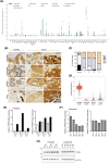
Fig. 1
Fig. 1
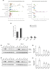
TRPM8 RNA and protein expression in solid cancers. (A) Landscape of TRPM8 transcript levels in primary tumor samples across different tissues (horizontal axis) was retrieved from The Cancer Genome Atlas (TCGA) datasets of cBioPortal. Panel display boxplots illustrating data distributions; boxes represent the median and interquartile range, while whiskers indicate variability beyond the quartiles, with individual data points shown above the plots. (B) Representative images of TRPM8 immunolocalization in healthy and malignant prostate, colorectal, breast, and lung tissue samples spotted on a commercial tissue microarray (TMA). TRPM8 immunostaining was scored as absent (0), weak (1), moderate (2) or high (3) (scale bar 20 μm). (C) TRPM8 score related to tumor stage (n = 48 cores per cancer type; n = 4 cores per normal tissue type). (D) Direct comparison of TRPM8 RNA expression across prostate, colorectal, breast, and lung cancers. Panel display boxplots illustrating data distributions; boxes represent the median and interquartile range, while whiskers indicate variability beyond the quartiles, with individual data points shown above the plots. (E, F) Quantification of TRPM8 RNA expression and protein amount in matched normal prostate tissue (N) and prostate cancer (T) samples isolated from n = 3 radical prostatectomies of prostate cancer (PCa) patients (E), and n = 5 independent colorectal cancer (CRC) samples (F). (G) Western blot of TRPM8 protein in the samples described in (E, F).
Fig. 2
Fig. 2
Variable amounts of TRPM8 channel in tumor cells with low levels of its coding transcript. (A) TRPM8 RNA expression in cancer cell lines described in the NCI‐60 cell lines and Cancer Cell Lines Encyclopedia projects (retrieved from cBioPortal). (B) Quantitative reverse transcription polymerase chain reaction (RT‐qPCR) comparative analysis of TRPM8 RNA expression in prostate (VCaP, LNCaP, PC3), colon (HCT116), breast (MCF7) and lung (A549) cancer cells. (C–F) Western blot replicas I and II of TRPM8 in VCaP, LNCaP, PC3, HCT116, MCF7, A549 cell lines with the Alomone ACC‐049 (C) and Abcam Ab3243 (E) antibodies, and relative quantification of TRPM8 protein (D, F) in the n = 4 independent replicas shown in C–E and Fig. S2C. β‐Actin is used as loading control and normalizer. Data are presented as mean ± standard deviation (sd) of three (Alomone ACC‐049 antibody) and four (Abcam Ab3243 antibody) independent experiments. ***P ≤ 0.001, **P ≤ 0.01, *P ≤ 0.05. Statistical analysis was performed using Student's t‐test.
Fig. 3
Fig. 3
Lethal synergy between TRPM8 agonist D‐3263 and chemotherapy in cancer cells. (A) Representative crystal violet staining of LNCaP, HCT116, MCF7, A549, and PC3 cells untreated or treated with the indicated drugs for 24 h (chemotherapy = Docetaxel for LNCaP, MCF7, A549, and PC3; 5‐fluorouracil (5‐FU + Oxaliplatin for HCT116). TRPM8 knock‐down (siRNA1 and siRNA2) confirmed D‐3263 specificity. (B) Quantification of the viability of treated cells compared with untreated controls. Data are presented as mean ± standard deviation (sd) of n = 3 independent experiments (A, Fig. S2C). ***P < 0.001. Statistical analysis was performed using Student's t‐test. (C, D) Western blot analysis of Caspase 3 and Parp cleavage in LNCaP, HCT116, MCF7, A549, and PC3 cells untreated or treated with the indicated drugs for 24 h (C). TRPM8 knock‐down (siRNA2) confirmed D‐3263 specificity (D). (E) Direct comparison of D‐3263 efficacy in wild type and TRPM8 knocked down (KD) cancer cells. Western blot analysis in C–E was repeated with n = 2 sets of biologically independent samples.
Fig. 4
Fig. 4
Efficacy of D‐3263 + 5‐FU + Oxaliplatin in CRC organoids raises with the amount of TRPM8. (A) TRPM8 immunolocalization in a dedicated homemade tissue microarray (TMA) of colorectal cancer (scale bar 100 μm; ACC‐049). Representative images of colorectal cancer (CRC) with different levels of TRPM8 staining and relative scores. Score 1: weak expression, score 2: moderate expression, score 3: high expression. (B) Distribution of TRPM8 immunostaining scores across the colorectal cancer (CRC) specimens (n = 79 independent cores). (C) Immunolocalization of TRPM8 in n = 5 different lines of patient‐derived colorectal cancer (CRC) organoids (scale bar 20 μm). (D) Western blot of TRPM8 from colorectal cancer (CRC) specimens (n = 5 independent samples) derived from the tissue bank of the Santa Chiara Hospital of Trento and—independent—n = 5 patient‐derived organoids (PDO). Western blot analysis in D was repeated twice. (E) Relative viability of colorectal cancer (CRC) organoids subjected to the indicated treatments or left untreated to serve as control pooled together based on TRPM8 protein amount (high (Hi): PDO #1 and PDO #4; low (Lo): PDO #2, PDO #3 and PDO #5). Distribution of viability measures across the different conditions and stratifications is shown using boxplots. Paired two‐sample t‐test was used to compare the viability of organoid lines. Data are presented as mean ± standard deviation (sd) of two independent biological experiments, each with six technical replicates.

References

    1. Heo YJ, Hwa C, Lee G‐H, Park J‐M, An J‐Y. Integrative multi‐omics approaches in cancer research: from biological networks to clinical subtypes. Molecules and Cells. 2021;44(7):433–443. - PMC - PubMed
    1. Raufaste‐Cazavieille V, Santiago R, Droit A. Multi‐omics analysis: paving the path toward achieving precision medicine in cancer treatment and immuno‐oncology. Frontiers in Molecular Biosciences. 2022;9:962743. - PMC - PubMed
    1. Sammut S‐J, Crispin‐Ortuzar M, Chin S‐F, Provenzano E, Bardwell HA, Ma W, et al. Multi‐omic machine learning predictor of breast cancer therapy response. Nature. 2022;601(7894):623–629. - PMC - PubMed
    1. Jiménez‐Santos MJ, Nogueira‐Rodríguez A, Piñeiro‐Yáñez E, López‐Fernández H, García‐Martín S, Gómez‐Plana P, et al. PanDrugs2: prioritizing cancer therapies using integrated individual multi‐omics data. Nucleic Acids Research. 2023;51(W1):W411–W418. - PMC - PubMed
    1. Labrie M, Brugge JS, Mills GB, Zervantonakis IK. Therapy resistance: opportunities created by adaptive responses to targeted therapies in cancer. Nature Reviews. Cancer. 2022;22(6):323–339. - PMC - PubMed