. 2025 May 21;6(7):1089-1099.
doi: 10.1039/d5cb00021a. eCollection 2025 Jul 2.

RaPID discovery of cell-permeable helical peptide inhibitors con-taining cyclic β-amino acids against SARS-CoV-2 main protease

Affiliations

RaPID discovery of cell-permeable helical peptide inhibitors con-taining cyclic β-amino acids against SARS-CoV-2 main protease

Marina Kawai et al. RSC Chem Biol. .

Abstract

Structurally constrained cyclic β-amino acids are attractive building blocks for peptide drugs because they induce unique and stable conformations. Introduction of (1S,2S)-2-aminocyclopentanecarboxylic acid [(1S,2S)-2-ACPC] into peptides stabilizes helical conformations, so improving proteolytic stability and cell membrane permeability. We report on the ribosomal synthesis of a helical peptide library incorporating (1S,2S)-2-ACPC at every third position and its application for the discovery of SARS-CoV-2 main protease (Mpro) inhibitors. We identified two peptide sequences containing multiple (1S,2S)-2-ACPC residues, which exhibit helical conformations and superior proteolytic stability compared with their α-Ala or β-Ala counterparts. Studies using the chloroalkane cell-penetration assay showed that their cell permeability values (CP50) are comparable with or even slightly better than that of the cell-penetrating nona-arginine (R9) peptide. The new approach is thus a highly efficient method that combines a helical peptide library containing structurally constrained cyclic β-amino acids with the classical RaPID discovery method, enabling de novo discovery of proteolytically stable and cell-penetrating bioactive peptides that target intracellular proteins.


Conflict of interest statement

There are no conflicts to declare.

Figures

Fig. 1
Fig. 1. Structure of (1S,2S)-2-aminocyclopentanecarboxylic acid and models of hydrogen-bond patterns that define 10/11/11-helix and 14-helix. Reported i, i + 3 CO⋯H–N hydrogen bonds are in blue (10/11/11-helix), and i, i + 4 CO⋯H–N hydrogen bonds are in green (14-helix).
Fig. 2
Fig. 2. Library designs of the (1S,2S)-2-ACPC-containing helical peptides. (A) Sequences of the mRNA libraries, I–III, coding α/β-helical peptide libraries containing random 4–10 NNU codons and the corresponding peptide sequences. The puromycin linker covalently links the respective mRNA to the translated peptide by ribosome catalysis. These mRNA libraries were combined into one mRNA library for the expression of a peptide library containing (1S,2S)-2-ACPC at the designated positions using the elongator AUG codon. (B) A model peptide, Pcβ5, and its mRNA template, mRcβ5. Pcβ5 was designed to contain five (1S,2S)-2-ACPCs at the designated positions assigned by the elongator AUG codon. (C) MALDI-TOF MS analysis of Pcβ5 expressed in the custom-made FIT system. Purple arrows indicate monovalent ([M + H]+, and [M + K]+) and divalent ([M + 2H]2+) ions of Pcβ5, respectively. “Obsd.” and “Calcd” denote observed and calculated m/z values of monovalent ion of Pcβ5, respectively. Asterisks (*) show unknown impurities, inherently present in the FIT system.
Fig. 3
Fig. 3. Far-UV circular dichroism spectra of Mph3, Mph3A, Mph3B (top), and Mph5, Mph5A, Mph5B (bottom). Measurements were performed using 75 μM peptide in 50%v/v trifluoroethanol in 10 mM phosphate buffer (pH 8.0) at room temperature. Red lines, peptides obtained from the RaPID selection; green lines, α-Ala mutants; and blue lines, β-Ala mutants.
Fig. 4
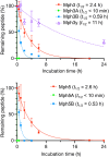
Fig. 4. Proteolytic stability of Mph3, Mph3A, Mph3B, Mph3y (top), and Mph5, Mph5A, Mph5B (bottom). The peptides were co-incubated with an internal standard peptide (NH2-PEG5-DWDSDTDNDDDWDSDTDNDD-PEG5-CONH2) in human serum for up to 20 h for (1S,2S)-2-ACPC mutants, and 24 h for Mph3y. The percentage of remaining peptides were estimated by LC/MS. Half-life (t1/2) values were calculated using Prism8 GraphPad software. n = 3.
Fig. 5
Fig. 5. Measurement of cell penetration using the chloroalkane penetration assay (CAPA). Each peptide with chloroalkane-tag (ct) was incubated with HaloTag-GFP-Mito cells in Opti-MEM for 24 h. The fluorescence of living cells and ct-TAMRA was detected and qualified by FACS. The data were normalized using peptide(−) control as 100% signal and peptide(−), ct-TAMRA(−) as 0% signal. CP50 values were calculated using Prism8 GraphPad software. n = 3.
Fig. 6
Fig. 6. Structural calculation of the conformation of (1S,2S)-2-ACPC-containing peptides Mph3, Mph3A, and Mph3B. (A) A MD-generated structure of Mph3. (1S,2S)-2-ACPC is shown in pink and the initiator AcTyr in orange. Polar contacts are shown as dotted lines. (B) Comparison of structural clusters and enriched structures of Mph3, Mph3A, and Mph3B.

References

    1. Katsu T. Kuroko M. Morikawa T. Sanchika K. Yamanaka H. Shinoda S. Fujita Y. Biochim. Biophys. Acta. 1990;1027:185–190. doi: 10.1016/0005-2736(90)90083-Z. - DOI - PubMed
    1. Hou K. K. Pan H. Lanza G. M. Wickline S. A. Biomaterials. 2013;34:3110–3119. doi: 10.1016/j.biomaterials.2013.01.037. - DOI - PMC - PubMed
    1. O’Neil K. T. DeGrado W. F. Trends Biochem. Sci. 1990;15:59–64. doi: 10.1016/0968-0004(90)90177-D. - DOI - PubMed
    1. Hait W. N. Grais L. Benz C. Cadman E. C. Cancer Chemother. Pharmacol. 1985;14:202–205. doi: 10.1007/BF00258116. - DOI - PubMed
    1. Moreno M. Giralt E. Toxins. 2015;7:1126–1150. doi: 10.3390/toxins7041126. - DOI - PMC - PubMed

LinkOut - more resources