. 2025 May 13;16(24):11067-11076.
doi: 10.1039/d5sc02453f. eCollection 2025 Jun 18.

Engineering of soluble bacteriorhodopsin

Affiliations

Engineering of soluble bacteriorhodopsin

Andrey Nikolaev et al. Chem Sci. .

Abstract

Studies and applications of membrane proteins remain challenging due to the requirement of maintaining them in a lipid membrane or a membrane mimic. Modern machine learning-based protein engineering methods offer a possibility of generating soluble analogs of membrane proteins that retain the active site structure and ligand-binding properties; however, clear examples are currently missing. Here, we report successful engineering of proteins dubbed NeuroBRs that mimic the active site (retinal-binding pocket) of bacteriorhodopsin, a light-driven proton pump and well-studied model membrane protein. NeuroBRs are soluble and stable, bind retinal and exhibit photocycles under illumination. The crystallographic structure of NeuroBR_A, determined at anisotropic resolution reaching 1.76 Å, reveals an excellently conserved chromophore binding pocket and tertiary structure. Thus, NeuroBRs are promising microbial rhodopsin mimics for studying retinal photochemistry and potential soluble effector modules for optogenetic tools. Overall, our results highlight the power of modern protein engineering approaches and pave the way towards wider development of molecular tools derived from membrane proteins.


Conflict of interest statement

The authors declare no competing interests.

Figures
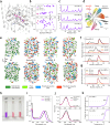
Fig. 1
Fig. 1. Sequence design and initial characterisation of soluble BR variants. (a) Chromophore-binding pocket in WT BR. (b) Backbone RMSD, pocket RMSD and pLDDT of AlphaFold-predicted models for WT BR and SolubleMPNN-generated sequences. (c) Amplitude of thermal fluctuations of Cα atoms of engineered BR variants in MD simulations. (d) Phylogenetic tree for NeuroBRs and representative microbial rhodopsins (based on the dataset by Rozenberg et al.). (e) Surface amino acid properties for WT and engineered BR variants. Histidines and cysteines are absent in WT and engineered variants, except for 6×His tags added to NeuroBRs for metal affinity purification (not shown). (f and g) SEC profiles for NeuroBRs reconstituted with retinal and for monomeric fractions of NeuroBR_A and C after one week of incubation at 4 °C, respectively. Chromatography was performed using a Superdex 75 Increase 10/300 GL column with a void volume V0 of approximately 8.2 ml. (h) Visual appearance of WT BR in purple membranes and NeuroBR_A and C in detergent-free buffer. (i) Absorbance spectra of trimeric WT BR in purple membranes and engineered monomeric BR in solution. (j) Comparison of absorption spectra of dark-adapted and illuminated samples of NeuroBR_A and C. (k) Thermal denaturation of engineered BR variants.
Fig. 2
Fig. 2. Photocycle and structure of soluble BR variant NeuroBR_A. (a) Changes in absorbance of NeuroBR_A in solution after flash illumination. M and O correspond to areas, where absorbance raises due to formation of putative M and O intermediates. GSB is the ground state bleaching area, where absorbance corresponding to the ground state is diminished due to formation of photocycle intermediates. (b) Model of the NeuroBR_A photocycle at pH 4.0, where the different photocycle intermediates are clearly distinguishable. (c) Recovered absorption spectra of NeuroBR_A photocycle intermediates. (d) Dependence of NeuroBR_A photocycle intermediate half-lives on pH. (e) Overlay of the backbone and retinal structures for WT BR (purple) and NeuroBR_A (yellow and green). (f) Weighted 2FoFc electron density map around the retinal contoured at the 2σ level. (g) Comparison of the retinal Schiff base environment in WT BR and NeuroBR_A. w406 is absent in NeuroBR_A and R82 changes its conformation.
Fig. 3
Fig. 3. Photocycle of soluble BR variant NeuroBR_C. (a) Changes in absorbance of NeuroBR_C in solution after flash illumination. M and O correspond to areas where absorbance raises due to formation of putative M and O photocycle intermediates. GSB is the ground state bleaching area where absorbance corresponding to the ground state is diminished due to formation of photocycle intermediates. (b) Recovered absorption spectra of NeuroBR_C photocycle intermediates at different buffer pH values. The intermediates are designated in accordance with the photocycle at low pH. Dotted spectra correspond to the ground state. (c) Dependence of NeuroBR_C photocycle intermediate half-lives on pH. (d) Model of NeuroBR_C photocycles at pH 4.0 and pH 6.8.

References

    1. Levental I. Lyman E. Nat. Rev. Mol. Cell Biol. 2023;24:107–122. doi: 10.1038/s41580-022-00524-4. - DOI - PMC - PubMed
    1. Privé G. G. Methods. 2007;41:388–397. doi: 10.1016/j.ymeth.2007.01.007. - DOI - PubMed
    1. Serebryany E. Zhu G. A. Yan E. C. Y. Biochim. Biophys. Acta. 2012;1818:225–233. doi: 10.1016/j.bbamem.2011.07.047. - DOI - PubMed
    1. Pandey A. Shin K. Patterson R. E. Liu X.-Q. Rainey J. K. Biochem. Cell Biol. 2016;94:507–527. - PMC - PubMed
    1. Zhang Q. Cherezov V. Curr. Opin. Struct. Biol. 2019;58:278–285. doi: 10.1016/j.sbi.2019.06.002. - DOI - PMC - PubMed

LinkOut - more resources