Published Erratum
. 2025 May 8:16:1597953.
doi: 10.3389/fpls.2025.1597953. eCollection 2025.

Corrigendum: A proteome-level investigation into Plasmodiophora brassicae resistance in Brassica napus canola

Affiliations
Published Erratum

Corrigendum: A proteome-level investigation into Plasmodiophora brassicae resistance in Brassica napus canola

Dinesh Adhikary et al. Front Plant Sci. .

Abstract

[This corrects the article DOI: 10.3389/fpls.2022.860393.].

Keywords: Brassica napus; calcium binding; clubroot; plant–pathogen interaction; proteomics.


Figures

Figure 1
Figure 1
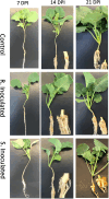
Clubroot gall development following inoculation with P. brassicae pathotype 3. Control at 7-, 14-, and 21-DPI, Clubroot resistant (CR) inoculated line at 7-, 14-, and 21-DPI Clubroot susceptible (CS) inoculated 7-, 14-, and 21-DPI.
Figure 2
Figure 2
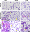
Histology images of root cross sections after P. brassicae infection at 7-, 14-, and 21-days post inoculation (DPI). Root tissues were stained with eosin and hematoxylin. Each column indicates DPI of the pathogen and the rows show the control and inoculated genotypes [clubroot- susceptible (CS) and resistant (CR) lines]. At 7 DPI, infected cells showed primary plasmodia with dark purple mass within cells indicated by the solid yellow arrow. At 14 DPI, CS inoculated line showed the presence of secondary plasmodia; however, the pathogen development on the CR inoculated line was not progressed to secondary plasmodia phase. At 21 DPI, pathogen clearly progressed to secondary plasmodia phase, maturing into developing resting spores in the CS line. However, the infection development was not progressed further at the same time point on the CR line.
Figure 3
Figure 3
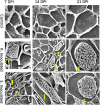
Scanning electron micrograph (SEM) of root cross sections after P. brassicae infection at 7-, 14-, and 21-DPI. Each column indicates days after inoculation of the pathogen and the rows show the control and inoculated genotypes (CR and CS). At 7 DPI, infected cells showed primary plasmodia within cells indicated by the solid yellow arrow. At 14 DPI, CS inoculated line showed the presence of secondary plasmodia; however, the pathogen development on the CR inoculated line was not progressed to secondary plasmodia phase. At 21 DPI, pathogen clearly progressed to secondary plasmodia phase, maturing into resting spores. However, the infection development was not progressed in the CR line at the timepoint.

Erratum for

Publication types

LinkOut - more resources