Coexistent Ankylosing Spondylitis and Ocular Toxocariasis in a Pediatric Patient Manifesting As Bilateral Panuveitis
- PMID: 40406765
- PMCID: PMC12096418
- DOI: 10.7759/cureus.82767
Coexistent Ankylosing Spondylitis and Ocular Toxocariasis in a Pediatric Patient Manifesting As Bilateral Panuveitis
Abstract
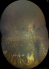
The coexistence of ankylosing spondylitis and ocular toxocariasis in the literature is rare and limited to a few case reports. Typically, such cases present as acute nongranulomatous anterior uveitis with Toxocara IgG seropositivity. A patient manifesting with findings of both ankylosing spondylitis and toxocariasis bilaterally has not been reported previously in the literature. We present a case of coexistent juvenile spondyloarthritis and ocular toxocariasis in a 16-year-old male presenting with generalized pustules, back pain, peripheral polyarthritis, and bilateral panuveitis. Both eyes displayed abnormalities in the anterior segments, including corectopia, seclusio pupillae, and occlusio pupillae. Posterior segment examination of the right eye showed vitritis, disc edema, and a retinochoroidal granuloma surrounded by infiltrates and perivascular sheathing. A B-scan of the left eye revealed vitritis and the presence of a hyperechoic band from the disc to the retinal periphery. Toxocara IgG and HLA-B27 were positive, and lumbosacral magnetic resonance imaging confirmed sacroiliitis. Treatment involved subtenon injections of triamcinolone and subcutaneous Etanercept injections, resulting in stabilization of visual acuity. This case highlights the rare co-occurrence of two diseases with overlapping symptoms and uncertain pathogenetic contributions from each to cause the observed manifestations. It supports studies proposing a connection between rheumatic disease and parasitosis.
Keywords: ankylosing spondylitis; bilateral uveitis; granulomatous panuveitis; ocular toxocariasis; pediatric uveitis.
Copyright © 2025, Tanchuling et al.
Conflict of interest statement
Human subjects: Consent for treatment and open access publication was obtained or waived by all participants in this study. Conflicts of interest: In compliance with the ICMJE uniform disclosure form, all authors declare the following: Payment/services info: All authors have declared that no financial support was received from any organization for the submitted work. Financial relationships: All authors have declared that they have no financial relationships at present or within the previous three years with any organizations that might have an interest in the submitted work. Other relationships: All authors have declared that there are no other relationships or activities that could appear to have influenced the submitted work.
Figures


References
-
- The eye and inflammatory rheumatic diseases: the eye and rheumatoid arthritis, ankylosing spondylitis, psoriatic arthritis. Murray PI, Rauz S. Best Pract Res Clin Rheumatol. 2016;30:802–825. - PubMed
-
- Human toxocariasis: diagnosis, worldwide seroprevalences and clinical expression of the systemic and ocular forms. Rubinsky-Elefant G, Hirata CE, Yamamoto JH, Ferreira MU. Ann Trop Med Parasitol. 2010;104:3–23. - PubMed
-
- Seroprevalence of Toxocara spp. infection in Southeast Asia and Taiwan. Chou CM, Fan CK. Adv Parasitol. 2020;109:449–463. - PubMed
-
- Prevalence, clinical features, and causes of vision loss among patients with ocular toxocariasis. Stewart JM, Cubillan LD, Cunningham ET Jr. Retina. 2005;25:1005–1013. - PubMed
-
- Adalimumab or etanercept as first line biologic therapy in enthesitis related arthritis (ERA) - a drug-survival single centre study spanning 10 years. Shipa MR, Heyer N, Mansoor R, et al. Semin Arthritis Rheum. 2022;55:152038. - PubMed
Publication types
LinkOut - more resources
Full Text Sources
Research Materials
