

Ficolin-3 Activates Complement and Triggers Necroptosis in Cholangiocarcinoma Cells via the RIPK1/RIPK3/MLKL Signaling Pathway
- PMID: 40407231
- PMCID: PMC12101045
- DOI: 10.1096/fj.202403313R
Ficolin-3 Activates Complement and Triggers Necroptosis in Cholangiocarcinoma Cells via the RIPK1/RIPK3/MLKL Signaling Pathway
Abstract
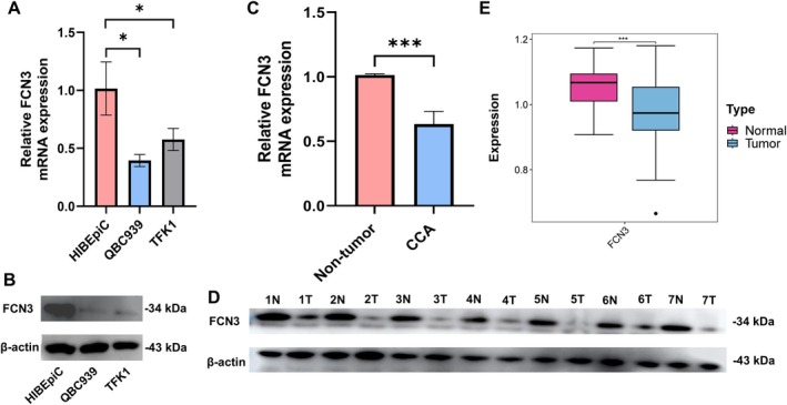
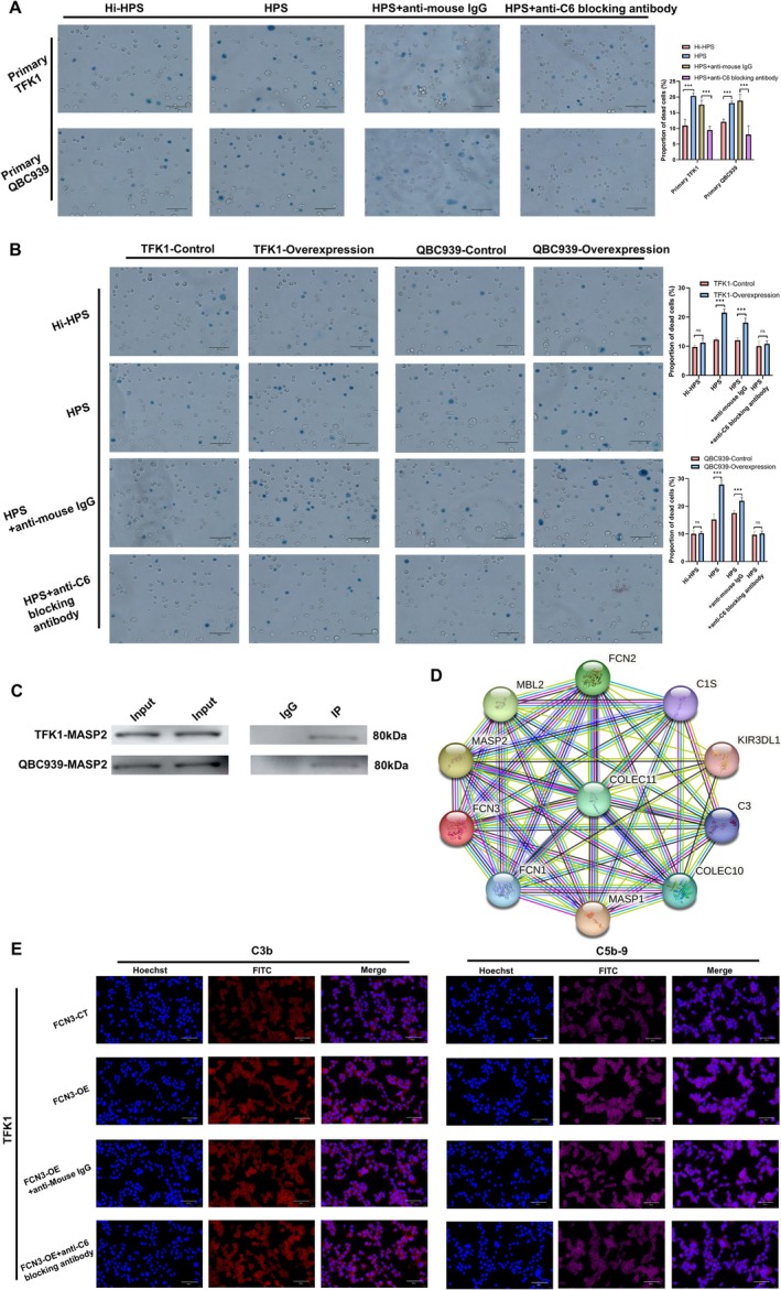
Ficolin 3 (FCN3) is a pattern recognition molecule that activates the complement system via the lectin pathway. While its immunological roles are known, the specific mechanisms by which FCN3 affects cholangiocarcinoma (CCA) pathogenesis remain unclear. In this study, we investigated FCN3 expression in CCA and benign cells, as well as tumor versus non-tumor tissues, using RT-qPCR and Western blotting analyses. The effects of FCN3 on CCA cell proliferation, migration, and invasion were analyzed through CCK-8, EdU, transwell, and wound-healing assays, with in vivo studies supporting these findings. The complement-mediated cytotoxicity of CCA cells was assessed using human serum with or without heat inactivation and an anti-C6 blocking antibody. Immunocytochemical staining was used to examine membrane attack complex (MAC) deposition, and an immunoprecipitation assay was adopted to evaluate the interaction between FCN3 and MASP family members. The role of FCN3 in inducing necroptosis was explored through transmission electron microscopy (TEM) and Western blotting analysis, focusing on the RIPK1/RIPK3/MLKL pathway. The results of the study demonstrate that FCN3 expression was significantly lower in CCA cells and tissues. Overexpressing FCN3 suppressed cell proliferation and migration, enhanced complement-mediated cytotoxicity via MASP2 binding, and increased MAC deposition. FCN3 also induced necroptosis through activating the RIPK1/RIPK3/MLKL pathway. These results highlight FCN3 as a tumor suppressor in CCA and suggest its potential as a therapeutic target for this malignancy.
Keywords: cholangiocarcinoma; complement activation; ficolin‐3; membrane attack complex; necroptosis.
© 2025 The Author(s). The FASEB Journal published by Wiley Periodicals LLC on behalf of Federation of American Societies for Experimental Biology.
Conflict of interest statement
The authors declare no conflicts of interest.
Figures


Similar articles
-
Ficolin-3 induces apoptosis and suppresses malignant property of hepatocellular carcinoma cells via the complement pathway.Life Sci. 2024 Nov 15;357:123103. doi: 10.1016/j.lfs.2024.123103. Epub 2024 Sep 30. Life Sci. 2024. PMID: 39357793
-
Receptor-interacting protein kinase 1 is a key mediator in TLR3 ligand and Smac mimetic-induced cell death and suppresses TLR3 ligand-promoted invasion in cholangiocarcinoma.Cell Commun Signal. 2020 Oct 9;18(1):161. doi: 10.1186/s12964-020-00661-3. Cell Commun Signal. 2020. PMID: 33036630 Free PMC article.
-
Key necroptotic proteins are required for Smac mimetic-mediated sensitization of cholangiocarcinoma cells to TNF-α and chemotherapeutic gemcitabine-induced necroptosis.PLoS One. 2020 Jan 8;15(1):e0227454. doi: 10.1371/journal.pone.0227454. eCollection 2020. PLoS One. 2020. PMID: 31914150 Free PMC article.
-
RIPK3 signaling and its role in the pathogenesis of cancers.Cell Mol Life Sci. 2021 Dec;78(23):7199-7217. doi: 10.1007/s00018-021-03947-y. Epub 2021 Oct 15. Cell Mol Life Sci. 2021. PMID: 34654937 Free PMC article. Review.
-
The regulation of necroptosis by post-translational modifications.Cell Death Differ. 2021 Mar;28(3):861-883. doi: 10.1038/s41418-020-00722-7. Epub 2021 Jan 18. Cell Death Differ. 2021. PMID: 33462412 Free PMC article. Review.
Cited by
-
Impact of Echinococcus granulosus Cyst Fluid on the Phenotypic Dynamics and Degranulation of RBL-2H3 Mast Cells.J Inflamm Res. 2025 Jul 24;18:9837-9848. doi: 10.2147/JIR.S522640. eCollection 2025. J Inflamm Res. 2025. PMID: 40727458 Free PMC article.
References
-
- Matsushita M., Endo Y., Hamasaki N., and Fujita T., “Activation of the Lectin Complement Pathway by Ficolins,” International Immunopharmacology 1, no. 3 (2001): 359–363. - PubMed
-
- Matsushita M. and Fujita T., “Ficolins and the Lectin Complement Pathway,” Immunological Reviews 180 (2001): 78–85. - PubMed
MeSH terms
Substances
Grants and funding
LinkOut - more resources
Full Text Sources
Medical
Miscellaneous
