. 2025 Jun 11;16(6):e0386524.
doi: 10.1128/mbio.03865-24. Epub 2025 May 23.

Identification of a series of pyrrolo-pyrimidine-based SARS-CoV-2 Mac1 inhibitors that repress coronavirus replication

Affiliations

Identification of a series of pyrrolo-pyrimidine-based SARS-CoV-2 Mac1 inhibitors that repress coronavirus replication

Jessica J Pfannenstiel et al. mBio. .

Abstract

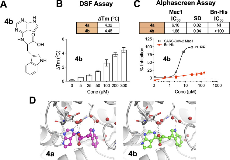
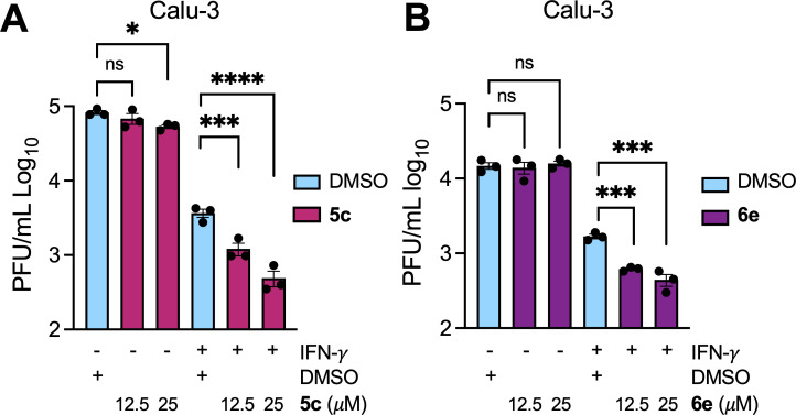
Coronaviruses (CoVs) can emerge from zoonotic sources and cause severe diseases in humans and animals. CoVs encode for a macrodomain (Mac1) that binds to and removes ADP-ribose from target proteins. SARS-CoV-2 Mac1 promotes virus replication in the presence of interferon (IFN) and blocks the production of IFN, although the mechanisms by which it mediates these functions remain unknown. Mac1 inhibitors could help elucidate these mechanisms and serve as therapeutic agents against CoV-induced diseases. We previously identified compound 4a (a.k.a. MCD-628), a pyrrolo-pyrimidine that inhibited Mac1 activity in vitro at low micromolar levels. Here, we determined the binding mode of 4a by crystallography, further defining its interaction with Mac1. However, 4a did not reduce CoV replication, which we hypothesized was due to its acidic side chain limiting permeability. To test this hypothesis, we developed several hydrophobic derivatives of 4a. We identified four compounds that both inhibited Mac1 in vitro and inhibited murine hepatitis virus (MHV) replication: 5a, 5c, 6d, and 6e. Furthermore, 5c and 6e inhibited SARS-CoV-2 replication only in the presence of IFNγ, similar to a Mac1 deletion virus. To confirm their specificity, we passed MHV in the presence of 5a to identify drug-resistant mutations and identified an alanine-to-threonine and glycine-to-valine double mutation in Mac1. Recombinant virus with these mutations had enhanced replication compared with the WT virus when treated with 5a, demonstrating the specificity of these compounds during infection. However, this virus is highly attenuated in vivo, indicating that drug resistance emerged at the expense of viral fitness.IMPORTANCECoronaviruses (CoVs) present significant threats to human and animal health, as evidenced by recent outbreaks of MERS-CoV and SARS-CoV-2. CoVs encode for a highly conserved macrodomain protein (Mac1) that binds to and removes ADP-ribose from proteins, which promotes virus replication and blocks IFN production, although the exact mechanisms remain unclear. Inhibiting Mac1 could provide valuable insights into these mechanisms and offer new therapeutic avenues for CoV-induced diseases. We have identified several unique pyrrolo-pyrimidine-based compounds as Mac1 inhibitors. Notably, at least two of these compounds inhibited both murine hepatitis virus (MHV) and SARS-CoV-2 replication. Furthermore, we identified a drug-resistant mutation in Mac1, confirming target specificity during infection. However, this mutant is highly attenuated in mice, indicating that drug resistance appears to come at a fitness cost. These results emphasize the potential of Mac1 as a drug target and the promise of structure-based inhibitor design in combating CoV infections.

Keywords: ADP-ribosylation; COVID-19; SARS-CoV2; coronavirus; murine hepatitis virus; nsp3 macrodomain.


Conflict of interest statement

A.R.F. was named as an inventor on a patent filed by the University of Kansas for a live-attenuated SARS-CoV-2 vaccine.

Figures

Fig 1
Fig 1
Crystal structure of 4a provides new insight into its interaction with Mac1. (A and B) Chemical synthesis plan to produce 4a-b (A), and the chemical structure of 4a (B). (C and D) Crystal structure of 4a in two different poses (PDB id. 9GUB). These poses include images where the pyrrolo-pyrimidine is oriented in the front left (C) or in the lower middle (D). Note that the carboxylate makes hydrogen bonds with three different water molecules, and the tryptophanate makes a hydrogen bond with the backbone of L126. The sigma-A weighted 2Fo-Fc electron density map is contoured at 1.0 σ. Waters are shown as red spheres, and hydrogen bonds are illustrated as black dashed lines.
Fig 2
Fig 2
Compound 4b interacts with Mac1 and inhibits Mac1-ADP-ribose binding. (A) Chemical structure of compound 4b. (B) Compound 4b was incubated with SARS-CoV-2 Mac1 at increasing concentrations, and the thermal stability of SARS-CoV-2 Mac1 was determined by a DSF assay. The ΔTm is the average of five experimental replicates. n = 5. (C) Competition assays were used to demonstrate that 4a and 4b block the interaction between Mac1 and ADP-ribosylated peptides in the AS assay. The IC50 represents the average value of 2 independent experiments, each done with three experimental replicates. The graphs are from one experiment representative of two independent experiments. (D) Compounds 4a and 4b were docked into Mac1 using PDB:9GUB. Hydrogen bonds are illustrated as dashed lines.
Fig 3
Fig 3
Compounds 5a and 5c interact with Mac1 and inhibit Mac1-ADP-ribose binding. (A) Modification of 4a-b to ester derivatives 5a and 5c. (B) Compounds 5a and 5c were incubated with SARS-CoV-2 Mac1 at increasing concentrations, and the thermal stability of SARS-CoV-2 Mac1 was determined by a DSF assay. The ΔTm is the average of five experimental replicates. n = 5. (C) Competition assays were used to demonstrate that 5a and 5c block the interaction between Mac1 and ADP-ribosylated peptides in the AS assay. The IC50 represents the average value of two independent experiments. n = 3 experimental replicates. (D) Compounds 5a and 5c were docked into Mac1 using PDB: 9GUB. Hydrogen bonds are illustrated as dashed lines. (E) The LogD values were experimentally determined using the shake-flask method for 4a, 5a, and 5c.
Fig 4
Fig 4
Compounds 5a and 5c, but not 4a, inhibit MHV replication. (A) In total, 17 Cl-1 cells were infected with JHMV-WT and JHMV-nLuc viruses at an MOI = 0.1. Cells and supernatants were collected at indicated time points, and progeny virus was determined by plaque assay; (B) 17 Cl-1 cells were infected as described in A. Lysates were collected at indicated times, and luciferase activity was determined using a nano-Glo luciferase assay kit measured as per the manufacturer’s instructions. The results in A and B are from one experiment representative of two independent experiments. N = 3 biological replicates. (C) DBT cells were infected with JHMV-nLuc at an MOI = 0.1, and at one hpi, the indicated concentration of each compound was added to the media. Lysates were collected at 20 hpi, and luciferase activity was measured as described in B. (D and E) DBT cells were infected with JHMV-WT, and at 1 hpi, the indicated concentration of each compound was added to the media. Cells and supernatants were collected at 20 hpi, and progeny virus was measured by plaque assay. The results in C-E are from one experiment representative of two independent experiments. n = 3 biological replicates. (F) The combined average % JHMV-WT inhibition by 5c on DBT cells over two independent experiments. (G and H) L929 cells were infected with JHMV-WT, and at 1 hpi, the indicated concentration of each compound was added to the media. Cells and supernatants were collected at 20 hpi, and progeny virus was measured by plaque assay. The results in G-H are from one experiment representative of two independent experiments. n = 3 biological replicates. (I) The combined average % JHMV-WT inhibition by 5c on L929 cells over three independent experiments. The results in C, D, E, G, and H are from one experiment representative of three independent experiments. n = 3 biological replicates.
Fig 5
Fig 5
Group 6 compounds interact with Mac1 and inhibit Mac1-ADP-ribose binding. (A) Modification of 4a-b to several new derivatives, 6a–6e. 6a–6d are derivatives of 4a, whereas 6e is a derivative of 4b. (B) Competition assays were used to demonstrate that 6a–6e block the interaction between Mac1 and ADP-ribosylated peptides in the AS assay. (C) Compounds 6a–6e were incubated with SARS-CoV-2 Mac1 at increasing concentrations, and the thermal stability of SARS-CoV-2 Mac1 was determined by a DSF assay. Quantification data in B and C represent the average value of 2 independent experiments. (D) Predicted cLogD values of 6a–6e. (E) Compounds 6a–6e were docked into Mac1. Hydrogen bonds are illustrated as dashed lines.
Fig 6
Fig 6
Compounds 6d and 6e inhibit JHMV replication. (A) DBT cells were infected with JHMV-nLuc at an MOI = 0.1, and at 1 hpi, the indicated concentration of each compound was added to the media. GS-441524 (active metabolite of remdesivir) was used as a positive control for inhibition at relative experimental concentrations. Lysates were collected at 20 hpi, and luciferase activity was measured. Luciferase activity was determined using a nano-Glo luciferase assay kit measured as per the manufacturer’s instructions. The results in A are from one experiment and are representative of two independent experiments. n = 3 biological replicates. (B) DBT cells were infected with JHMV-WT, and at one hpi, the indicated concentration of each compound was added to the media. Cells and supernatants were collected at 20 hpi, and progeny virus was measured by plaque assay. (C) L929 cells were infected with JHMV-WT, and at one hpi, the indicated concentration of each compound was added to the media. Cells and supernatants were collected at 20 hpi, and progeny virus was measured by plaque assay. The results in B-C are from one experiment representative of two independent experiments. n = 3 biological replicates. (D and E) The combined average % JHMV-WT inhibition by 6d and 6e, respectively, on DBT cells over two independent experiments.
Fig 7
Fig 7
Compounds 5c and 6e inhibit SARS-CoV-2 replication. (A and B) Calu-3 cells were mock or IFN-γ pre-treated (100 U) for 18 h, then were infected with SARS-CoV-2, and at 1 h post-infection (hpi), the indicated concentration of 5c (A) or 6e (B) was added to the media. Cells and supernatants were collected at 20 hpi, and progeny virus was measured by plaque assay. The results in A-B are from one experiment representative of 3 independent experiments. n = 3 biological replicates.
Fig 8
Fig 8
Identification of a Mac1 drug-resistant mutation. (A) Cartoon depiction of the passaging method for creating the drug-resistant virus. Three separate wells of DBT cells were initially infected with 0.1 MOI JHMV in the presence of DMSO or 5a. Each well was then passaged by taking 100 µL of cells/supernatants from the prior passage and infecting a new well of DBT cells. Image was created using BioRender.com. (B) Cells and supernatants were collected at 18–20 hpi at each passage, and progeny virus was measured by plaque assay. (C) Progeny virus at passage three was sequenced, which identified a two amino acid A1439T/G1439V mutation. (D) DBT cells were infected with WT or A1438T/G1439V recombinant virus at an MOI of 0.1 in the presence of DMSO, 5a, or 5c. Cells and supernatants were collected at 20 hpi, and progeny virus was measured by plaque assay. The data in D is from one experiment representative of three independent experiments. n = 3 biological replicates.

Update of

References

    1. World Health Organization . 2025. Number of COVID-19 cases reported to WHO (cumulative total). Available from: https://data.who.int/dashboards/covid19/cases. Retrieved 16 Mar 2025.
    1. World Health Organization . 2020. Impact of COVID-19 on people's livelihoods, their health and our food systems. World Health Organization.
    1. Zhao J, Wan W, Yu K, Lemey P, Pettersson JH-O, Bi Y, Lu M, Li X, Chen Z, Zheng M, Yan G, Dai J, Li Y, Haerheng A, He N, Tu C, Suchard MA, Holmes EC, He W-T, Su S. 2024. Farmed fur animals harbour viruses with zoonotic spillover potential. Nature 634:228–233. doi: 10.1038/s41586-024-07901-3 - DOI - PMC - PubMed
    1. Minkoff JM, tenOever B. 2023. Innate immune evasion strategies of SARS-CoV-2. Nat Rev Microbiol 21:178–194. doi: 10.1038/s41579-022-00839-1 - DOI - PMC - PubMed
    1. Yao T, Foo C, Zheng G, Huang R, Li Q, Shen J, Wang Z. 2023. Insight into the mechanisms of coronaviruses evading host innate immunity. Biochim Biophys Acta Mol Basis Dis 1869:166671. doi: 10.1016/j.bbadis.2023.166671 - DOI - PMC - PubMed

LinkOut - more resources