. 2025 Jun 11;16(6):e0085125.
doi: 10.1128/mbio.00851-25. Epub 2025 May 23.

Unraveling the molecular mechanisms of DNA capture by the Com pilus in naturally transformable monoderm bacteria

Affiliations

Unraveling the molecular mechanisms of DNA capture by the Com pilus in naturally transformable monoderm bacteria

Jérémy Mom et al. mBio. .

Abstract

Transformation is a mechanism of horizontal gene transfer widespread in bacteria. The first step in transformation-capture of exogenous DNA-is mediated by surface-exposed filaments belonging to the type 4 filament (T4F) superfamily. How these protein polymers, composed of major and minor pilin subunits, interact with DNA remains poorly understood. Here, we address this question for the Com pilus, a widespread T4F mediating DNA capture in competent monoderm species. Our functional analysis, performed in Streptococcus sanguinis, was guided by a complete structural model of the Com pilus. We show that the major pilin ComGC does not bind DNA. In contrast, a systematic mutational analysis of electropositive residues exposed at the filament surface in the four minor pilins (ComGD, ComGE, ComGF, and ComGG) reveals that the interface between ComGD and ComGF is important for DNA capture. Sequential mutations in these two interacting subunits lead to complete abolition of transformation, without affecting piliation. We further demonstrate the physical interaction between ComGD and ComGF using disulfide crosslinking, upon mutagenesis of two strategically positioned residues into cysteines. A structural model of the Com pilus tip interacting with DNA recapitulates all these findings and highlights a novel mode of DNA-binding, conserved in hundreds of monoderm species.

Importance: Bacteria are capable of evolving and diversifying very rapidly by acquiring new genetic material via horizontal gene transfer (HGT). Transformation is a widespread mechanism of HGT, which results from the capture of extracellular DNA by surface-exposed pili belonging to the type 4 filament (T4F) superfamily. How T4F-composed of major and minor pilins-interact with DNA remains poorly understood, especially in monoderm species that use a unique T4F for DNA capture, known as Com pilus or T4dP. The significance of this work is in characterizing a novel mode of DNA-binding by showing that the interface between two minor pilins, part of a tip-located complex of four pilins-found in different T4F-has been functionalized in monoderms to capture DNA. This is an evolutionary mechanism promoting the exceptional functional versatility of T4F.

Keywords: DNA-binding proteins; genetic competence; gram-positive bacteria; natural transformation systems; type 4 pili.


Conflict of interest statement

The authors declare no conflict of interest.

Figures
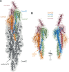
Fig 1
Fig 1
Modeling of T4dP reveals a canonical T4P, with a ComGC filament capped by a complex of four minor pilins. (A) Structural T4dP model in S. sanguinis predicted by AlphaFold 3 (25) with 10 copies of ComGC and one copy of each of the four minor pilins. The cartoon representation with surfaces shown in transparency reveals that T4dP display a canonical T4P structure, where a filament of ComGC (gray) is capped by a complex of four minor pilins ComGD (orange), ComGE (blue), ComGE (green), and ComGG (maroon). The same color code is used throughout the manuscript. As in available near-atomic resolution structures of T4aP (26–28), helical packing of ComGC subunits via their α1 helices is accompanied by “melting” of a portion of α1N that becomes non-helical. (B) Focus on the tip-located complex of ComGD-ComGF-ComGE-ComGG minor pilins. Two different sides are shown (180° views). As for ComGC, packing of ComGD in the pilus is accompanied by partial melting of its α1N. Intriguingly, the local confidence for the last 30 residues of ComGG, which was very low when this pilin was modeled on its own (see Fig. S2), remained very low, and the corresponding α-helix still protrudes from the tip of the pilus.
Fig 2
Fig 2
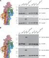
ComGC has no intrinsic DNA-binding activity. The DNA-binding propensity of purified MBP-ComGC was assessed by agarose EMSA. A standard amount (120 ng) of pUC19 plasmid was incubated with increasing concentrations of purified MBP-ComGC and resolved by electrophoresis on a 0.8% agarose gel (upper panel). As a positive control, we used purified MBP-ComEA (lower panel). ComEA is a conserved DNA receptor involved in the late stages of DNA uptake (5).
Fig 3
Fig 3
The charged C-terminal tail in ComGG is dispensable for S. sanguinis piliation and transformation. (A) Tip-located complex of four minor pilins with the last 30 residues of ComGG, which form an α-helical tail with very low local modeling confidence, highlighted in black. Two different sides are shown (180° views). (B) The ComGG tail is highly charged, as could be seen from the sequence logo generated from MSA of 31 S. sanguinis IPR047665 entries (Streptococcus-type ComGG) in InterPro (31). Charged residues are colored in blue (electropositive) or red (electronegative). (C) An unmarked tail-less S. sanguinis mutant expressing ComGGΔ94–122 is piliated. Piliation was assessed by immunoblotting using an anti-ComGC antibody on pilus preparations made from equal volumes of culture. The WT strain is included as a control. (D) The S. sanguinis comGGΔ94–122 mutant is transformable. The WT strain is included as a control. Transformation frequencies (%)—mean ± SD from six independent experiments—are the ratio of transformants relative to number of viable bacteria. The comGGΔ94–122 mutant is as transformable as the WT strain as assessed by a two-tailed t-test. ns, not statistically different.
Fig 4
Fig 4
Electropositive residues in ComGD and ComGF exposed on the filament surface are key for transformation. (A–D) Quantifying transformation in S. sanguinis mutants expressing minor pilins in which we altered surface-exposed electropositive residues potentially contributing to DNA binding. The targeted residues are highlighted in black on the corresponding structures in surface representation (the C-terminal tail in ComGG is not shown). Transformation frequencies (%) were the mean ± SD from at least three independent experiments. We used Dunnett’s one-way ANOVA to compare the means to the WT. ns, not statistically different; *, P < 0.0332; **, P < 0.0021; ***, P < 0.0002; ****, P < 0.0001. (E) Assessing piliation in the mutants affected for transformation. This was done by immunoblotting using an anti-ComGC antibody on pilus preparations made from equal volumes of culture. The WT strain is included as a control.
Fig 5
Fig 5
The interface between ComGD and ComGF is key for DNA capture by T4dP. (A) ComGD-ComGF interface at the tip of the pilus. The electropositive residues shown to be important for transformation in Fig. 4 are highlighted in black. We constructed a quintuple S. sanguinis ComGDK101Q/K121Q/K123Q ComGFR73Q/R93Q mutant—named 5Q—in which all these electropositive residues were altered simultaneously. (B) The 5Q mutant is piliated as assessed by immunoblotting using an anti-ComGC antibody on pilus preparations made from equal volumes of culture. The WT strain is included as a control. (C) Pili in the 5Q mutant are morphologically normal as assessed by TEM on purified filaments. The scale bar represents 100 nm. (D) Transformation is abolished in the 5Q mutant. Transformation frequencies (%) are the mean ± SD from three independent experiments. Statistical significance assessed by a two-tailed t-test. *** 0.0002 < P < 0.0021.
Fig 6
Fig 6
Probing the interface between ComGD and ComGF by Cys crosslinking. We constructed S. sanguinis single and double mutants in a strain that constitutively expresses T4dP (17), with Cys substitutions in two different pairs of residues. (A) ComGDL52C/ComGFD47C and (B) ComGDG118C/ComGFQ96C. Disulfide crosslinking was tested in the presence of 4-DPS oxidizer (33). Disulfide-bonded ComGD-ComGF adducts were detected by immunoblotting on pilus purifications. The adducts were not detected in the presence of β-ME reducing agent.
Fig 7
Fig 7
A structural model of the DNA/tip complex in S. sanguinis supports that the ComGD-ComGF interface binds DNA. (A) Structural model of the pilus tip in S. sanguinis interacting with DNA predicted by AlphaFold 3 (25). Since DNA uptake shows no sequence specificity in monoderms, we used for modeling a random 20 bp portion of pUC19 with 50% GC content. The surface representation clearly shows that DNA interacts only with the ComGD-ComGF interface on one side of the tip-located complex, involving the electropositive residues in these minor pilins that we identified as important for DNA (highlighted in black). (B) DNA-binding site as defined by analysis of the DNA/tip complex using PISA (34) and DNAproDB (35). Left panel, the residues involved in DNA-binding according to PISA and DNAproDB highlighted in black on a cartoon representation, or in yellow when they were also identified in our functional analysis. Right panel, summary of the residues involved in DNA-binding identified in the different analyses, displayed on the sequence of mature ComGD and ComGF. Bold, identified by DNAproDB. Red, identified by PISA. Underlined, identified in our functional analysis. Relevant structural features (α-helix or β-strand) are shown under the sequences.
Fig 8
Fig 8
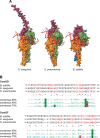
The novel mode of DNA binding we have identified in S. sanguinis is broadly conserved in monoderms expressing T4dP. (A) Structural models, predicted by AlphaFold 3 (25), of the Com pilus tips with bound DNA in S. pneumoniae and B. subtilis compared to S. sanguinis. The DNA always interacts exclusively with the ComGD-ComGF interface. (B) Comparison of the DNA-binding sites as defined by PISA (34) and DNAproDB (35) analyses of the DNA/tip complexes in B. subtilis, S. pneumoniae, and S. sanguinis. The residues involved in DNA-binding as defined by DNAproDB, PISA, and/or our functional analysis were identified in red, bold, and/or underlined, respectively. The sequences were aligned using Clustal Omega (36), with minor corrections for ComGF. The three bottom rows represent the 90%, 80%, and 70% consensus sequences—aligned to B. subtilis—formatted with MView (37) from MSA computed from the 1,794 ComGD and 2,580 ComGF entries in InterPro. h, hydrophobic (A, C, F, G, H, I, K, L, M, R, T, V, W, Y). t, turn-like (A, C, D, E, G, H, K, N, Q, R, S, T). a, aromatic (F, H, W, Y). p, polar (C, D, E, H, K, N, Q, R, S, T). s, small (A, C, D, G, N, P, S, T, V). o, alcohol (S, T). l, aliphatic (I, L, V). u, tiny (A, G, S). +, positive (H, K, R).

References

    1. Thomas CM, Nielsen KM. 2005. Mechanisms of, and barriers to, horizontal gene transfer between bacteria. Nat Rev Microbiol 3:711–721. doi: 10.1038/nrmicro1234 - DOI - PubMed
    1. Johnston C, Martin B, Fichant G, Polard P, Claverys JP. 2014. Bacterial transformation: distribution, shared mechanisms and divergent control. Nat Rev Microbiol 12:181–196. doi: 10.1038/nrmicro3199 - DOI - PubMed
    1. Dubnau D, Blokesch M. 2019. Mechanisms of DNA uptake by naturally competent bacteria. Annu Rev Genet 53:217–237. doi: 10.1146/annurev-genet-112618-043641 - DOI - PubMed
    1. Ellison CK, Dalia TN, Vidal Ceballos A, Wang J-Y, Biais N, Brun YV, Dalia AB. 2018. Retraction of DNA-bound type IV competence pili initiates DNA uptake during natural transformation in Vibrio cholerae. Nat Microbiol 3:773–780. doi: 10.1038/s41564-018-0174-y - DOI - PMC - PubMed
    1. Provvedi R, Dubnau D. 1999. ComEA is a DNA receptor for transformation of competent Bacillus subtilis. Mol Microbiol 31:271–280. doi: 10.1046/j.1365-2958.1999.01170.x - DOI - PubMed

MeSH terms

LinkOut - more resources