

Skin-Colored Papules on the Neck of a Postmenopausal Woman: A Diagnostic Challenge
- PMID: 40407483
- PMCID: PMC12101269
- DOI: 10.3390/dermatopathology12020015
Skin-Colored Papules on the Neck of a Postmenopausal Woman: A Diagnostic Challenge
Abstract
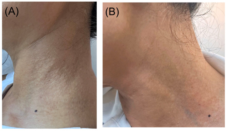
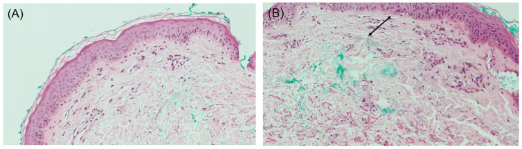
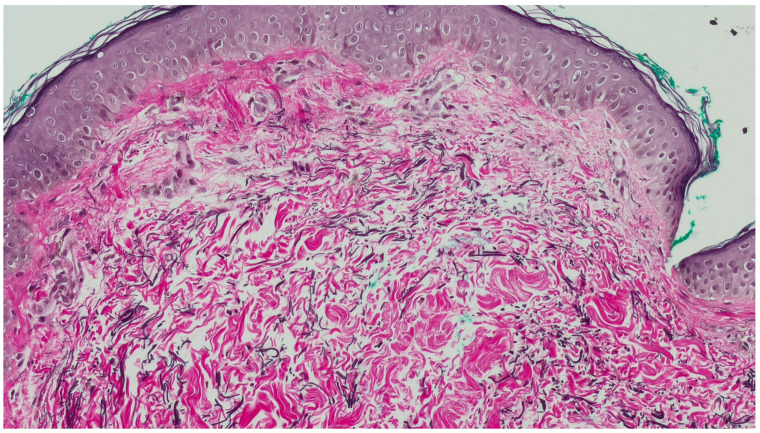
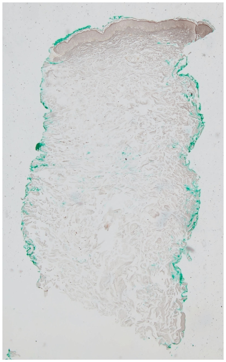
A 64-year-old patient presented for management of symptomatic skin-colored papules symmetrically distributed over the lateral neck over the past two years, which failed to improve on multiple topical corticosteroids, antifungal creams, and topical calcineurin inhibitor. Histopathologic examination showed a regular epidermis with increased melanophages in the papillary dermis, without vacuolar degeneration of the basement membrane. Verhoeff Van Gieson stain highlighted a band-like zone of attenuated elastic fibers in the papillary dermis, while Von Kossa stain was negative for calcified fibers. PAS staining was negative for fungal organisms and Alcian blue showed no increase in dermal mucin.
Keywords: acquired elastic tissue disorder; dermatopathology; histopathology; pseudoxanthoma elasticum-like papillary dermal elastolysis; trifarotene therapy.
Conflict of interest statement
The authors declare no conflicts of interest.
Figures
Similar articles
-
Pseudoxanthoma Elasticum-Like Papillary Dermal Elastolysis: A Single Case Report.J Cutan Med Surg. 2017 Jul/Aug;21(4):345-347. doi: 10.1177/1203475417699407. Epub 2017 Mar 10. J Cutan Med Surg. 2017. PMID: 28282240
-
Pseudoxanthoma elasticum-like papillary dermal elastolysis in non-exposed skin.An Bras Dermatol. 2020 Mar-Apr;95(2):247-249. doi: 10.1016/j.abd.2019.08.024. Epub 2020 Feb 12. An Bras Dermatol. 2020. PMID: 32111413 Free PMC article.
-
Pseudoxanthoma elasticum-like papillary dermal elastolysis: A mimicker of genetic pseudoxanthoma elasticum.Clin Dermatol. 2021 Mar-Apr;39(2):206-210. doi: 10.1016/j.clindermatol.2020.10.018. Epub 2020 Oct 16. Clin Dermatol. 2021. PMID: 34272011
-
Pseudoxanthoma elasticum-like papillary dermal elastolysis: immunohistochemical study using elastic fiber cross-reactivity with an antibody against amyloid P component.Am J Dermatopathol. 2012 Aug;34(6):637-43. doi: 10.1097/DAD.0b013e318257be63. Am J Dermatopathol. 2012. PMID: 22722462 Review.
-
Pseudoxanthoma elasticum-like papillary dermal elastolysis; A report of two cases and a literature review.Dermatol Online J. 2024 Oct 15;30(5). doi: 10.5070/D330564429. Dermatol Online J. 2024. PMID: 39680970 Review.
References
-
- Orlandi A., Bianchi L., Nini G., Spagnoli L.G. Familial occurrence of pseudoxanthoma-elasticum-like papillary dermal elastolysis. J. Eur. Acad. Dermatol. Venereol. 1998;10:175–178. - PubMed
Publication types
LinkOut - more resources
Full Text Sources
Miscellaneous
