Review
. 2025 May 18;15(3):61.
doi: 10.3390/audiolres15030061.

Vestibular Atelectasis: A Narrative Review and Our Experience

Affiliations
Review

Vestibular Atelectasis: A Narrative Review and Our Experience

Andrea Tozzi et al. Audiol Res. .

Abstract

Vestibular atelectasis (VA) is a rare clinical entity characterized by a collapse of the endolymphatic space resulting in vestibular loss with the possible onset of positional and/or sound/pressure-induced vertigo. It could be idiopathic or secondary to other inner-ear diseases including Meniere's disease (MD). A collapse of the membranous labyrinth involving the semicircular canals (SCs) and the utricle represents its distinctive histopathological feature. While specific radiological patterns consistent with VA have been described on contrast-enhanced MRI with delayed acquisitions, an impairment of the blood-labyrinthine barrier (BLB) could be detected in several disorders leading to vestibular loss. We conducted a narrative review of the literature on VA focusing on the putative pathomechanisms accounting for positional and sound/pressure-induced nystagmus despite unilateral vestibular loss (UVL) in this condition, providing two novel cases of VA. Both patients presented with a clinical picture consistent with unilateral MD that rapidly turned into progressive UVL and positional and/or sound/pressure-induced vertigo. In both cases, the posterior SC was initially impaired at the video-head impulse test (vHIT) and both cervical and ocular VEMPs were initially reduced. Progressively, they developed unsteadiness with paretic spontaneous nystagmus, an impairment also for the lateral and anterior SCs, caloric hypo/areflexia and VEMPs areflexia. They both exhibited ipsilesional nystagmus to sound/pressure stimuli and in one case a persistent geotropic direction-changing positional nystagmus consistent with a "light cupula" mechanism involving the lateral SC of the affected side. A collapse of the membranous labyrinthine walls resulting in contact between the vestibular sensors and the stapes footplate could explain the onset of nystagmus to loud sounds and/or pressure changes despite no responses to high- and low-frequency inputs as detected by caloric irrigations, vHIT and VEMPs. On the other hand, the onset of positional nystagmus despite UVL could be explained with the theory of the "floating labyrinth". Both patients received contrast-enhanced brain MRI with delayed acquisition exhibiting increased contrast uptake in the pars superior of the labyrinth, suggesting an impairment of the BLB likely resulting in secondary VA. A small intralabyrinthine schwannoma was detected in one case. VA should always be considered in case of positional and/or sound/pressure-induced vertigo despite UVL.

Keywords: Hennebert sign; MRI; Menière disease; Tullio phenomenon; endolymphatic hydrops; intralabyrinthine schwannoma; vestibular atelectasis.


Conflict of interest statement

The authors declare no conflicts of interest.

Figures

Figure 1
Figure 1
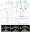
Early instrumental assessment of patient n. 1. (A) Pure-tone audiometry with slight low-frequency SNHL on the right side. (B) vHIT with selective slight VOR-gain impairment for the right PSC. Blue lines represent head impulses exciting left canals, orange lines correspond to impulses for right canals, green lines represent eye movements induced by the activation of the VOR following each impulse and red lines correspond to corrective saccades. Mean value of VOR gain (eye velocity/head velocity) is reported for each canal. The hexagonal plot in the center of the figure summarizes mean VOR gains for each canal; normal gains are shown in green and deficient gains are shown in red. Gains are considered normal if more than 0.8 for lateral SCs and more than 0.7 for vertical SCs. (C) cVEMPs (above) and oVEMPs (below) with a significant reduction in amplitudes on the right side (AR > 35%).
Figure 2
Figure 2
vHIT data of patient n. 1 at the relapse of dizziness. Reduced VOR gain values for the right LSC and PSC with corrective saccades can be detected.
Figure 3
Figure 3
Final instrumental follow up and radiological data of in patient n. 1. (A) Pure-tone audiometry with slight low-frequency SNHL on the right side. (B) vHIT with severe reduction in the VOR gain for all the right SCs. (C) cVEMPs (above) and oVEMPs (below) showing no detectable responses on the right side. (D) BCT with caloric areflexia on the right side. (E) FLAIR images from 1.5 T brain MRI with delayed acquisitions: sequences from the lower to the upper part of the inner-ear (a–f) highlighting on the right side an intralabyrinthine enhancement (yellow arrow) of the vestibule, utricle, semicircular canals and basal turn of the cochlea.
Figure 4
Figure 4
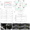
Early instrumental assessment of patient n. 2. (A) Pure tone audiometry with slight high-frequency SNHL on the right side and mild–severe flat SNHL on the left side. (B) vHIT with selective slight VOR gain impairment for the left PSC. (C) cVEMPs (above) and oVEMPs (below) with significant reduction in amplitude on the left side (AR > 35%).
Figure 5
Figure 5
vHIT data of patient n. 2 at the onset of both positional and sound/pressure-induced vertigo. Reduced VOR gain values for the left SCs (in particular for the PSC and the LSC) with a slight hypofunction also for the contralateral PSC can be detected.
Figure 6
Figure 6
Final instrumental follow up and radiological data of in patient n. 2. (A) Pure-tone audiometry with a slight impairment of the flat SNHL on the left side. (B) vHIT with severe reduction in the VOR gain for all the left SCs and a slight impairment for the contralesional vertical SC. (C) cVEMPs (above) and oVEMPs (below) showing no detectable responses on the left side. (D) BCT with caloric areflexia on the left side. (E) Brain MRI: T2 (a) and T1 with Gadolinium (b) highlighting a small ILS on the left side (yellow arrow). (F) FLAIR images from 1.5 T brain MRI with delayed acquisitions: sequences from the lower to the upper part of the inner-ear (ad) highlighting intralabyrinthine enhancement (yellow arrow) involving all portions of the perilymphatic space of the left side.

References

    1. Merchant S.N., Schuknecht H.F. Vestibular atelectasis. Pt 1Ann. Otol. Rhinol. Laryngol. 1988;97:565–576. doi: 10.1177/000348948809700601. - DOI - PubMed
    1. Wenzel A., Ward B.K., Schubert M.C., Kheradmand A., Zee D.S., Mantokoudis G., Carey J.P. Patients with vestibular loss, Tullio phenomenon, and pressure-induced nystagmus: Vestibular atelectasis? Otol. Neurotol. 2014;35:866–872. doi: 10.1097/MAO.0000000000000366. - DOI - PubMed
    1. Finn S., Dietzek M., Karvouniari P., Klingner C.M., Neumann R., Guntinas-Lichius O., Witte O.W., Axer H. Bilateral vestibulopathy with positive Tullio phenomenon. Laryngoscope. 2018;128:1223–1225. doi: 10.1002/lary.26690. - DOI - PubMed
    1. Roy S., Kumarasamy G., Salim R. Bilateral Vestibular Atelectasis with Tullio’s Phenomenon. Indian J. Otolaryngol. Head Neck Surg. 2019;71((Suppl. 2)):1599–1602. doi: 10.1007/s12070-019-01685-z. - DOI - PMC - PubMed
    1. Maslovara S., Butkovic-Soldo S., Pajic-Matic I., Sestak A. Vestibular atelectasis: Decoding pressure and sound-induced nystagmus with bilateral vestibulopathy. Laryngoscope. 2019;129:1685–1688. doi: 10.1002/lary.27724. - DOI - PubMed

LinkOut - more resources