. 2025 Dec;14(1):2511134.
doi: 10.1080/22221751.2025.2511134. Epub 2025 Jun 19.

Respiratory transmission potential of severe fever with thrombocytopenia syndrome bunyavirus: evidence from intranasal exposure in a humanized mouse model

Affiliations

Respiratory transmission potential of severe fever with thrombocytopenia syndrome bunyavirus: evidence from intranasal exposure in a humanized mouse model

Dafeng Lu et al. Emerg Microbes Infect. 2025 Dec.

Abstract

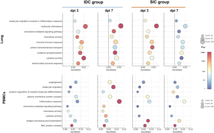
Severe Fever with Thrombocytopenia Syndrome Bunyavirus (SFTSV) is a highly lethal pathogen with expanding endemic regions in Asia. While primarily transmitted by ticks, recent evidence suggests potential airborne transmission, raising significant public health concerns. This study investigates the potential for respiratory transmission and pathogenesis using humanized NCG mice inoculated with SFTSV via subcutaneous injection challenge (SIC) or intranasal drop challenge (IDC). Both groups demonstrated rapid systemic dissemination, marked by viremia, weight loss, and multi-organ injury, with hemorrhagic manifestations observed in high-dose infection groups. Histopathological evaluations revealed lung pathology in the intranasal drop challenge mice, including extensive alveolar disruption and inflammatory cell infiltration. Transcriptomic analyses further confirmed that respiratory route inoculation resulted in heightened expression of inflammatory signalling pathways such as IL-17 and NF-κB, potentially contributing to severe local immunopathology. Subcutaneous infection provoked an earlier systemic immune response, with significant upregulation of antigen-processing genes in peripheral blood mononuclear cells. Nevertheless, both routes ultimately culminated in widespread injury to the liver, spleen, kidney, highlighting the systemic nature of SFTSV pathogenesis. These findings underscore the need for preventive strategies addressing respiratory spread.

Keywords: SFTSV; humanized mouse model; intranasal drop challenge; next-generation sequencing; respiratory transmission potential.


Conflict of interest statement

No potential conflict of interest was reported by the author(s).

Figures

Figure 1.
Figure 1.
Progression of SFTSV Infection in Mice: Survival, Viral Load, and Immune Response Dynamics Across Infection Groups. A. Distribution of total, infected, and deceased mice across experimental groups. B. Survival probability following SFTSV infection across different infection routes and doses (log-rank test, p = 0.11). C. Longitudinal changes in body weight during infection. D. Infection status of individual mice over time by group. E. Plasma viral load (copies/mL) measured at multiple time points post-infection. F. SFTSV-specific antibody levels (pg/mL) in plasma at days 0, 7, and 14. G. Plasma IL-6 concentrations (pg/mL) in individual mice over time. H. Number of mice with elevated TNF-α levels above 117 pg/mL detection threshold. Groups: Control (n = 12), SIC (subcutaneous injection challenge group, n = 10), IDC-High (high dose intranasal drop challenge group: 5×10⁵ TCID₅₀, n = 10), IDC-Moderate (moderate dose: 5×10⁴ TCID₅₀, n = 10), IDC-Low (low dose: 5×10³ TCID₅₀, n = 10)
Figure 2.
Figure 2.
Histopathology and Viral Dynamics in Tissues of SFTSV-Infected Mice. A: Comparative Histopathology of Lung Tissues Control Group: Lung Tissue: Lung tissue exhibits intact pulmonary architecture, well-defined bronchioles, tightly arranged blood vessels, and clearly structured alveoli. Alveolar spaces show moderate and uniform expansion, with mild perivascular inflammatory cell infiltration (long arrows) observed. Lung IDC Group: a: Lung sections display moderate histopathological changes, characterized by focal mononuclear cell infiltration (long arrows) extending into alveolar and perivascular regions. b: Severe structural disruptions in alveolar integrity are observed, accompanied by fibrin-like exudates and necrotic cellular debris. c: Numerous infiltrating cells with eosinophilic cytoplasm (long arrows) and abundant foamy cells (arrowheads) are observed. The overall pulmonary structure shows severe damage indicative of exacerbated viral pathogenicity. Lung SIC Group: a and b: Lung tissues demonstrate severe pathological alterations, including extensive mononuclear cell infiltration (long arrows), pronounced alveolar structural disruption, and indistinct vascular boundaries. c: Multiple focal infarctions (pentagrams) occur within alveolar regions, characterized by extensive epithelial and endothelial cell necrosis, fibrin-like remnants (long arrows), and prominent inflammatory exudates. B: Inflammatory Scores of Lung Tissue Lung injury was evaluated using a semi-quantitative scoring system (1-5) based on key histopathological features. Shown are the average scores for four representative indicators: macrophage infiltration, lymphocyte infiltration, perivascular inflammation, and interstitial inflammation, across control, SIC, and IDC groups. The boxplots reveal distinct inflammatory patterns among the groups, with IDC lungs exhibiting the most pronounced changes. C: Comparative Histopathology of Liver, Kidney, Spleen, and Brain Control Group: Liver Tissue: Neat hepatocyte arrangements with no abnormalities. Mild focal infiltration of mononuclear cells is seen around the central vein, with clustered distribution (pentagrams). Spleen Tissue: Clearly defined red and white pulp regions, with a normal lymphocyte count. Occasional clusters of extramedullary hematopoietic cells are observed in the red pulp (pentagrams). Kidney Tissue: Orderly structures, with well-organized renal tubules and glomeruli. Brain Tissue: Healthy neuronal integrity, with normal neuron size, Nissl bodies, and moderate glial cells. Infection Group: Liver Tissue: Pericentral hepatocyte necrosis with inflammatory cell infiltration (pentagrams). Histological structure is disrupted, with hepatic cords replaced by inflammatory cells. Necrotic hepatocytes display strong eosinophilia with nuclear pyknosis, karyorrhexis, or karyolysis (long arrows). Spleen Tissue: Loosely organized structure; blurred red and white pulp boundaries; shrunken lymphocytes detached from tissues (long arrows). Kidney Tissue: Dilated renal tubules (long arrows); mononuclear cell clusters with eosinophilic cytoplasm around glomeruli (pentagrams). Brain Tissue: Disorganized structure; reduced neurons with eosinophilic cytoplasm, Nissl body loss, satellite cells (long arrows), activated microglia (arrowheads), and neutrophils (green long arrows). D: Tissue-Specific Detection Rates of SFTSV in Infected Mice. E: Tissue Viral Loads Across Experimental Groups.
Figure 3.
Figure 3.
Overlap and Expression Patterns of Differentially Expressed Genes (DEGs) in Lung Tissues and PBMCs During SFTSV Infection. Venn diagrams (left) show the number of differentially expressed genes (DEGs) identified in the lung tissues and PBMCs at dpi3 and dpi7, across subcutaneous injection challenge (SIC) and intranasal drop challenge (IDC) routes compared to uninfected controls. Heatmaps (right) display representative DEGs from the overlapping regions, reflecting transcriptional responses in both compartments.
Figure 4.
Figure 4.
GO Enrichment Analysis of Differentially Expressed Genes in Lung Tissues and PBMCs at dpi3 and dpi7. Gene Ontology (GO) enrichment analysis was performed on differentially expressed genes (DEGs) identified in lung tissues (top panels) and PBMCs (bottom panels) at dpi3 and dpi7, in both subcutaneous injection challenge (SIC) and intranasal drop challenge (IDC). Dot plots display significantly enriched GO biological processes and molecular functions, with dot size representing the number of enriched genes and color indicating the adjusted p-value.
Figure 5.
Figure 5.
Comparative KEGG Pathway Enrichment Analysis Between IDC and SIC Groups in Lung Tissues and PBMCs. KEGG pathway enrichment analysis was performed to compare the transcriptomic differences between the subcutaneous injection challenge (SIC) and intranasal drop challenge (IDC) in both lung tissues (left) and PBMCs (right) at dpi3 and dpi7. Enriched pathways were separated into relatively upregulated (top) and downregulated (bottom) in the IDC group compared to the SIC group. Dot size represents the number of enriched genes per pathway, while color indicates the adjusted p-value.

Similar articles

References

    1. Yu X, Liang M, Zhang S, et al. Fever with thrombocytopenia associated with a novel bunyavirus in China. N Engl J Med. 2011;364(16):1523–1532. doi: 10.1056/NEJMoa1010095 - DOI - PMC - PubMed
    1. Shao L, Pang Z, Fu H, et al. Identification of recently identified tick-borne viruses (Dabieshan tick virus and SFTSV) by metagenomic analysis in ticks from Shandong Province, China. J Infect. 2020;81(6):973–978. doi: 10.1016/j.jinf.2020.10.022 - DOI - PubMed
    1. Xie J, Li H, Zhang X, et al. Akkermansia muciniphila protects mice against an emerging tick-borne viral pathogen. Nat Microbiol. 2023;8(1):91–106. doi: 10.1038/s41564-022-01279-6 - DOI - PubMed
    1. Li H, Lu Q, Xing B, et al. Epidemiological and clinical features of laboratory-diagnosed severe fever with thrombocytopenia syndrome in China, 2011-17: a prospective observational study. Lancet Infect Dis. 2018;18(10):1127–1137. doi: 10.1016/S1473-3099(18)30293-7 - DOI - PubMed
    1. Takahashi T, Maeda K, Suzuki T, et al. The first identification and retrospective study of severe fever with thrombocytopenia syndrome in Japan. J Infect Dis. 2014;209(6):816–827. doi: 10.1093/infdis/jit603 - DOI - PMC - PubMed

MeSH terms

LinkOut - more resources