. 2025 May 23;6(1):32.
doi: 10.1186/s43556-025-00269-4.

Chemoproteomics unveils Sofalcone targeting ribosomal proteins to inhibit protein synthesis in Staphylococcus aureus

Affiliations

Chemoproteomics unveils Sofalcone targeting ribosomal proteins to inhibit protein synthesis in Staphylococcus aureus

Lirun Zhou et al. Mol Biomed. .

Abstract

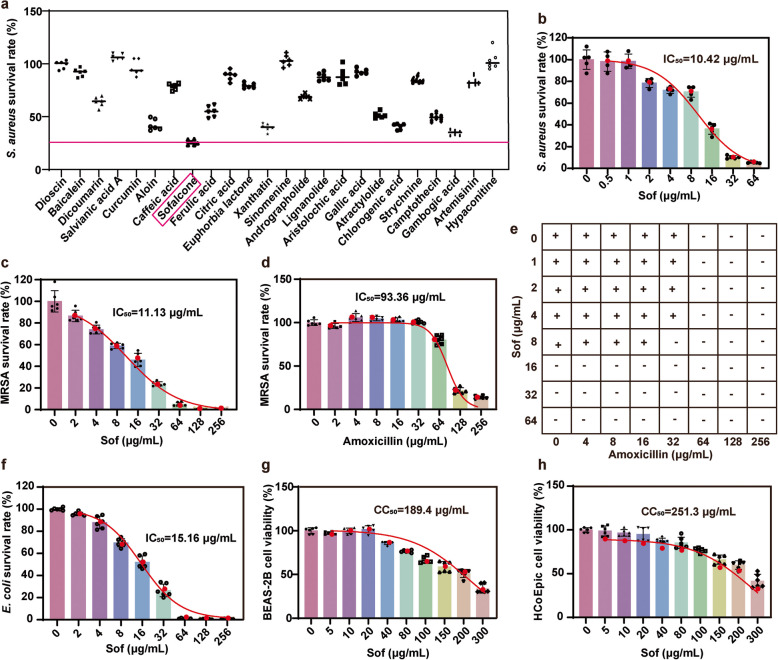
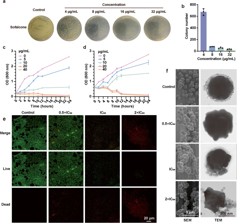
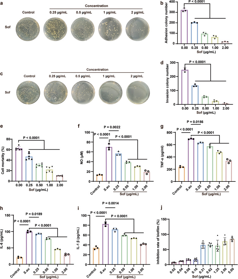
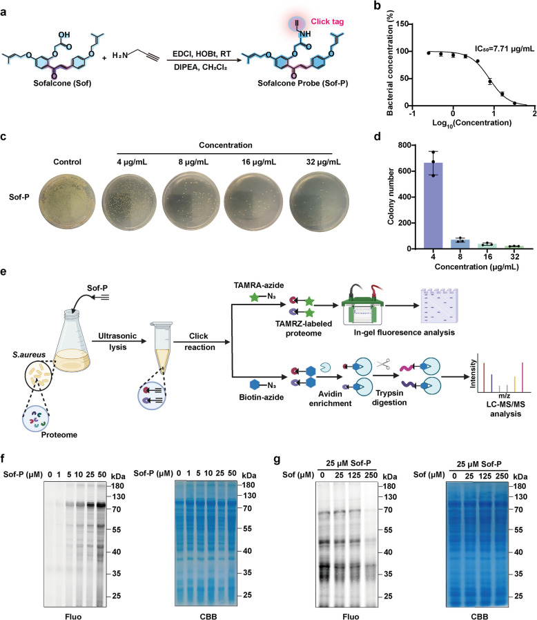
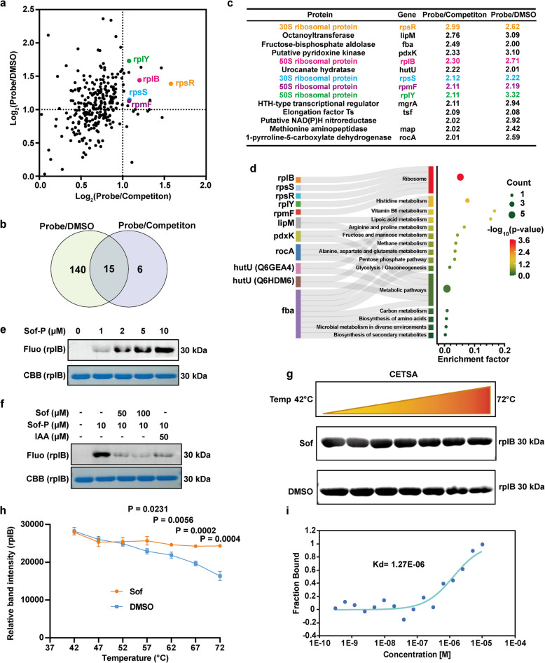
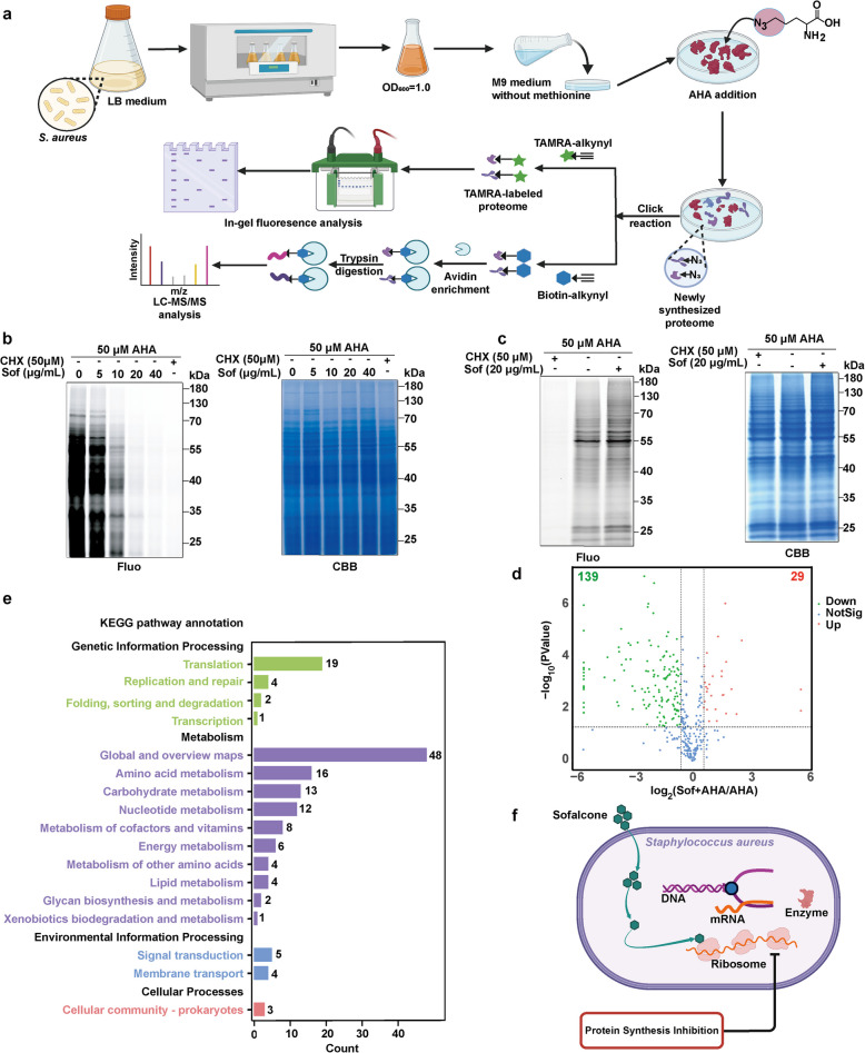
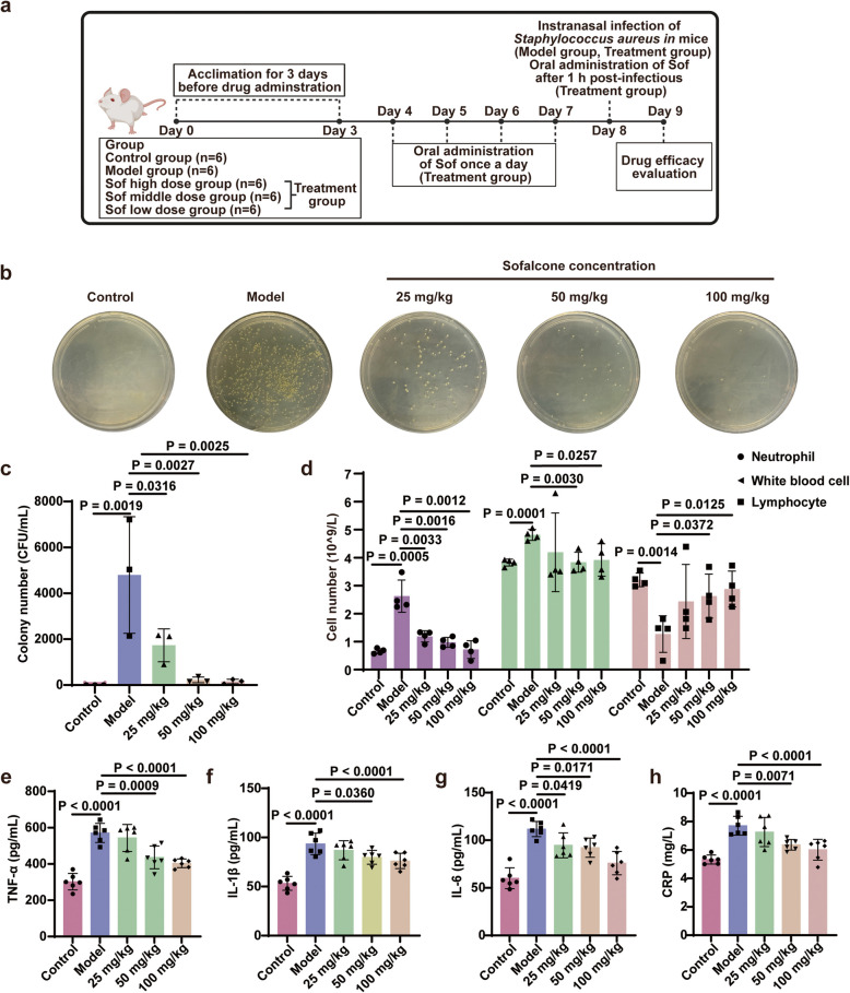
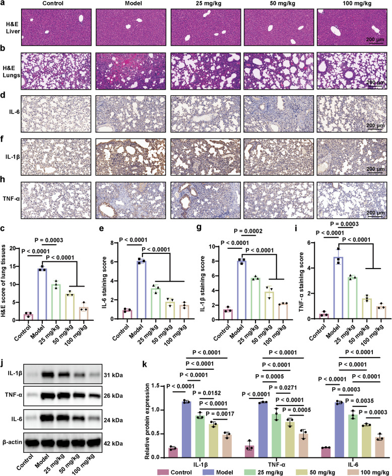
The escalating threat of antibiotic resistance, particularly in Staphylococcus aureus (including methicillin-resistant strains, MRSA), underscores the urgent need for novel therapeutics. Sofalcone (Sof), a chalcone derivative from Sophora subprostrata with established anti-inflammatory and anti-ulcer properties, exhibits promising yet underexplored antibacterial activity. Here, we demonstrate that Sof potently inhibits S. aureus and MRSA while showing minimal cytotoxicity in human cells. Notably, Sof synergized with amoxicillin, and significantly reduced the pathogenicity of S. aureus through inhibiting biofilm formation addressing key virulence factors. Through chemoproteomic profiling using a clickable Sof-derived probe, ribosomal proteins, specifically the 50S subunit protein rplB, were identified as primary targets. Sof covalently binds to rplB via cysteine residues, as validated by cellular thermal shift assays, microscale thermophoresis, and competition assays. Bio-orthogonal noncanonical amino acid tagging revealed that Sof disrupts bacterial protein synthesis by impairing ribosomal function, a mechanism distinct from conventional antibiotics. In a murine model of S. aureus-induced acute lung injury, Sof greatly reduced bacterial load in lungs, attenuated systemic inflammation, and mitigated histopathological damage. Its dual antibacterial and anti-inflammatory efficacy, coupled with activity against Gram-negative Escherichia coli, highlights broad-spectrum potential. This study unveils a covalent ribosomal-targeting strategy, positioning Sof as a multifaceted candidate against multidrug-resistant infections. Our findings bridge natural product pharmacology and mechanistic antimicrobial discovery, offering a template for combating the global antibiotic resistance crisis.

Keywords: Staphylococcus aureus; Antibiotics; Chemoproteomics; Drug-resistance; Ribosomal proteins; Sofalcone.


Conflict of interest statement

Declarations. Ethics approval and consent to participate: Animal experimentation and the corresponding protocol were approved by the Animal Ethics Committee of the Institute of Chinese Materia Medica China Academy of Chinese Medical Sciences (Ethical Number: 2023B299). Consent for publication: Not applicable. Competing interests: The authors declare no conflicts of interest to disclose.

Figures

Fig. 1
Fig. 1
Sof selectively killed bacteria with limited cytotoxicity on human cells. a Bacterial survival rate of S. aureus following 12 h of treatment with natural products at 20 μg/mL (n = 6). b Bacterial survival rate of S. aureus following 12 h of treatment with Sof at different concentrations (n = 6). c Bacterial survival rate of Methicillin-resistant S. aureus (MRSA) following 12 h of treatment with Sofalcone at different concentrations (n = 6). d Bacterial survival rate of MRSA following 12 h of treatment with amoxicillin at different concentrations (n = 6). e The checkerboard method showing the synergy of Sof and amoxicillin combination. “ + ”, none- sterile; “-”, sterile. f Bacterial survival rate of E. coli following 12 h of treatment with Sofalcone at different concentrations (n = 6). g Cell viability of BEAS-2B cells following 12 h of treatment with Sofalcone at different concentrations (n = 6). h Cell viability of HCoEpic cells following 12 h of treatment with Sofalcone at different concentrations (n = 6)
Fig. 2
Fig. 2
Sof effectively inhibited the growth of S. aureus. a Optical images of S. aureus colony following 6 h of treatment with Sofalcone at different concentrations. b Colony counting number of S. aureus following 6 h of treatment with Sofalcone at different concentrations (n = 3). c Growth curve of S. aureus within 24 h of treatment with Sof at different concentrations (n = 3). d Killing curve of S. aureus within 24 h of treatment with Sofalcone at different concentrations (n = 3). e Fluorescence imaging showing the live and dead status of S. aureus following 6 h of treatment with Sof at different concentrations. Live and dead cells are indicated by green and red fluorescence, respectively. f Morphological characterization of S. aureus following 6 h of treatment with Sof at different concentrations by scanning electron microscope (SEM) and transmission electron microscope (TEM)
Fig. 3
Fig. 3
Sof significantly reduced the pathogenicity of S. aureus. a Optical images of S. aureus colonies adhering to BEAS-2B cells following 6 h of treatment with Sofalcone at different concentrations (n = 3). b Colony count of S. aureus adhering to BEAS-2B cells following 6 h of treatment with Sofalcone at different concentrations (n = 3). c Optical images of S. aureus colonies invading BEAS-2B cells following 6 h of treatment with Sofalcone at different concentrations (n = 3). d Colony count of S. aureus invading BEAS-2B cells following 6 h of treatment with Sofalcone at different concentrations (n = 3). e Cell mortality of BEAS-2B cells after S. aureus infection following 6 h of treatment with Sofalcone at different concentrations (n = 3). f-i Secretion of NO (f), TNF-α (g), IL-6 (h), and IL-1β (i) by BEAS-2B cells after S. aureus infection following 6 h of treatment with Sofalcone at different concentrations (n = 3). j Biofilm formation of S. aureus following 6 h of treatment with Sofalcone at different concentrations (n = 6). Data are expressed as the mean ± SEM
Fig. 4
Fig. 4
Antibacterial effect of Sof-P against S. aureus and the proteome labeling efficiency. a Synthesis scheme of sofalcone probe bearing alkynyl (Sof-P). b The half inhibitory concentrations (IC50) of Sof-P against S. aureus after 12 h of treatment. c Optical images of S. aureus colony following 6 h of treatment with Sof-P at different concentrations. d Colony counting number of S. aureus following 6 h of treatment with Sof-P at different concentrations (n = 3). e Overall workflow for profiling protein targets of Sof in S. aureus. f Fluorescent labelling of proteins in S. aureus by Sof-P after in situ treatment. Coomassie brilliant blue (CBB) staining was used to normalize the amount of whole protein. g Competitive fluorescent labelling of proteins in S. aureus by Sof-P after in situ treatment in the presence of excess Sof. Coomassie brilliant blue (CBB) staining was used to normalize the amount of whole protein
Fig. 5
Fig. 5
Protein target identification of Sof in S. aureus via quantitative chemoproteomic profiling. a Volcano plot depicting differential protein profiles captured in the Sof-P group versus the DMSO and competition groups. b Venn diagram displaying the number and overlap of protein targets in the Sof-P group versus the DMSO and competition groups c The table list including the protein name and the quantitative ratios of the 15 overlapped proteins. d KEGG pathway enrichment analysis of Sof-targeted proteins. e Fluorescence labeling of the Sof probe on purified recombinant rplB protein (Sof 1 μM = 0.45 μg/mL). f Competition assay of the Sof probe on purified recombinant rplB protein in the presence of Sof and IAA. g-h Cellular thermal shift assays showing the thermal ability of rplB in the absence and presence of Sof (13 μM). i Binding affinity of rplB for Sof, measured using microscale thermophoresis (MST)
Fig. 6
Fig. 6
Sof suppressed protein synthesis in S. aureus by targeting ribosome proteins. a Schematic workflow for detecting newly synthesized proteins in S. aureus through AHA metabolic labelling. b Fluorescence labeling of proteins bearing AHA incorporation in S. aureus in the presence of Sof at different concentrations. c Fluorescence labeling of proteins bearing AHA incorporation in BEAS-2B cells in the absence and presence of Sof. d Volcano plot for differential protein profiles in the groups of AHA + Sof versus AHA. e KEGG pathway enrichment analysis of proteins whose synthesis inhibited by Sof. f Illustration of mechanism of actions of Sof against S. aureus by hampering protein synthesis
Fig. 7
Fig. 7
Sof inhibited bacterial reproduction and reduced inflammation level in S. aureus infected mice. a Schematic illustration of the animal experiment. (Mice were divided into five groups: Control, Model, and Sof treatment group (25, 50, and 100 mg/kg). b-c In vivo antimicrobial activity assessment of Sof in S. aureus-infected mice by plate colony counting method, where the samples were collected from bronchoalveolar lavage fluid (n = 3). d Inflammatory cells including neutrophil, white blood cell, and lymphocyte counted in peripheral anticoagulation blood (n = 4). eh Serum level of TNF-α, IL-1β, IL-6, and CRP in mice (n = 6). Data are expressed as the mean ± SEM
Fig. 8
Fig. 8
Sof ameliorated acute lung injury in mice induced by S. aureus. a-b Representative hematoxylin and eosin (H&E) staining of liver (a) and lung (b) tissues. c Quantitative scoring of lung histopathological changes (n = 3). d-i Immunohistochemical staining (d, f, h) and staining scores (e, g, i) for IL-6 (d-e), IL-1β (f-g), and TNF-α (h-i) in lung tissues. j-k Relative expression level of IL-1β, TNF-α, and IL-6 in lung tissues examined by western blot. Data are expressed as the mean ± SEM

Similar articles

References

    1. Cheung GYC, Bae JS, Otto M. Pathogenicity and virulence of S. aureus. Virulence. 2021;12(1):547–69. 10.1080/21505594.2021.1878688. - PMC - PubMed
    1. Zhu J, Xie R, Gao R, Zhao Y, Yodsanit N, Zhu M, et al. Multimodal nanoimmunotherapy engages neutrophils to eliminate S. aureus infections. Nat Nanotechnol. 2024;19(7):1032–43. 10.1038/s41565-024-01648-8. - PubMed
    1. Rybak MJ. The efficacy and safety of daptomycin: first in a new class of antibiotics for Gram-positive bacteria. Clin Microbiol Infect. 2006;12(Suppl 1):24–32. 10.1111/j.1469-0691.2006.01342.x. - PubMed
    1. Kato H, Hagihara M, Kato M, Yamagishi Y, Umemura T, Asai N, et al. A Retrospective Study on the Effectiveness and Safety of Vancomycin versus Daptomycin in Hemodialysis Patients. Antibiotics (Basel). 2022;11(6). 10.3390/antibiotics11060710. - PMC - PubMed
    1. Zhang J, Guo H, Liu M, Tang K, Li S, Fang Q, et al. Recent design strategies for boosting chemodynamic therapy of bacterial infections. Exploration (Beijing). 2024;4(2):20230087. 10.1002/EXP.20230087. - PMC - PubMed

MeSH terms

LinkOut - more resources