Assessment of Corneal Endothelial Barrier Function Based on "Y-Junctions": A Finite Element Analysis
- PMID: 40408094
- PMCID: PMC12118505
- DOI: 10.1167/iovs.66.5.33
Assessment of Corneal Endothelial Barrier Function Based on "Y-Junctions": A Finite Element Analysis
Abstract
Purpose: Corneal endothelial cell dysfunction is a major contributor to corneal edema, opacity, and, in severe cases, corneal blindness. Currently, no direct and reliable clinical indicator is available for evaluating the function of corneal endothelial cells. This study aimed to identify new noninvasive indicators for the clinical assessment of corneal endothelial barrier function.
Methods: This study established a finite element simulation model of a monolayer full-corneal endothelium to screen for sensitive indicators that reflect the barrier function of the corneal endothelium. Scanning electron microscopy (SEM) was used to observe the morphology of endothelial junctions, and immunofluorescence was used to examine the expression of fluorescent particles.
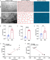
Results: The "Y-junctions" area was identified as the parameter most sensitive to changes in intraocular pressure when considering the different analytical indices obtained from the finite element model. SEM of Corneal endothelial dysfunction models in rabbits and mice further confirmed a substantial increase in the "Y-junctions" relative to control groups. Additionally, functional in vitro experiments provided further evidence of a positive relationship between larger "Y-junctions" and enhanced permeability to fluorescent particles. Finally, clinical analysis of measurements related to "Y-junctions" in patients suffering from various corneal endothelial disorders consistently revealed that these junctions were significantly larger compared to those observed in healthy control subjects.
Conclusions: The "Y-junctions" area serves as a potentially sensitive indicator for assessing endothelial barrier integrity. Consistent observation of alterations in this area may facilitate the early identification of dysfunction in circulating endothelial cells.
Conflict of interest statement
Disclosure:
Figures


References
-
- Armitage WJ, Dick AD, Bourne WM. Predicting endothelial cell loss and long-term corneal graft survival. Invest Ophthalmol Vis Sci. 2003; 44: 3326–3331. - PubMed
-
- Price MO, Mehta JS, Jurkunas UV, Price FW Jr.. Corneal endothelial dysfunction: evolving understanding and treatment options. Prog Retin Eye Res. 2021; 82: 100904. - PubMed
-
- Gain P, Jullienne R, He Z, et al.. Global survey of corneal transplantation and eye banking. JAMA Ophthalmol. 2016; 134: 167–173. - PubMed
-
- Kaur K, Gurnani B. Specular microscopy. In: StatPearls. Treasure Island, FL: StatPearls Publishing; 2024. - PubMed
MeSH terms
LinkOut - more resources
Full Text Sources
Medical

