. 2025 Jul 3;112(7):1681-1698.
doi: 10.1016/j.ajhg.2025.04.016. Epub 2025 May 22.

Bi-allelic variants in POPDC2 cause an autosomal recessive syndrome presenting with cardiac conduction defects and hypertrophic cardiomyopathy

Michele Nicastro  1 Alexa M C Vermeer  2 Pieter G Postema  1 Rafik Tadros  3 Forrest Z Bowling  4 Hildur M Aegisdottir  5 Vinicius Tragante  6 Lukas Mach  7 Alex V Postma  8 Elisabeth M Lodder  9 Karel van Duijvenboden  10 Rob Zwart  11 Leander Beekman  1 Lingshuang Wu  4 Sean J Jurgens  12 Paul A van der Zwaag  13 Mariëlle Alders  11 Mona Allouba  14 Yasmine Aguib  15 J Luis Santome  16 David de Una  16 Lorenzo Monserrat  17 Antonio M A Miranda  18 Kazumasa Kanemaru  19 James Cranley  19 Ingeborg E van Zeggeren  20 Eleonora M A Aronica  21 Michela Ripolone  22 Simona Zanotti  22 Gardar Sveinbjornsson  6 Erna V Ivarsdottir  6 Hilma Hólm  6 Daníel F Guðbjartsson  6 Ástrós Th Skúladóttir  6 Kári Stefánsson  6 Lincoln Nadauld  23 Kirk U Knowlton  24 Sisse Rye Ostrowski  25 Erik Sørensen  26 Ole Birger Vesterager Pedersen  27 Jonas Ghouse  28 Søren A Rand  29 Henning Bundgaard  29 Henrik Ullum  30 Christian Erikstrup  31 Bitten Aagaard  32 Mie Topholm Bruun  33 Mette Christiansen  34 Henrik K Jensen  35 Deanna Alexis Carere  36 Christopher T Cummings  37 Kristen Fishler  38 Pernille Mathiesen Tørring  39 Klaus Brusgaard  40 Trine Maxel Juul  39 Lotte Saaby  41 Bo Gregers Winkel  29 Jens Mogensen  42 Francesco Fortunato  43 Giacomo Pietro Comi  44 Dario Ronchi  44 J Peter van Tintelen  45 Michela Noseda  46 Michael V Airola  4 Imke Christiaans  13 Arthur A M Wilde  1 Ronald Wilders  10 Sally-Ann Clur  47 Arie O Verkerk  48 Connie R Bezzina  1 Najim Lahrouchi  49
Affiliations

Bi-allelic variants in POPDC2 cause an autosomal recessive syndrome presenting with cardiac conduction defects and hypertrophic cardiomyopathy

Michele Nicastro et al. Am J Hum Genet. .

Abstract

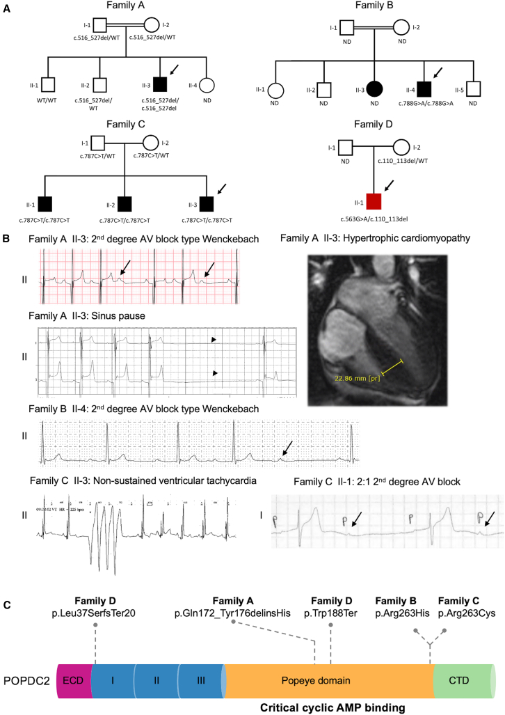
POPDC2 encodes the Popeye domain-containing protein 2, which has an important role in cardiac pacemaking and conduction, due in part to its cyclic AMP (cAMP)-dependent binding and regulation of TREK-1 potassium channels. Loss of Popdc2 in mice results in sinus pauses and bradycardia, and morpholino-mediated knockdown of popdc2 in zebrafish results in atrioventricular (AV) block. We identified bi-allelic variants in POPDC2 in four families with a phenotypic spectrum consisting of sinus node dysfunction, AV conduction defects, and hypertrophic cardiomyopathy. Using homology modeling, we show that the identified variants are predicted to diminish the ability of POPDC2 to bind cAMP. In in vitro electrophysiological studies, we demonstrated that, in contrast with wild-type POPDC2, variants found in affected individuals failed to increase TREK-1 current density. While muscle biopsy of an affected individual did not show clear myopathic disease, it showed significantly reduced abundance of both POPDC1 and POPDC2, suggesting that stability and/or membrane trafficking of the POPDC1-POPDC2 complex is impaired by pathogenic variants in either protein. Single-cell RNA sequencing from human hearts demonstrated that co-expression of POPDC1 and POPDC2 was most prevalent in AV node, AV node pacemaker, and AV bundle cells. Using population-level genetic data of more than 1 million individuals, we show that none of the familial variants were associated with clinical outcomes in heterozygous state, suggesting that heterozygous family members are unlikely to develop clinical manifestations and therefore might not necessitate clinical follow-up. Our findings provide evidence for bi-allelic variants in POPDC2 causing a Mendelian autosomal recessive cardiac syndrome.

Keywords: AV conduction defects; cardiac arrhythmia; hypertrophic cardiomyopathy; population genetics; sinus node disease.


Conflict of interest statement

Declaration of interests L. Monserrat is a shareholder in Dilemma Solutions SL. D.A.C. is an employee of and may own stock in GeneDx. H.M.A., V.T., G.S., E.V.I., H.H., D.F.G., A.T.S., and K.S. report employment at deCODE Genetics during the conduct of the study. C.E. reports grants from Abbott Diagnostics and Novo Nordisk outside the submitted work. K.U.K. reports research support from Intermountain Foundation during the conduct of the study. L.N. reports a stock option grant from Culmination Bio. H.B. reports lecture fees from Amgen, MSD, Sanofi Avensis, Bristol Myers Squibb, and Pfizer; grants from Novo Nordic Foundation; and another from Novo Nordic Foundation (shares) outside the submitted work.

Figures

None
Graphical abstract
Figure 1
Figure 1
Bi-allelic variants in POPDC2 cause a recessive syndrome with sinus node disease and atrioventricular conduction defects with HCM (A) Pedigrees of families A–D. Closed symbols indicate affected individuals. Males are indicated by squares and females by circles. A double line indicates a consanguineous relationship. The arrows point to the probands. Affected individual 6 from family D (highlighted in the pedigree with a filled red box) was diagnosed with bradycardia resulting in an arrest and first-degree AV block during an episode of fulminant myocarditis. (B) Selection of ECG abnormalities: (1) affected individual II-3 from family A (second-degree AV block type Wenckebach and sinus pause [indicated by an arrowhead]; see Figure S1 for longer Holter registration), (2) affected individual II-4 from family B (second-degree AV block type Wenckebach), affected individual II-3 (non-sustained ventricular tachycardia), and affected individual II-1 (2:1 second-degree AV block) from family C. Arrows point to a non-conducted P wave. Upper right panel: cardiac MRI at the age of 11–15 years showing marked hypertrophy of the interventricular septum (23 mm, Z score: 16.43; height, 160 cm; weight, 49 kg) in the proband of family A. (C) POPDC2 protein domain structure and location of variants found in affected individuals. AV, atrioventricular; CTD, carboxy-terminal domain; ECD, extracellular domain; ND, genotype not determined; regions I/II/II, transmembrane region 1–3; WT, wild type.
Figure 2
Figure 2
Functional characterization of the POPDC2 variants (A) Structural model of POPDC2 bound to cAMP generated using AlphaFold2 Multimer and SWISS-MODEL. Dimer subunits are shown in green and cyan; cAMP molecules, green and cyan sticks. N′ and C′ indicate the N and C termini. The positions of the transmembrane and Popeye domains are indicated by the labels. The intrinsically disordered C termini (residues 275–364) are shown as dotted lines. (B) Zoom-in of the predicted cAMP-binding pocket of POPDC2. Dimer subunits are shown in green and cyan; cAMP, green sticks; residues p.Gln172_Tyr176delinsHis, orange; p.Arg263, cyan sticks. (C) Structural models of the POPDC2 variants p.Leu37Serfs20 and p.Trp188Ter, which would both generate truncated proteins that would lack the ability to bind cAMP. (D) Homology model of POPDC2 by AlphaFold Multimer color coded by the average pathogenicity score for each residue as predicted by AlphaMissense. (E) Heatmap of predicted effects of amino-acid substitutions on POPDC2. AlphaMissense (AM) scores range from zero to one, with higher scores corresponding to increased pathogenicity. (F) Homology model of POPDC2 by AlphaFold Multimer color coded by the average pathogenicity score for each residue as predicted by AlphaMissense. The position of non-disease-associated variants found in general population are shown as blue spheres, indicating predicted AlphaMissense pathogenicity scores <0.1. (G) Typical examples of TREK-1 currents upon 500-ms voltage-clamp steps to membrane potentials ranging from −100 to +50 mV from a holding potential of −80 mV in absence or presence of wild-type (WT) and mutant POPDC2. (H) Average current-voltage relationships of TREK-1 currents in absence or presence of WT and mutant POPDC2. (I) TREK-1 current amplitude at +50 mV in absence or presence of WT and mutant POPDC2. p < 0.05 with one-way ANOVA. Error bars indicate the standard error of the mean (SEM).
Figure 3
Figure 3
Functional effects of the POPDC2 variants from in silico modeling (A) Fits to the experimental data on TREK-1 currents in absence or presence of wild-type (WT) and mutant POPDC2. The fit to the mutant data were obtained by scaling the fit to the WT data by a factor of 0.59. (B) Membrane potential (top) and associated TREK-1 current (bottom) of a single human sinus nodal pacemaker cell as simulated using the comprehensive mathematical model developed by Fabbri et al. ITREK-1 was introduced into the original model cell using the fits of (A). ITREK-1 magnitude was set to 0.12 pA/pF at a membrane potential of +30 mV (C) Cycle length of the simulated single human sinus nodal pacemaker cell as a function of ITREK-1 magnitude. (D) Membrane potential (top) and associated TREK-1 current (bottom) of a single human atrial cell as simulated using the comprehensive mathematical model developed by Maleckar et al. ITREK-1 was introduced into the original model cell using the fits of (A). ITREK-1 magnitude was set to 4.0 pA/pF at a membrane potential of +30 mV. Action potentials were elicited at a rate of 1 Hz with a 1-ms, 20% suprathreshold stimulus current. (E) Diastolic potential of the simulated single human atrial cell as a function of ITREK-1 magnitude. (F) Threshold stimulus current of the simulated single human atrial cell as a function of ITREK-1 magnitude.
Figure 4
Figure 4
Evaluation of muscle biopsy from the proband in family A (A and B) (A) Hematoxylin and eosin (H&E) stain and (B) modified Gomori trichrome (MGT) staining of affected individual and, in the inset, control muscle. Scale bar, 50 μm. (C) Immunofluorescent staining for caveolin-3 (red), POPDC1 (green), and merge for affected individual. The inset shows the corresponding immunofluorescence staining for control. Scale bar, 50 μm. (D) Immunofluorescent staining for caveolin-3 (red), POPDC2 (green), and merge for affected individual. The corresponding control staining is shown in the inset. Scale bar, 50 μm. (E–G) Ultrastructural findings. (E) Tubular aggregates in subsarcolemmal region. (F) Sarcolemma alteration (asterisks). (G) Increase in lipid droplets. Scale bar (E and F), 0.84 μm; (G), 3.33 μm.
Figure 5
Figure 5
Expression of POPDC1-3 in human hearts (A) Overview of single-nucleus and spatial-transcriptomics data analysis from a previously published human heart cell atlas.. (B) Spatial-transcriptomics (Visium) analysis of POPDC1 (BVES), POPDC2, and POPDC3 expression across different anatomical regions and histological microstructures in adult human hearts. The anatomical sites sampled included AVN, SAN, left ventricle free wall, left ventricular apex, interventricular septum, left atrium, and right atrium. (C) Percentage of spatial spots where co-expression of both POPDC1/BVES and -2 was detected is shown for each histological feature. (D) POPDC family gene expression across cell types in adult human heart profiled by snRNA-seq (10× Genomics). (E) POPDC1/BVES, -2, and -3 gene expression in cardiomyocyte cell states in adult human hearts. (F) Percentage of cells that co-express POPDC1/BVES and POPDC2 in the same single cardiomyocyte. Figure created with BioRender. Error bars indicate the 95% confidence interval.

Update of

  • Biallelic variants in POPDC2 cause a novel autosomal recessive syndrome presenting with cardiac conduction defects and variable hypertrophic cardiomyopathy.
    Nicastro M, Vermeer AMC, Postema PG, Tadros R, Bowling FZ, Aegisdottir HM, Tragante V, Mach L, Postma AV, Lodder EM, van Duijvenboden K, Zwart R, Beekman L, Wu L, van der Zwaag PA, Alders M, Allouba M, Aguib Y, Santomel JL, de Una D, Monserrat L, Miranda AMA, Kanemaru K, Cranley J, van Zeggeren IE, Aronica EMA, Ripolone M, Zanotti S, Sveinbjornsson G, Ivarsdottir EV, Hólm H, Guðbjartsson DF, Skúladóttir ÁT, Stefánsson K, Nadauld L, Knowlton KU, Ostrowski SR, Sørensen E, Vesterager Pedersen OB, Ghouse J, Rand S, Bundgaard H, Ullum H, Erikstrup C, Aagaard B, Bruun MT, Christiansen M, Jensen HK, Carere DA, Cummings CT, Fishler K, Tøring PM, Brusgaard K, Juul TM, Saaby L, Winkel BG, Mogensen J, Fortunato F, Comi GP, Ronchi D, van Tintelen JP, Noseda M, Airola MV, Christiaans I, Wilde AAM, Wilders R, Clur SA, Verkerk AO, Bezzina CR, Lahrouchi N. Nicastro M, et al. medRxiv [Preprint]. 2024 Jul 5:2024.07.04.24309755. doi: 10.1101/2024.07.04.24309755. medRxiv. 2024. Update in: Am J Hum Genet. 2025 Jul 3;112(7):1681-1698. doi: 10.1016/j.ajhg.2025.04.016. PMID: 39006410 Free PMC article. Updated. Preprint.

References

    1. Park D.S., Fishman G.I. The cardiac conduction system. Circulation. 2011;123:904–915. doi: 10.1161/CIRCULATIONAHA.110.942284. - DOI - PMC - PubMed
    1. Wilde A.A.M., Semsarian C., Márquez M.F., Sepehri Shamloo A., Ackerman M.J., Ashley E.A., Sternick E.B., Barajas-Martinez H., Behr E.R., Bezzina C.R., et al. European Heart Rhythm Association (EHRA)/Heart Rhythm Society (HRS)/Asia Pacific Heart Rhythm Society (APHRS)/Latin American Heart Rhythm Society (LAHRS) Expert Consensus Statement on the state of genetic testing for cardiac diseases. Europace. 2022;24 doi: 10.1093/europace/euac030. 1307–136. - DOI - PubMed
    1. Schindler R.F.R., Scotton C., Zhang J., Passarelli C., Ortiz-Bonnin B., Simrick S., Schwerte T., Poon K.L., Fang M., Rinné S., et al. POPDC1S201F causes muscular dystrophy and arrhythmia by affecting protein trafficking. J. Clin. Investig. 2016;126:239–253. doi: 10.1172/JCI79562. - DOI - PMC - PubMed
    1. De Ridder W., Nelson I., Asselbergh B., De Paepe B., Beuvin M., Ben Yaou R., Masson C., Boland A., Deleuze J.F., Maisonobe T., et al. Muscular dystrophy with arrhythmia caused by loss-of-function mutations in BVES. Neurol. Genet. 2019;5 doi: 10.1212/NXG.0000000000000321. - DOI - PMC - PubMed
    1. Froese A., Breher S.S., Waldeyer C., Schindler R.F.R., Nikolaev V.O., Rinné S., Wischmeyer E., Schlueter J., Becher J., Simrick S., et al. Popeye domain containing proteins are essential for stress-mediated modulation of cardiac pacemaking in mice. J. Clin. Investig. 2012;122:1119–1130. doi: 10.1172/JCI59410. - DOI - PMC - PubMed

LinkOut - more resources