. 2025 May 23;16(1):413.
doi: 10.1038/s41419-025-07736-4.

Mechanical signal modulates prostate cancer immune escape by USP8-mediated ubiquitination-dependent degradation of PD-L1 and MHC-1

Affiliations

Mechanical signal modulates prostate cancer immune escape by USP8-mediated ubiquitination-dependent degradation of PD-L1 and MHC-1

Zhi-Bin Ke et al. Cell Death Dis. .

Abstract

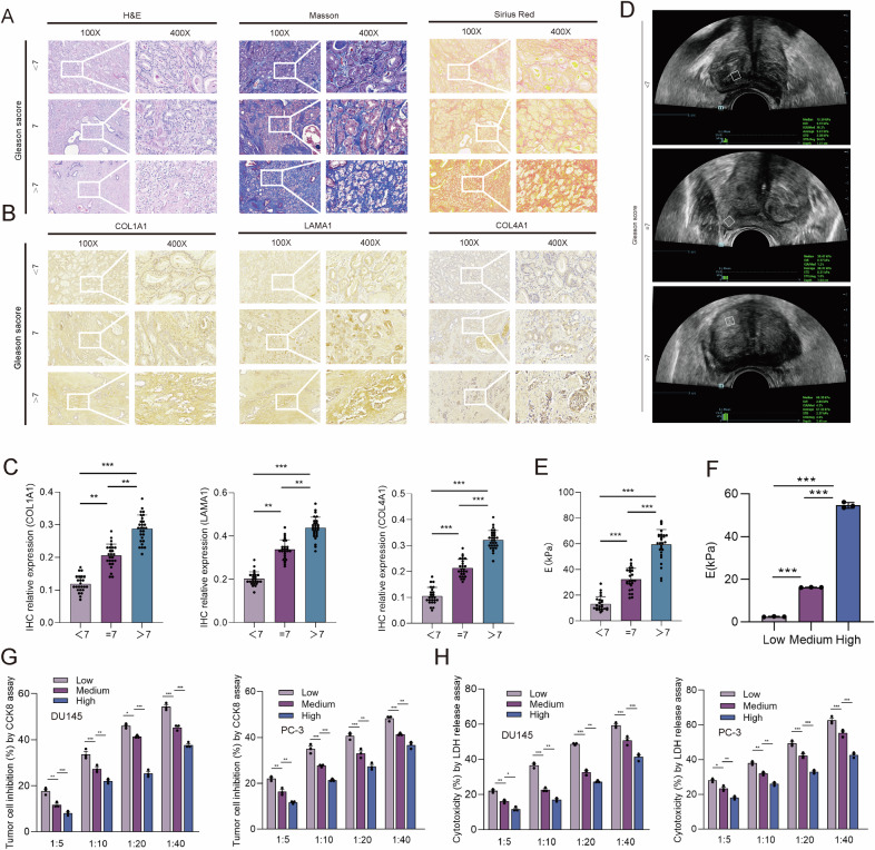
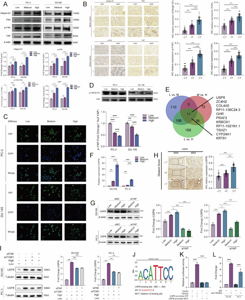
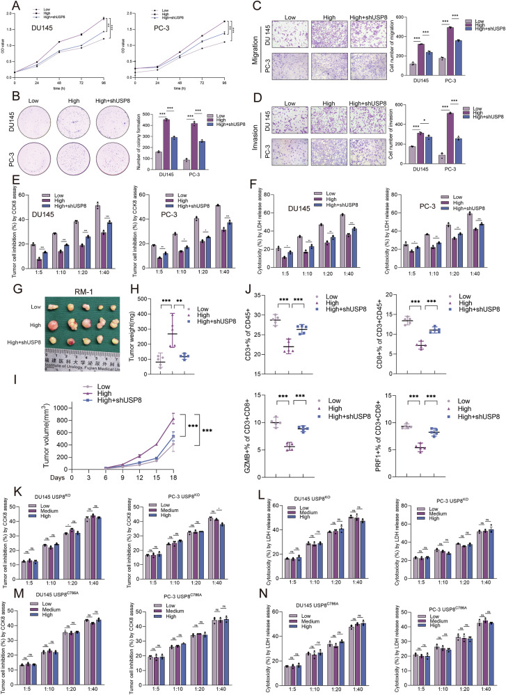
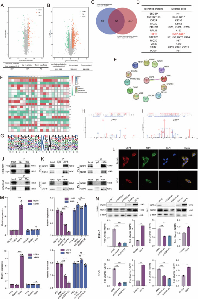
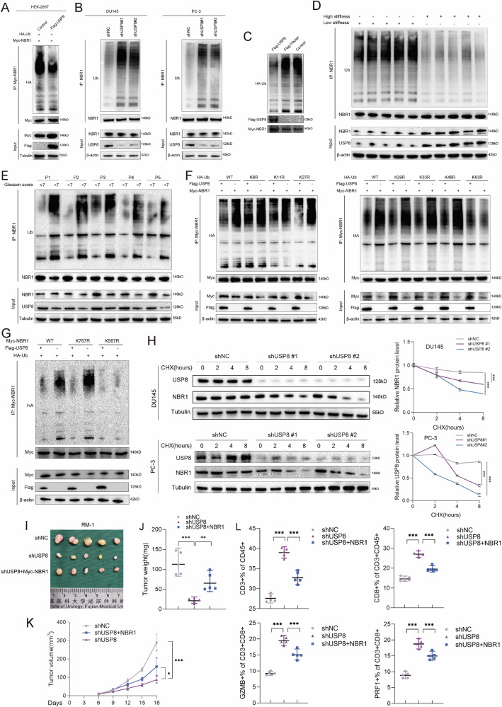
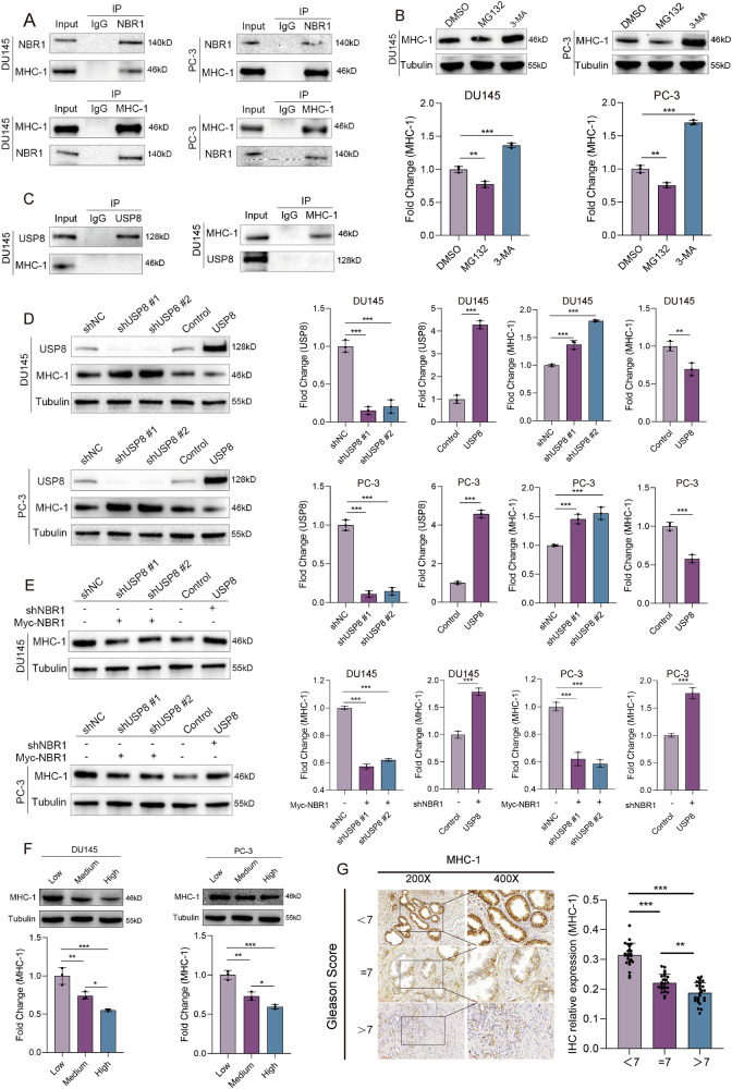
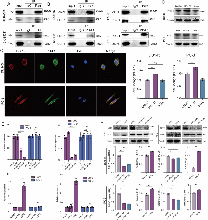
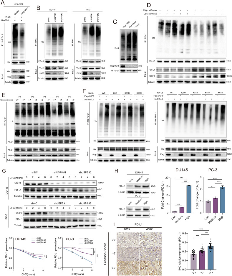
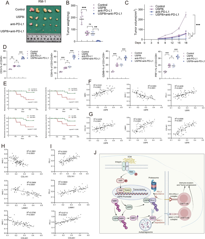
The tumor environment of prostate cancer (PCa) tissues of high Gleason score has been proved to be more immune suppressive and has higher extracellular matrix (ECM) stiffness, but whether ECM mechanical stiffness is the cause of higher ability of invasiveness and immune escape of PCa with high Gleason score remains uncertain. In this study, we showed that higher polyacrylamide hydrogels (PAAG) stiffness resulted in the progression and immune escape of PCa via integrin β1/FAK/YAP axis. The translocation of YAP into cell nucleus to bind to TEAD2 promoted the transcriptional activation of USP8. NBR1 could be ubiquitinated, and then degraded, via interacting with P62/SQSTM1 and through autophagy-lysosome pathway. Increased expression of USP8 promoted the abundance of NBR1 via K63-linked de-ubiquitination and PD-L1 via K48-linked de-ubiquitination in response to high PAAG stiffness. NBR1-mediated selective autophagy accelerated the degradation of MHC-1 of PCa. The USP8 inhibitor presented a potential application value in sensitizing immunotherapy of PCa. Taken together, we identified a USP8-mediated de-ubiquitination mechanism that involves in the process of high PAAG stiffness-mediated high expression of PD-L1 and low expression of MHC-1 of PCa cells, which provided a rationale of immunotherapy sensitization of PCa via USP8 inhibition.


Conflict of interest statement

Competing interests: The authors declare no competing interests. Ethical approval and consent to participate: All experiments were performed according to the written confirmation that this study was in accordance with relevant institutional guidelines and regulations of Fujian Medical University and national guidelines and regulations of China. All animal experiments were approved by the Experimental Animal Ethics Committee of Fujian Medical University (Approved No. of ethic committee: IACUC FJMU 2023-Y-0722). Consent for publication: Informed consent for publication of the manuscript was obtained from all authors.

Figures

Fig. 1
Fig. 1. High matrix stiffness facilitates the immune evasion of PCa cells.
A H&E staining, Masson staining, and Sirius Red staining indicating the different deposition of collagen fiber in PCa tissues of Gleason score < 7, or =7, or >7. B IHC staining and (C) the quantitative results indicating the different expression of COL1A1, LAMA1, and COL4A1 in PCa tissues of Gleason score < 7, or =7, or >7. D A representative image for the measure of prostate tissue stiffness via ultrasonic elastography and (E) the quantitative results showing the different stiffness in PCa tissues of Gleason score < 7, or =7, or >7. We used a cognitive fusion image of ultrasound, mpMRI and PSMA PET/CT to locate the region of interest (ROI), and the same ROI was measured at least 3 times and the mean value was regarded as the final value of prostate stiffness. The Gleason score was obtained from the pathological report of ROI biopsy or postoperative large pathological section of prostate. F The quantitative results of elastic moduli for different PAAG stiffness. G, H The effect of different PAAG stiffness on the cytotoxic T cell killing ability to DU145/PC-3 cells. Data are presented as the mean ± SD of at least three independent experiments and were analyzed with one-way ANOVA test unless otherwise stated, *P < 0.05, **P < 0.01, ***P < 0.001.
Fig. 2
Fig. 2. High matrix stiffness upregulates the expression of USP8 via integrin β1/FAK/YAP axis.
A Western blot and its quantitative results indicating the relative expression of integrin β1, p-FAK, FAK, and YAP protein in DU145/PC-3 cells cultured in different PAAG stiffness. B IHC staining and its quantitative results indicating the different expression of integrin β1, p-FAK, FAK, and YAP protein in PCa tissues of Gleason score < 7, or =7, or >7. C Immunofluorescence detecting the subcellular localization of YAP protein in DU145/PC-3 cells cultured in different PAAG stiffness. D Western blot and relative quantification of p-YAP (S127) and YAP protein. E Venn diagram showing the differentially expressed genes (DEGs) detected by RNA-seq of DU145 cultured in different PAAG stiffness. F RT-qPCR showing the relative transcript level of USP8 in DU145/PC-3 cells cultured in different PAAG stiffness. G Western blot indicating the relative expression of USP8 protein in DU145/PC-3 cells cultured in different PAAG stiffness or in combination with siYAP transfection. H IHC staining and its quantitative results indicating the different expression of USP8 protein in PCa tissues of Gleason score < 7, or, =7, or >7. I Western blot indicating the relative expression of USP8 protein in DU145/PC-3 cells cultured in different PAAG stiffness or in combination with siITGB1 or siFAK transfection. J, K Relative luciferase activity in HEK-293T cells co-transfected with either TEAD2 plasmid or empty vector and either USP8-promoter-WT, or USP8-promoter-MUT vectors. L ChIP-qPCR was used to detect the binding of TEAD2 to the USP8 promoter in DU145 and PC-3 cells using TEAD2 primary antibody, and IgG was applied as a negative control. Data are presented as the mean ± SD of at least three independent experiments and were analyzed with unpaired two-tailed student’s t-test or one-way ANOVA test unless otherwise stated, *P < 0.05, **P < 0.01, ***P < 0.001.
Fig. 3
Fig. 3. High matrix stiffness promotes the progression and immune evasion of PCa via upregulating USP8.
A CCK8 assay showing the cell viability of DU145/PC-3 cells cultured in different PAAG stiffness or in combination with USP8 shRNA transduction. B Representative images and quantitative results of colony formation assay of DU145/PC-3 cells cultured in different PAAG stiffness or in combination with USP8 shRNA transduction. C, D Representative images and quantitative results of Transwell assay of DU145/PC-3 cells cultured in different PAAG stiffness or in combination with USP8 shRNA transduction. E, F The cytotoxic T cell killing ability to DU145/PC-3 cells cultured in different PAAG stiffness or in combination with USP8 shRNA transduction. GI RM-1 cells, cultured in different PAAG stiffness or in combination with USP8 shRNA transduction, were injected into the flank of mice. Tumor volumes were measured every 3 days. Tumor images, weight and growth curves were obtained at day 18 after dissection. J Flow cytometry was used to quantitatively analyze the proportion of CD3+ T lymphocytes in CD45+ cells, CD8+ T lymphocytes in CD45+CD3+ cells, and the proportion of GZMB+ cells or PRF1+ cells in CD8+ T lymphocytes in the subcutaneous tumor samples of mice. The X axis represents the different group. K, L USP8KO DU145/PC-3 cells were constructed and the effect of PAAG stiffness on the cytotoxic T cell killing ability to USP8KO DU145/PC-3 cells was explored. M, N We transfected USP8 C786A (dead USP8) plasmid into USP8KO DU145/PC3 cells. The effect of PAAG stiffness on the cytotoxic T cell killing ability to USP8C786A DU145/PC-3 cells was explored. Data in A and I were analyzed by two-way repeated measures ANOVA test. Data are presented as the mean ± SD of at least three independent experiments and were analyzed with unpaired two-tailed student’s t-test or one-way ANOVA test unless otherwise stated, *P < 0.05, **P < 0.01, ***P < 0.001.
Fig. 4
Fig. 4. USP8 specifically interacts with autophagy cargo receptor NBR1.
A Volcano plot showing the differentially expressed protein detected by 4D label free quantitative proteomics of DU145 cells transduced with USP8 shRNA or shNC. B Volcano plot showing the differentially ubiquitinated protein detected by 4D label free quantitative ubiquitinome of DU145 cells transduced with USP8 shRNA or shNC. C Venn diagram showing the number of down-regulated proteins and ubiquitinated proteins with up-regulated ubiquitin sites in response to shUSP8. D The list of 12 expressed down-regulated protein with up-regulated ubiquitin sites and the corresponding ubiquitin sites. E STRING database indicating the interaction of USP8 and 12 down-regulated protein with up-regulated ubiquitin sites. F, G Ubiquitination motifs consisting of 20 residues surrounding the modified lysine site analyzed by Motif-x. H, I The possible ubiquitin sites of NBR1 through mass spectrometry analysis. J The exogenous interaction between Flag-USP8 and Myc-NBR1 was detected in HEK-293T cells by performing coimmunoprecipitation and western blot. K The endogenous interaction between USP8 and NBR1 was detected in DU145 and PC-3 cells by coimmunoprecipitation and western blot. LCo-localization of USP8 and NBR1 was visualized in DU145 and PC-3 cells via immunofluorescence. M RT-qPCR assay showing the effect of USP8 knockdown or overexpression on the expression levels of NBR1 mRNA. NWestern blot showing the effect of USP8 knockdown or overexpression on the expression levels of NBR1 protein. Data are presented as the mean ± SD of at least three independent experiments and were analyzed with unpaired two-tailed student’s t-test or one-way ANOVA test unless otherwise stated, ***P < 0.001.
Fig. 5
Fig. 5. USP8 deubiquitinates and stabilizes NBR1.
A Exogenous ubiquitination assay of Myc-NBR1 in HEK-293T cells transduced with Flag-USP8 together with Myc-NBR1 and HA-Ub plasmid, and treated with 3-MA for 6 h. B Endogenous ubiquitination assay of NBR1 in DU145 and PC-3 cells treated with 3-MA for 6 h. C In vitro de-ubiquitination assay of ubiquitinated Myc-NBR1 protein with purified Flag-USP8. D Ubiquitination assay of NBR1 in subcutaneous tumors derived from RM-1 cells in high or low PAAG stiffness. E Ubiquitination assay of NBR1 in PCa tissues of Gleason score < 7 or >7. F Ubiquitination assay of Myc-NBR1 in HEK-293T cells co-transfected with Myc-NBR1, Flag-USP8 together with HA-Ub, HA-Ub-K6R, HA-Ub-K11R, HA-Ub-K27R, HA-Ub-K29R, HA-Ub-K33R, HA-Ub-K48R, HA-Ub-K63R, and treated with 3-MA for 6 h. G Ubiquitination assay of Myc-NBR1 in HEK-293T cells co-transfected with HA-Ub, Flag-USP8 together with Myc-NBR1, Myc-NBR1-K767R, Myc-NBR1-K887R, and treated with 3-MA for 6 h. H Stability analysis of NBR1 protein in DU145 and PC-3 cells transfected with shNC or shUSP8, and treated with 40 μM cycloheximide (CHX) for indicated times. IK RM-1 related stable cells (shNC, shUSP8, and shUSP8+Myc-NBR1) were injected into the flanks of mice, respectively. Tumor volumes were measured every 3 days. Tumor images, growth curves and weight were obtained at day 18 after dissection. L Flow cytometry was used to quantitatively analyze the proportion of CD3+ T lymphocytes in CD45+ cells, CD8+ T lymphocytes in CD45+CD3+ cells, and the proportion of GZMB+ cells or PRF1+ cells in CD8+ T lymphocytes. The X axis represents the different group. Data in H and K were analyzed by two-way repeated measures ANOVA test. Data are presented as the mean ± SD of at least three independent experiments and were analyzed with one-way ANOVA test unless otherwise stated, *P < 0.05, **P < 0.01, ***P < 0.001.
Fig. 6
Fig. 6. High matrix stiffness downregulates MHC-I expression through USP8/NBR1 axis.
A The endogenous interaction between MHC-1 and NBR1 was detected in DU145 and PC-3 cells by coimmunoprecipitation and western blot. B Western blot analysis of MHC-1 expression in DU145 and PC-3 cells treated with 3-MA or MG132. C Coimmunoprecipitation and western blot indicating the endogenous non-interaction between USP8 and MHC-1 in DU145 cells. D Western blot showing the effect of USP8 knockdown or overexpression on the expression levels of MHC-1 protein in DU145 and PC-3 cells. E Western blot indicating that the effect of USP8 on MHC-1 expression could be rescued by NBR1. F Western blot and its quantitative results indicating the expression levels of MHC-1 of DU145 and PC-3 cultured in different PAAG stiffness. G IHC staining and its quantitative results indicating the different expression of MHC-1 protein in PCa tissues of Gleason score < 7, or =7, or >7. Data are presented as the mean ± SD of at least three independent experiments and were analyzed with unpaired two-tailed student’s t-test or one-way ANOVA test unless otherwise stated, *P < 0.05, **P < 0.01, ***P < 0.001.
Fig. 7
Fig. 7. USP8 specifically interacts with PD-L1.
A The exogenous interaction between Flag-USP8 and His-PD-L1 was detected in HEK-293T cells by performing coimmunoprecipitation and western blot. B The endogenous interaction between USP8 and PD-L1 was detected in DU145 and PC-3 cells by coimmunoprecipitation and western blot. C Co-localization of USP8 and PD-L1 was visualized in DU145 and PC-3 cells via immunofluorescence. D Western blot analysis of PD-L1 expression in DU145 and PC-3 cells treated with 3-MA or MG132. E RT-qPCR assay showing the effect of USP8 knockdown or overexpression on the expression levels of PD-L1 mRNA. F Western blot showing the effect of USP8 knockdown or overexpression on the expression levels of PD-L1 protein. Data are presented as the mean ± SD of at least three independent experiments and were analyzed with unpaired two-tailed student’s t-test or one-way ANOVA test unless otherwise stated, *P < 0.05, **P < 0.01, ***P < 0.001.
Fig. 8
Fig. 8. USP8 deubiquitinates and stabilizes PD-L1.
A Exogenous ubiquitination assay of His-PD-L1 in HEK-293T cells transduced with Flag-USP8 together with His-PD-L1 and HA-Ub, and treated with MG132 for 6 h. B Endogenous ubiquitination assay of PD-L1 in DU145 and PC-3 cells treated with MG132 for 6 h. C In vitro de-ubiquitination assay of ubiquitinated His-PD-L1 protein with purified Flag-USP8. D Ubiquitination assay of PD-L1 in subcutaneous tumors derived from RM-1 cells in high or low PAAG stiffness. E Ubiquitination assay of PD-L1 in PCa tissues of Gleason score < 7 or >7. F Ubiquitination assay of His-PD-L1 in HEK-293T cells co-transfected with His-PD-L1, Flag-USP8 together with HA-Ub, HA-Ub-K6R, HA-Ub-K11R, HA-Ub-K27R, HA-Ub-K29R, HA-Ub-K33R, HA-Ub-K48R, HA-Ub-K63R, and treated with MG132 for 6 h. G Stability analysis of PD-L1 protein in DU145 and PC-3 cells transfected with shNC or shUSP8, and treated with 40 μM cycloheximide (CHX) for indicated times. H Western blot indicating the relative expression of PD-L1 protein in DU145 and PC-3 cells cultured in different PAAG stiffness. I IHC staining and its quantitative results indicating the different expression of PD-L1 protein in PCa tissues of Gleason score < 7, or =7, or >7. Data in G were analyzed by two-way repeated measures ANOVA test. Data are presented as the mean ± SD of at least three independent experiments and were analyzed with unpaired two-tailed student’s t-test or one-way ANOVA test unless otherwise stated, *P < 0.05, **P < 0.01, ***P < 0.001.
Fig. 9
Fig. 9. Exploration of USP8 inhibitor/anti-PD-L1 combination therapy and clinical sample validation of molecular mechanism.
AC RM-1 cells were injected into the flanks of C57BL/6J mice and randomly divided into four group: the untreated control group, the USP8 inhibitor group, the anti-PD-L1 therapy group, the USP8 inhibitor and anti-PD-L1 combination therapy group. Tumor volumes were measured every 3 days. Tumor images, growth curves and weight were obtained at day 18 after dissection. D Flow cytometry was used to quantitatively analyze the proportion of CD3+ T lymphocytes in CD45+ cells, CD8+ T lymphocytes in CD45+CD3+ cells, and the proportion of GZMB+ cells or PRF1+ cells in CD8+ T lymphocytes in the subcutaneous tumor samples of mice. E The effect of the expression levels of COL1A1, LAMA1, COL4A1, and USP8 on biochemical recurrence-free survival. F Correlation analysis of the IHC relative expression levels of NBR1, MHC-1, and PD-L1 with USP8. G Correlation analysis of the IHC relative expression levels of COL1A1, LAMA1, and COL4A1 with USP8. H, I Correlation analysis of the IHC relative expression levels of COL1A1, LAMA1, COL4A1 with PD-L1 and MHC-1. J The model showing the extracellular matrix-derived mechanical stiffness promotes the progression and immune evasion of PCa via USP8-mediated MHC-1 degradation and PD-L1 abundance. Data in C were analyzed by two-way repeated measures ANOVA test. Data are presented as the mean ± SD of at least three independent experiments and were analyzed with one-way ANOVA test unless otherwise stated, *P < 0.05, **P < 0.01, ***P < 0.001.

Similar articles

References

    1. Miller KD, Nogueira L, Devasia T, Mariotto AB, Yabroff KR, Jemal A, et al. Cancer treatment and survivorship statistics, 2022. CA Cancer J Clin. 2022;72:409–36. - PubMed
    1. Wang G, Zhao D, Spring DJ, DePinho RA. Genetics and biology of prostate cancer. Genes Dev. 2018;32:1105–40. - PMC - PubMed
    1. O’Donnell JS, Teng MWL, Smyth MJ. Cancer immunoediting and resistance to T cell-based immunotherapy. Nat Rev Clin Oncol. 2019;16:151–67. - PubMed
    1. Achard V, Putora PM, Omlin A, Zilli T, Fischer S. Metastatic prostate cancer: treatment options. Oncology. 2022;100:48–59. - PubMed
    1. Adorno Febles VR, Hao Y, Ahsan A, Wu J, Qian Y, Zhong H, et al. Single-cell analysis of localized prostate cancer patients links high Gleason score with an immunosuppressive profile. Prostate. 2023;83:840–9. - PubMed