. 2025 May 23;16(1):4802.
doi: 10.1038/s41467-025-60073-0.

Enhancing the potency of in vivo lentiviral vector mediated gene therapy to hepatocytes

Affiliations

Enhancing the potency of in vivo lentiviral vector mediated gene therapy to hepatocytes

Cesare Canepari et al. Nat Commun. .

Abstract

In vivo gene therapy to the liver using lentiviral vectors (LV) may represent a one-and-done therapeutic approach for monogenic diseases. Increasing LV gene therapy potency is crucial for reducing the effective doses, thus alleviating dose-dependent toxicities and facilitating manufacturing. LV-mediated liver transduction may be enhanced by positively selecting LV-transduced hepatocytes after treatment (a posteriori) or by augmenting the initial fraction of LV-targeted hepatocytes (a priori). We show here that the a posteriori enhancement increased transgene output without expansion of hepatocytes bearing LV genomic integrations near cancer genes, in mouse models of hemophilia, an inherited coagulation disorder. Furthermore, we enhanced hepatocyte transduction a priori in mice by transiently inhibiting antiviral pathways and/or through a fasting regimen. The most promising transduction-enhancer combination synergized with phagocytosis-shielded LV, resulting in a remarkable 40-fold increase in transgene output. Overall, our work highlights the potential of minimally invasive, cost-effective treatments capable of improving the potency of in vivo LV gene therapy to hepatocytes, in order to expand its applicability and ease clinical translation.


Conflict of interest statement

Competing interests: C.C. and A.C. are inventors on patent applications submitted by Fondazione Telethon, San Raffaele Scientific Institute, on LV technology related to the work presented in this manuscript. Fondazione Telethon and San Raffaele Scientific Institute, through SR-Tiget, have established a research collaboration on liver-directed LV gene therapy of hemophilia with GeneSpire. The remaining authors declare no competing interests.

Figures
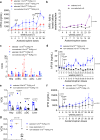
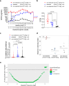
Fig. 1
Fig. 1. Positive selection of transduced hepatocytes allows achieving therapeutically relevant FVIII and FIX amounts.
a Mean with standard error of the mean (SEM) of hFIX concentration in the plasma of adult WT mice, treated with LV-shCypor.FIX (1.3 × 1010 or 2.6 × 1010 transducing units, TU/kg) and subjected (blue, red dots), or not (light blue, pink dots) to positive selection of transduced hepatocytes. b Mean with SEM of the fold change of hFIX concentration in selected mice (purple dots) and in non-selected controls (black dots), over time, normalized to the hFIX amounts detected on each mouse at the 4 week post-LV time point. Two-tailed Mann-Whitney test (selected vs non-selected at last timepoint) (c) Single values and mean with SEM of VCN measured in the fractioned or FACS-sorted liver cell (Hep hepatocytes, LSEC liver sinusoidal endothelial cells, pDC plasmacytoid dendritic cells, KC Kupffer cells) and in the total liver of mice in (a), 10 months post-LV. d Mean with SEM of hFVIII concentration in the plasma of hemophilia A mice, treated as neonates with LV-shCypor.FVIII (3.6 × 1010 TU/Kg) and subjected (blue dots), or not (black dots) to positive selection of transduced hepatocytes. 10 ng/mL of FVIII (10% of FVIII) is considered the threshold to turn a severe hemophilia phenotype into a mild phenotype. Two-tailed Mann-Whitney test on time points before selection or at the last time point in selected vs non-selected mice. e Single values and mean with SEM of VCN measured in the fractioned or FACS-sorted liver cell types and in the total liver of mice in (d), 8 months post-LV. Two-tailed Mann-Whitney test. f Mean with SEM of hFIX concentration in the plasma of WT mice, treated with LV-shCypor.FIX as 2 week-old (1 × 1010 TU/kg) and subjected (blue dots), or not (black dots), to positive selection of transduced hepatocytes. Two-tailed Mann-Whitney test on time points before selection or at the last time point in selected vs non-selected mice. g Single values and mean with SEM of VCN measured in the total liver of LV-treated mice, 1.3 years post-LV. Two-tailed Mann-Whitney test. Source data are provided as a Source Data file.
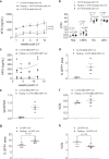
Fig. 2
Fig. 2. Positive selection of LV-transduced hepatocytes is most effective when starting from very low transgene amounts, and does not result in expansion of integrations nearby oncogenes.
a Mean with SEM of hFIX concentration in the plasma of adult WT mice, treated with LV-shCypor.FIX (6.8 × 109 or 3.4 × 1010 TU/kg) and subjected (blue dots), or not (black and red dots) to positive selection of transduced hepatocytes. Kruskal-Wallis test with Dunn’s multiple comparisons test on time point before selection or at the last time point in selected vs non-selected mice. b Single values and mean with SEM of VCN measured in the total liver of LV-treated mice. Kruskal-Wallis test with Dunn’s multiple comparisons test. c The relative abundance of the integration sites from all mice, split by the specific treatment group, is shown. Bars indicate mean relative abundance values per group. Kruskal–Wallis test with Dunn’s multiple comparisons test. d The Shannon diversity index of each mouse per specific treatment group is shown as a dot. The average value per group is indicated. e Volcano plot of the common IS (CIS), performed on the IS identified in mice treated with 6.8 × 109 TU/kg and subjected to positive selection of transduced hepatocytes. All genes targeted by IS were tested and plotted as dots; gene integration frequency normalized by gene length was placed on the x-axis, while the y-axis shows the p-value of the CIS Grubbs test for outliers (–log10 of p-value). Tumor suppressor genes are annotated in blue, protooncogenes in red, and a generic “other” in green for the remaining genes. Dots with significant p-values (α threshold of 0.05) are above the dashed horizontal line. Source data are provided as a Source Data file.
Fig. 3
Fig. 3. Transient proteasome inhibition increases gene transfer to hepatocytes in vivo.
a Single values with connecting means of human FIX (hFIX) concentration in the plasma of hemophilia B mice, treated with (blue dots) or without (black dots) Bortezomib (Bort, 1 mg/kg, i.v.) 1 h before LV-FIX (2.5 × 1010 TU/kg). b Single values with mean and SEM of VCN measured in the liver of mice in (a) 7 months post-LV. c Single values with connecting means of human FVIII (hFVIII) concentration in the plasma of immune tolerant hemophilia A mice, treated with (blue squares) or without (black squares) Bortezomib (1 mg/kg, i.v.) 1 h before LV-FVIII (4.5 × 1010 TU/kg). d Single values and mean with SEM of VCN measured in fractionated and FACS-sorted liver cell types or in total liver of mice in (c) 3 months after LV administration. e Single values and mean with SEM of the percentage of GFP-positive liver area in mice treated with (blue triangles) or without (black triangles) Bortezomib (1 mg/Kg, i.v.) 1 h before LV-GFP (2.5 × 1010 TU/kg), analyzed 1 month post-LV. Two-tailed Mann–Whitney test. f Single values and mean with SEM of VCN measured in the liver of mice in (e) 1 month post-LV. Two-tailed Mann-Whitney test. Source data are provided as a Source Data file.
Fig. 4
Fig. 4. Transient IFN signaling inhibition increases gene transfer to hepatocytes in vivo without additional advantage if combined with Bortezomib.
a Mean with SEM of hFVIII concentration in the plasma of immune tolerant hemophilia A mice, treated with (red squares) or without (black squares) anti-IFNAR1 Ab (50 mg/kg, i.v.) or with a control Ab (immunoglobulin G control, IgG control, pink squares) 3 h before LV-FVIII (4–7 × 1010 TU/kg). Pool of two independent experiments. Kruskal-Wallis test with Dunn’s multiple comparisons test over LV-FVIII control group, performed at the last time-point. b Single values and mean with SEM of VCN measured in the indicated fractionated or FACS-sorted liver cell types of mice in (a) 5 months post-LV. Two-tailed Mann-Whitney test. c Mean with SEM of hFIX concentration in the plasma of WT mice, treated with Bortezomib (1 mg/kg, blue dots) and/or Anti-IFNAR1 Ab (50 mg/kg, purple or red dots, as indicated) or saline (black dots), 1 h or 3 h before LV-FIX (2.75 × 1010 TU/kg). Two-tailed linear-mixed effects (LME) model (for complete analyses see Supplementary Table 1) over LV-FIX group. d Single values and mean with SEM of VCN measured in fractionated and FACS-sorted liver cell types of mice in (c) 3 months post-LV. Kruskal-Wallis test with Dunn’s multiple comparisons test over LV-FIX control group. e Single values and mean with SEM of the percentage of GFP-positive hepatocytes (7 fields/sample) transduced with PGK.GFP LV (multiplicity of infection (MOI): 2), in the presence (blue triangles) or absence (black triangles) of Bortezomib (100 nM) at the time of transduction, and analyzed 7 days after transduction. As a control for inhibition of GFP degradation, Bortezomib (100 nM) was added 2 days after LV administration (condition Bort control, gray triangles). f Single values and mean with SEM of VCN measured in the above-described experiment. Kruskal-Wallis test with Dunn’s multiple comparisons test. Source data are provided as a Source Data file.
Fig. 5
Fig. 5. One day of fasting increases gene transfer to hepatocytes in vivo.
a Mean with SEM of hFIX concentration in the plasma of adult WT mice, fasted 24 h (empty black dots) or fed ad libitum (6 week-old, gray dots, 8 week-old, black dots) before LV-FIX (4 × 1010 TU/kg). Two-tailed linear-mixed effects (LME) model (for complete analyses see Supplementary Table 3) over Fasting + LV-FIX 8 week-old group. b Single values and mean with SEM of VCN measured in fractionated and FACS-sorted liver cell types of mice in (a) 3 months after LV administration. Kruskal-Wallis test with Dunn’s multiple comparisons test over fasted mice. c Mean with SEM of hFIX concentration in the plasma of adult WT mice, fasted 24 h (empty black dots) or fed ad libitum (black dots) before LV-FIX IRES GFP (2 × 1010 TU/kg). d Single values and mean with SEM of the percentage of GFP-positive liver area of mice in (c), 3 months post-LV. e Single values and mean with SEM of wpre expression (proxy of LV transgene), normalized on the endogenous Hprt gene, in the livers of mice in (c). f Single values and mean with SEM of VCN measured in the total liver of mice in (c). g Single values and mean with SEM of the percentage of GFP-positive liver area of mice adult WT mice, fasted 24 h (empty triangles) or fed ad libitum (black triangles) before LV-GFP (2.5 × 1010 TU/kg), 1 week post-LV. Two-tailed Mann-Whitney test. h Single values and mean with SEM of VCN measured in the total liver of mice in (g). Two-tailed Mann–Whitney test. Source data are provided as a Source Data file.
Fig. 6
Fig. 6. Ldlr−/− hepatocytes are transduced by LV at higher efficiency compared to WT mice in vivo.
a Mean with SEM of hFIX concentration in the plasma of adult WT (black and gray dots) or Ldlr−/− mice (dark and light green dots) treated with LV-FIX (3.5 × 1010 TU/kg or 1.2 × 1010 TU/kg). Two-tailed linear-mixed effects (LME) model (for complete analyses see Supplementary Table 4). b Single values and mean with SEM of VCN measured in fractioned or FACS-sorted liver cell types or in the total liver of mice in (a), 3 months post-LV. Two-tailed Mann–Whitney test. c Single values and mean with SEM of WPRE expression (proxy of LV transgene), normalized on the endogenous Hprt gene, in sorted hepatocytes or sorted LSEC of mice in (a). Two-tailed Mann-Whitney test. d Single values and mean with SEM of expression of Ldlr and Ldlr family members genes, normalized on the endogenous Hprt gene, in the livers of adult WT or Ldlr−/− mice. Two-tailed Mann-Whitney test. Source data are provided as a Source Data file.
Fig. 7
Fig. 7. Combinations of the previously identified transduction enhancers further improve gene transfer efficiency to hepatocytes in vivo.
a Single values and mean with SEM of hFIX concentration in the plasma of LV-FIX treated mice (average of 4 time points: 15, 20, 26, 46 weeks post-LV). WT and Ldlr−/− mice were either administered with LV-FIX (black dots, 2.75 × 1010 TU/kg) or pre-treated with Anti-IFNAR1 Ab (50 mg/kg, i.v., red dots) or with Bortezomib (1 mg/kg, blue dots) or fasted for 24 h (empty black dots), or fasted and pre-treated with Anti-IFNAR1 Ab (empty red dots), or fasted and pre-treated with Bortezomib (empty blue dots), or fasted and pre-treated with Anti-IFNAR1 and Bortezomib, before LV-FIX (2.75 × 1010 TU/kg). Kruskal-Wallis test with Dunn’s multiple comparisons test over LV-FIX control group. b Single values and mean with SEM of VCN measured in the total liver of mice in (a). Kruskall-Wallis test with Dunn’s multiple comparisons test over LV-only group. c Mean with SEM of hFIX concentration in the plasma of LV-FIX treated mice. WT mice were either administered with LV-FIX (black dots) or pre-treated with Bortezomib (blue dots) or fasted for 24 h (empty black dots), or fasted and pre-treated with Bortezomib (empty blue dots), before LV-FIX (1.8 × 1010 TU/kg). Two-tailed linear-mixed effects (LME) model (for complete analyses see Supplementary Table 5), over LV-FIX group. d Single values and mean with SEM of VCN measured in the sorted hepatocytes of mice in (c). Kruskal–Wallis test with Dunn’s multiple comparisons test over LV-only group. e Single values and mean with SEM of VCN measured in the sorted LSEC of mice in (c). Kruskal–Wallis test with Dunn’s multiple comparisons test over LV-only group. f Mean with SEM of hFIX concentration in the plasma of CD47hi-LV-FIX treated mice. NOD mice were either administered with CD47hi-LV-FIX (black dots), fasted and pre-treated with anti-IFNAR1 Ab (empty red dots) or fasted and pre-treated with Bortezomib (empty blue dots), before CD47hi-LV-FIX (1.4 × 1010 TU/kg). Two-tailed linear-mixed effects (LME) model (for complete analyses see Supplementary Table 6), over LV-FIX group. Source data are provided as a Source Data file.

References

    1. Zabaleta, N., Unzu, C., Weber, N. D. & Gonzalez-Aseguinolaza, G. Gene therapy for liver diseases - progress and challenges. Nat. Rev. Gastroenterol. Hepatol.20, 288–305 (2023). - PubMed
    1. Mahlangu, J. et al. Two-year outcomes of valoctocogene roxaparvovec therapy for hemophilia A. N. Engl. J. Med388, 694–705 (2023). - PubMed
    1. Pipe, S. W. et al. Gene therapy with etranacogene dezaparvovec for hemophilia B. N. Engl. J. Med388, 706–718 (2023). - PubMed
    1. Ronzitti, G., Gross, D. A. & Mingozzi, F. Human immune responses to adeno-associated virus (AAV) vectors. Front Immunol.11, 670 (2020). - PMC - PubMed
    1. Cantore, A. et al. Liver-directed lentiviral gene therapy in a dog model of hemophilia B. Sci. Transl. Med7, 277ra228 (2015). - PMC - PubMed

LinkOut - more resources