. 2025 May 23;16(1):4799.
doi: 10.1038/s41467-025-59489-5.

Fusogenic lipid nanoparticles for rapid delivery of large therapeutic molecules to exosomes

Affiliations

Fusogenic lipid nanoparticles for rapid delivery of large therapeutic molecules to exosomes

Gamsong Son et al. Nat Commun. .

Abstract

Exosomes, as cell-derived lipid nanoparticles, are promising drug carriers because they can traverse challenging physiological barriers such as the blood-brain barrier (BBB). However, a major obstacle in utilizing exosomes as drug carriers is loading large therapeutic molecules without compromising the structural integrity of embedded biomolecules. Here, we introduce a membrane fusion method utilizing fusogenic lipid nanoparticles, cubosomes, to load large molecules into exosomes in a non-destructive manner. When the drug-loaded cubosome and exosome solutions are simply mixed, membrane fusion is completed in just 10 min. Our method effectively loads doxorubicin and immunoglobulin G into exosomes. Moreover, even the most challenging molecule-mRNA-is loaded with nearly 100% efficiency, demonstrating the versatility of our approach. In terms of biological behavior, the resulting hybrid exosomes preserve the functional behavior of exosomes in BBB uptake and penetration. Surprisingly, controlling exosome-to-cubosome ratios allows precise control over BBB uptake and transport. Furthermore, these hybrid exosomes retain cell-specific delivery properties, preserving the targeted delivery functions dictated by their exosomal origin. This study demonstrates the feasibility of a mix-and-load method for rapid and efficient drug loading into exosomes, with significant potential for the treatment of neurological diseases.


Conflict of interest statement

Competing interests: The Korea Institute of Science and Technology is in the process of applying for a Korean invention patent (10-2025-0058245) related to the subject matter of this paper.

Figures

Fig. 1
Fig. 1. Rapid loading of large therapeutic molecules into exosomes.
a Schematic of the membrane fusion process between fusogenic lipid nanoparticle (cubosome), and exosome for rapid and efficient drug loading into exosomes. The rapid membrane fusion allows a simple workflow, mix-and-load. b Blood-brain barrier permeability test of hybrid exosomes. Exosome / Cubosome ratio can modulate BBB uptake and transport. b Created in BioRender. Song, J. (2025) https://BioRender.com/6p849jp.
Fig. 2
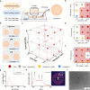
Fig. 2. Optimize cubosome fabrication for mass production.
a Schematic of cubosome fabrication using microfluidic mixer. When the solvent polarity changed abruptly upon mixing of two solutions, lipids precipitate into droplets. Afterwards, annealing of droplets induces the self-assembly into cubosomes. Lipid concentration (0.15, 0.3, 0.45 M), Flow rate ratio (FRR; lipid:water = 1:1, 1:2, 1:3), and total flow rate (TFR; 2, 3, 4 ml/min) were controlled independently. b Preferred lipid phases according to lipid concentration, FRR, and TFR. c 3D phase diagram representing the phases of 27 samples at varied lipid concentrations, FRR, and TFR. Red circle indicates a cubic phase. Gray circle represents coexistence of cubic and hexagonal phases, but cubic phase is dominant. Blue circle, on the other hand, represent the hexagonal phase dominant. Pure hexagonal phases are marked with green circles. d–f 2D projection diagrams of concentration-FRR, concentration-TFR, and FRR-TFR plane. Each circle is divided into three parts to indicate the overlapping color results of three replicate  samples. Areas where the cubic phase appeared at least once are marked with a red background. g DLS results under the final condition (0.45 M, lipid:water = 1:1.5, 4 ml/min). h SAXS results and 2D-SAXS image under the final conditions revealing a well-ordered primitive cubic phase. i Cryo-TEM image under the final conditions.
Fig. 3
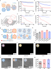
Fig. 3. Bottom-up approach for drug encapsulation in cubosome.
a Bottom-up strategy of drug encapsulation in cubosome using a microfluidic mixer. Drug-encapsulating droplets are fused to form a cubosome during the annealing process. b Encapsulation efficiency in cubosome for three different types of drugs; doxorubicin, immunoglobulin G, and mRNA. Data are presented as mean ± SE, n = 3. c–e Fluorescence optical microscope images showing mRNA loaded cubosomes. Cubosomes were labeled by Texas Red c, mRNAs were labeled by FAM d. Merged images of cubosome and mRNA e show yellow particles indicating co-localization of mRNA and cubosome. f DLS results for cubosome and mRNA-cubosome, showing the decrease of particle size upon mRNA encapsulation. g 1D and 2D SAXS scan results of cubosome and mRNA-cubosome samples, showing an mRNA specific peak that is only observed in mRNA-cubosome sample. h Cryo-TEM image of mRNA-cubosome showing the uniform size distribution and well-ordered internal structure as seen in SAXS and DLS studies. i Illustration of cubosome and mRNA conformation (folding degree is defined as domain size / mRNA d-spacing) in cubosome. j Unit cell size of cubosome and mRNA folding degree were maintained even after storing for 3 weeks at room temperature. k Optical images of mRNA-cubosome solution after 0 day and 3 weeks at room temperature.
Fig. 4
Fig. 4. Membrane fusion between cubosome and exosome.
a Scheme of describing the Fluorescence Resonance Energy Transfer (FRET) experiment for understanding fusion kinetics at different exosome: cubosome ratio and buffer. As fusion progresses, the energy transfer between FRET pair is decreased. The fluorescence intensity decrease of acceptor, Liss Rhod dye, was used as a lipid mixing indicator. Cubosomes were used as a control for comparison before fusion occurred. b Changes in fluorescent intensity of Liss Rhod intensity in Deionized water (D.W.) and PBS when the exosome: cubosome number ratio is 1:1. c Changes in Liss Rhod intensity in D.W. and PBS when the exosome: cubosome number ratio is 10:1. d SAXS scan results for solution of cubosome and exosome mixture. Under D.W. conditions, measurements were taken every 10 s for 10 min when the exosome: cubosome concentration ratio was 10:1. As fusion progresses, the intensity of the first Bragg peak indicating cubic phase is decreased. e Changes in the first peak of the cubic phase in D.W. and PBS when the exosome: cubosome concentration number ratio is 1:1. f Changes in the first peak of the cubic phase in D.W. and PBS when the exosome: cubosome concentration number ratio is 10:1. g Scheme showing the difference in the membrane fusion kinetics in D.W. and PBS due to charge screening. h DLS results in D.W. and PBS when the exosome: cubosome concentration ratio is 1:1 and 10:1. Data are presented as mean ± SE, n = 3. i Fluorescence image of hybrid exosome showing only the exosome labeled by DiI. j Fluorescence image of hybrid exosome showing only the cubosome labeled by NBD-PE. k Fluorescence image of hybrid exosome. Exosome and cubosome overlap and yellow signal observed, supporting a completion of membrane fusion. l Cryo-TEM image of the fusion process between exosome and cubosome. m Loading efficiency of doxorubicin, immunoglobulin G, and mRNA into exosomes. Data are presented as mean ± SE, n = 3.
Fig. 5
Fig. 5. Reconstruction of the in vitro BBB model.
a A scheme showing a 3D BBB model with three-phase guide channels. b Entire image of the microfluidic device and the process of creating an in vitro 3D BBB model and permeability test. c Confocal image showing cells cultured in the 3D BBB model. d In the 3D BBB model, human brain microvascular endothelial cells (HBMEC), human astrocytes (HA), and human brain vesicular pericytes (HBVP) are labeled with CD31(green), GFAP(red), and αSMA(orange), respectively. e Visualization of tight junction proteins in the endothelial cell layer, confirming the integrity and maturity of the endothelial barrier: CD31 (green), ZO-1 (yellow), VE-cadherin (purple), and Claudin-5 (cyan). a Created in BioRender. Song, J. (2025) https://BioRender.com/o9v2cps.
Fig. 6
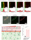
Fig. 6. Uptake and transport behavior in 3D BBB model.
ad Confocal image showing the BBB permeability results for each particle. The bar graphs illustrate the fluorescence intensity at various positions in the 3D BBB model. Endothelial cells are labeled with Lectin (green), and HBMEC-exosomes are labeled with DiI (red). HBMEC-hybrid exosomes and HA-hybrid exosomes are labeled with TexasRed (red). e Confocal images demonstrating the uptake of hybrid exosomes by human astrocytes labeled with Qtracker 565 (green). Texas Red-labeled hybrid exosomes (red) colocalize with astrocytes, confirming effective delivery across the BBB. Scale bars: 20 μm. f A scheme showing the BBB uptake and transport behavior of each particle. g Area of fluorescence intensity below the BBB boundary. Data are presented as mean values with SD and n = 3–4. Statistical significance: ****p < 0.0001. Statistical significance was determined using an ordinary one-way ANOVA followed by Tukey’s multiple comparisons test. f Created in BioRender. Song, J. (2025) https://BioRender.com/sw5wxrk.
Fig. 7
Fig. 7. Biomedical application of hybrid exosomes for drug delivery across the BBB.
a Confocal microscopy images showing the permeability of doxorubicin, IgG, and mRNA when delivered via cubosomes (top) and hybrid exosomes (bottom) across the BBB model. Scale bar: 200 μm. b Quantitative analysis of the area of fluorescence intensity for doxorubicin (Dox), IgG, and mRNA delivered using cubosomes and hybrid exosomes. Hybrid exosomes significantly enhance the transport of IgG and mRNA across the BBB compared to cubosomes. Data are presented as mean values with SD. Statistical significance: ****p < 0.0001 (mRNA: n = 6, Dox and IgG: n > 8; n represents the number of independent experiments). Statistical significance was determined using an ordinary one-way ANOVA followed by Tukey’s multiple comparisons test,. c Confirmation of intact mRNA transport across the BBB. Texas Red-labeled hybrid exosomes (red) successfully encapsulate and deliver mRNA (yellow) across the endothelial barrier. The merged image shows the colocalization of hybrid exosomes and mRNA beyond the BBB boundary. Scale bar: 20 μm.

References

    1. Zhang, Y., Liu, Y., Liu, H. & Tang, W. H. Exosomes: biogenesis, biologic function and clinical potential. Cell Biosci.9, 19 (2019). - PMC - PubMed
    1. Doyle, L. M. & Wang, M. Z. Overview of extracellular vesicles, their origin, composition, purpose, and methods for exosome isolation and analysis. Cells8, 727 (2019). - PMC - PubMed
    1. Kim, H. I. et al. Recent advances in extracellular vesicles for therapeutic cargo delivery. Exp. Mol. Med.56, 836–849 (2024). - PMC - PubMed
    1. Herrmann, I. K., Wood, M. J. A. & Fuhrmann, G. Extracellular vesicles as a next-generation drug delivery platform. Nat. Nanotechnol.16, 748–759 (2021). - PubMed
    1. Bang, S. et al. Exosome-inspired lipid nanoparticles for enhanced tissue penetration. ACS Nano19, 8882–8894 (2025). - PubMed

LinkOut - more resources