. 2025 May 23;15(1):17903.
doi: 10.1038/s41598-025-01695-8.

Pedunculopontine-stimulation obstructs hippocampal theta rhythm and halts movement

Affiliations

Pedunculopontine-stimulation obstructs hippocampal theta rhythm and halts movement

Jaspreet Kaur et al. Sci Rep. .

Abstract

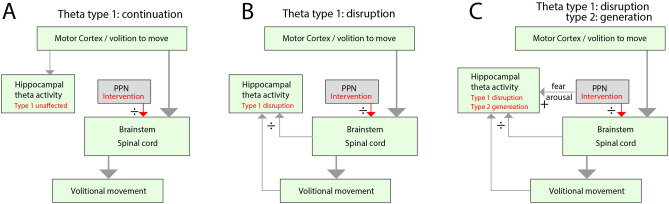
While the movement of rodents can be paused by optogenetic stimulation of a brainstem nucleus, the pedunculopontine nucleus (PPN), it is unknown whether this response has a functional purpose. The arrest appears conspicuously similar to fear-induced freezing behavior and could constitute a general halting mechanism for movement without an emotional component. Further, it is unclear to what extent brain activity is affected by the evoked motor arrest. Here, we investigate this phenomenon by engaging a distinct brain activity, the hippocampal theta rhythm. The theta rhythm is prominent during locomotor activity, absent under normal immobile situations, yet present under vigilant states like fear-induced freezing. Specifically, we ask whether the PPN-induced motor arrest has the same effect on the theta rhythm as if the animal would perform a volitional arrest, which results in the disappearance of the theta rhythm, or whether it would cause a continuation of the theta rhythm as would be expected by a fear-induced motor arrest. An alternative hypothesis is that the theta rhythm represents an ongoing intention to move rather than the movement itself. To distinguish between these two possibilities, we recorded the hippocampal brain rhythm before and during movement arrest induced by optogenetic stimulation of the PPN in rats. The PPN-induced motor arrest was associated with a clear obstruction of the ongoing theta activity. The timescale of movement arrest was less than 200 ms, similar to the obstruction of the theta rhythm. Since fear and behavioral freezing are associated with hippocampal theta rhythm, which we did not see during PPN stimulation, we suggest that induced motor arrest occurs without an associated emotional component. Further, our experiments reveal that the theta rhythm during motor activity does not represent an intention, but rather the ongoing sensory-motor state.


Conflict of interest statement

Declarations. Competing interests: The authors declare no competing interests. Declaration of AI Assistance in writing: Artificial intelligence (AI) in the form of generative Large Language Models (ChatGPT-4) assisted in writing this manuscript. The use of ChatGPT was done by rewriting a section for improving clarity. The output was reviewed and edited and adapted for meaning. All authors have read the manuscript and take full responsibility of the content.

Figures

Fig. 1
Fig. 1
Possible effects on hippocampal theta rhythm when internally inducing motor arrest by stimulating PPN. (A) Null-hypothesis: the motor-related theta activity (type 1) in the hippocampus is generated as a consequence of the intention to move rather than the movement itself. Therefore, theta rhythm continues despite the PPN-induced intervention (red arrows from PPN). (B) Alternative hypothesis: The hippocampal theta rhythm is closely tied to the movement via efference copy (from brainstem and spinal cord) and sensory feedback, rather than the intention of movement. Hence, the internally induced motor arrest also disrupts the hippocampal theta rhythm. (C) Alternative hypothesis 2: The motor arrest is linked to an alert state, e.g. fear and arousal, and therefore the PPN-stimulation is generating type 2 theta, besides inducing motor arrest and disruption of type 1 theta rhythm. Therefore, hippocampal theta activity will still be present during the PPN stimulation, but of a different type.
Fig. 2
Fig. 2
Optogenetic stimulation of PPN arrests ongoing movement. (A) The PPN was targeted in rats using optogenetics via a virus, which expresses an opsin (ChrimsonR) and an implanted optical fiber. A histology section (scale bar= 1000 μm) of the caudal mesencephalon shows the viral reporter (mScarlet) in red and DAPI staining in blue and the corresponding atlas section with injection sites in red. (B) Accelerometer readings of movement in 3 directions (anterior–posterior, mediolateral, and dorsoventral), and optical stimulation (“PPN stim”, blue regions) induced arrest and eliminated acceleration. (C) Chronophotography of rat locomotion on a treadmill. Top: While not stimulating PPN, the rat is moving faster than the belt speed, hence advancing forward. When optically stimulating the PPN, the locomotion is halted (bottom) while keeping its pose). Flexed paw pose is indicated (yellow arrows) while moving backward due to belt movement. (D) The stimulation-triggered and averaged rectified accelerometry (black line). The average is integrated across the shaded gray regions across trials as “control” and “PPN stimulation” (E). (F) The median values across animals (n=7) have a significant decrease when PPN is stimulated. Illustration in (A) was adapted with permission. (C) Wires from animal (except one) was removed in the picture for simplicity.
Fig. 3
Fig. 3
PPN activation obstructs the hippocampal theta rhythm. (A) Sagittal view of the location of the hippocampal LFP electrode and the optical fiber in PPN. (B) Coronal histological section showing the LFP electrode across the dentate gyrus and CA3 (dorsal up, fluorescent DAPI stain). (C) The arrest of movement of the rat is evident by a drop in acceleration following the onset of PPN stimulation (blue-shaded regions). (D) Concurrent LFP activity in the hippocampus with prominent theta rhythm, which evaporates with the PPN stimulation. (E) The spectrogram of the hippocampal LFP displays power in theta range which ceases after PPN stimulation. (F) The integrated theta band power (6–9 Hz). The histological section in (B) is a fluorescent DAPI stain. Illustrations in (A-B) were adapted with permission.
Fig. 4
Fig. 4
Decay dynamics of movement and theta rhythm following PPN stimulation are similar. (A) The mean movement before and during PPN stimulation (accelerometer measurement ± standard deviation, SD, as the grey lines) for three sample animals. An exponential decay was fitted to the mean (orange line). (B) Whisker plot of time constants of the exponential decay fit for individual instances and animals. The mean time constant was 157 ms across animals. (C) A sample trial of hippocampal LFP spectral content (top) during movement and evoked arrest (blue regions), which was measured by accelerometry (bottom). (D) Mean ± SD for the hippocampal theta power (cyan) and accelerometry (grey) for four sample animals before and after the onset of PPN stimulation (blue region). (E) Histogram of time constants of decay of the theta rhythm (cyan) and movement (grey) across animals. There was no significant difference (n.s.) in the mean of time constants for theta rhythm vs. movement in 3 out of 4 animals. (F) Comparison of the mean of the time constants over the cohort shows no significant difference. (G) The integrated theta power in the hippocampal LFP during movement compared with during PPN stimulation in each animal. All show signifcant decrease in power. Wilcoxon signed-rank test, p<0.05, p<0.01.
Fig. 5
Fig. 5
The transfected cells in PPN. (A) The injection of virus and implantation of optical fiber in sagittal stereotaxic coordinates. (B) Post-mortem reconstruction of the location of the optical fiber (MRI-structural scan, track shown in black). (C) histological section of the same tissue as in (B) with the viral reporter (mScarlet using immunohistochemistry) in red, Cholinergic cells (ChAT) in green, and nuclei (DAPI) in blue (scale bar = 1000 μm, cerebellum missing). (D) Zoomed-in view of the injection site in (C) shows mScarlet reporter in red (detected using RNAscope) and ChAT cells in green (scale bar = 200 μm). (E) Among the transfected cells were cholinergic cells (ChAT immunohistochemistry, upper and lower left, and middle) and glutamatergic (VGluT2, upper and lower right, in cyan using RNAscope). DAPI was used as a nuclear stain (shown in blue,scale bar = 20 μm). (F) Colocalization was quantified using Manders correlation of a fraction of ChAT (left) overlapping with mScarlet out of all ChAT (M1) and all transfected (M2) and glutamatergic cells (right) overlapping with mScarlet out of all glutamatergic cells (M1) and all transfected cells (M2). (G) Manual counting of colocalization of VGluT2, ChAT, and GluA2 out of all infected cells in percent. N = 3 rat brains, n = 9–10 sections. Illustrations in (A) were adapted with permission from.

Similar articles

Cited by

References

    1. Roseberry, T. et al. Cell-type-specific control of brainstem locomotor circuits by basal ganglia. Cell164, 526–537. 10.1016/j.cell.2015.12.037 (2016). - PMC - PubMed
    1. Wessel, J. R. & Aron, A. R. On the globality of motor suppression: Unexpected events and their influence on behavior and cognition. Neuron93, 259–280. 10.1016/j.neuron.2016.12.013 (2017). - PMC - PubMed
    1. Roelofs, K. Freeze for action: Neurobiological mechanisms in animal and human freezing. Philos. Trans. R. Soc. B Biol. Sci.372, 20160206. 10.1098/rstb.2016.0206 (2017). - PMC - PubMed
    1. Goñi-Erro, H., Selvan, R., Caggiano, V., Leiras, R. & Kiehn, O. Pedunculopontine chx10+ neurons control global motor arrest in mice. Nat. Neurosci.26, 1516–1528. 10.1038/s41593-023-01396-3 (2023). - PMC - PubMed
    1. Josset, N. et al. Distinct contributions of mesencephalic locomotor region nuclei to locomotor control in the freely behaving mouse. Curr. Biol.28, 884–901. 10.1016/j.cub.2018.02.007 (2018). - PubMed

LinkOut - more resources