

7-T MRI-based surrogate for histopathology examination of liver fibrosis
- PMID: 40410512
- PMCID: PMC12102402
- DOI: 10.1186/s41747-025-00589-8
7-T MRI-based surrogate for histopathology examination of liver fibrosis
Abstract
Background: To demonstrate that 7-T magnetic resonance imaging (MRI) provides a surrogate for histopathology of fresh ex vivo liver tissue, using the case study of liver fibrosis.
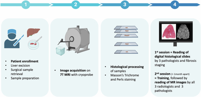
Methods: We prospectively enrolled 20 patients undergoing surgical liver resection between November 2021 and April 2023. Each ex vivo fresh liver tissue specimen (~ 1 cm3) was sectioned in half. The first half, stained using Masson's Trichrome and Perls, was assessed by three pathologists using the METAVIR score (reference standard). The second half was imaged with 7-T MRI using a cryoprobe (fat-suppressed T2-weighted turbo/fast spin-echo sequence, spatial resolution 75 × 75 × 200 µm3) and assessed by three radiologists and the same three pathologists, using a newly developed MRI-METAVIR score.
Results: Five patients were excluded from the final analysis (one patient due to poor specimen quality, two due to surgery cancellation, and two previously published used for reader training). Of the remaining 15 patients, 10 (67%) presented with chronic liver diseases and 8/15 (53%) with advanced (F3 or F4) fibrosis. Radiologists achieved 88% sensitivity, 100% specificity, 93% accuracy (95% confidence interval 68-100%) and 0.94 Harrell's c-index (0.86-1.00). Pathologists achieved 88% sensitivity, 86% specificity, 87% accuracy (60-98%) and 0.87 Harrell's c-index (0.74-0.99). There were no statistically significant differences between MRI-based and pathologic reference standard stage (p ≥ 0.655).
Conclusion: With an in-plane spatial resolution of ~ 75 × 75 µm2, MRI paralleled low-magnification histology, enabling the assessment of micro-architectural liver changes, and provided a surrogate for histopathology examination of fresh ex vivo liver tissue samples at a microscopic level.
Relevance statement: 7-T MRI provides a surrogate for histopathology visualisation of fresh ex vivo liver tissue, opening new research perspectives for clinical high-field MRI of the liver.
Key points: Using the newly developed MRI-METAVIR score, 7-T MRI data strongly correlated with histopathology, achieving excellent agreement and accuracy. 7-T MRI accurately differentiated advanced from minimal liver fibrosis. 7-T MRI visualises liver micro-architecture, enabling pathology-like, noninvasive three-dimensional imaging.
Keywords: Hepatitis; Histopathology; Liver diseases; Liver fibrosis; Magnetic resonance imaging.
© 2025. The Author(s).
Conflict of interest statement
Declarations. Ethics approval and consent to participate: This prospective project was approved by the Research Ethics Board (Protocol RIPH2, LivMod N°IDRCB 2019-A00738-49 ClinicalTrial NCT04690972) and followed ethical principles of the Declaration of Helsinki. Consent for publication: All patients provided written informed consent. Competing interests: AV is a member of the Scientific Editorial Board of European Radiology Experimental (section: Abdomen/gastrointestinal) and, as such, did not participate in the selection or review processes for this article. The remaining authors declare no conflicts of interest.
Figures
References
-
- Volmar KE, Idowu MO, Souers RJ et al (2015) Turnaround time for large or complex specimens in surgical pathology: a College of American Pathologists Q-Probes study of 56 institutions. Arch Pathol Lab Med 139:171–177. 10.5858/arpa.2013-0671-CP - PubMed
-
- Novis DA, Zarbo RJ (1997) Interinstitutional comparison of frozen section turnaround time. A College of American Pathologists Q-Probes study of 32868 frozen sections in 700 hospitals. Arch Pathol Lab Med 121:559–567. 10.5858/arpa.2013-0671-cp - PubMed
-
- Regev A, Berho M, Jeffers LJ et al (2002) Sampling error and intraobserver variation in liver biopsy in patients with chronic HCV infection. Am J Gastroenterol 97:2614–2618. 10.1002/hep.21346 - PubMed
-
- Merriman RB, Ferrell LD, Patti MG et al (2006) Correlation of paired liver biopsies in morbidly obese patients with suspected nonalcoholic fatty liver disease. Hepatology 44:874–480. 10.1002/hep.21346 - PubMed
-
- Potretzke TA, Saling LJ, Middleton WD et al (2018) Bleeding complications after percutaneous liver biopsy: do subcapsular lesions pose a higher risk? AJR Am J Roentgenol 211:204–210. 10.2214/ajr.17.18726 - PubMed
MeSH terms
Grants and funding
LinkOut - more resources
Full Text Sources
Medical
Miscellaneous