. 2025 May 23;75(2):69.
doi: 10.1007/s12031-025-02360-5.

Extracellular Vesicle Glial Fibrillary Acidic Protein as a Circulating Biomarker of Traumatic Brain Injury Severity

Affiliations

Extracellular Vesicle Glial Fibrillary Acidic Protein as a Circulating Biomarker of Traumatic Brain Injury Severity

Ayad Babaee et al. J Mol Neurosci. .

Abstract

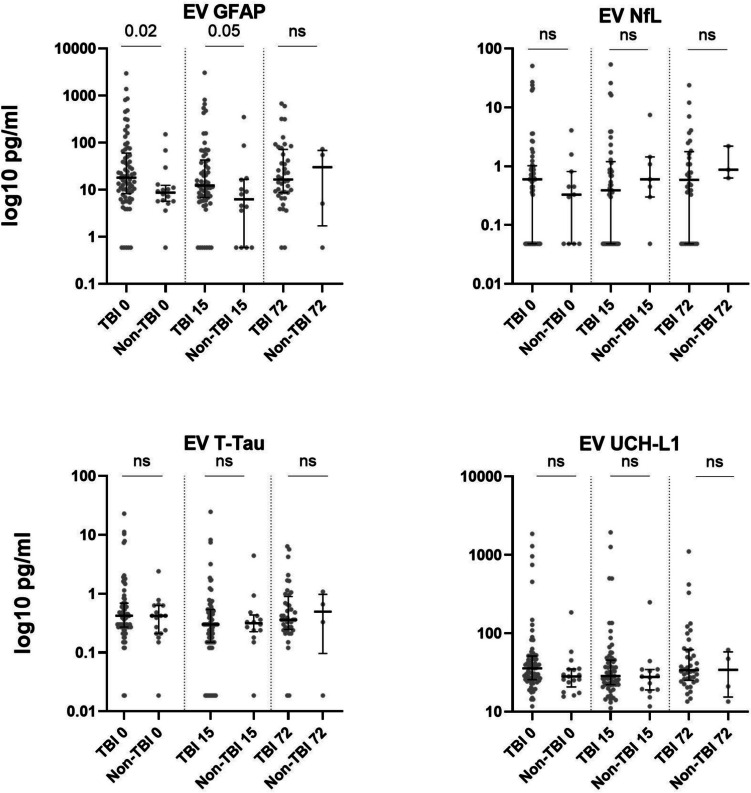
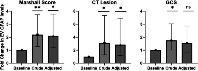
Traumatic brain injury (TBI) remains a major global health challenge with a need for improved diagnostic and prognostic biomarkers. This study aimed to evaluate the biomarker potential of extracellular vesicle (EV)-encapsulated glial fibrillary acidic protein (EV-GFAP), neurofilament light chain (EV-NfL), total tau (EV-T-Tau), and ubiquitin carboxy-terminal hydrolase L1 (EV-UCH-L1) in TBI. A cohort of 93 trauma patients (75 with TBI and 18 without TBI) was analyzed. Patients were sampled on admission, as well as 15 and 72 h post-injury. Following initial method validation, EVs were isolated from plasma using size exclusion chromatography (SEC), and plasma levels and EV cargo levels of biomarkers were measured using an ultra-sensitive Single Molecule Array. EV-GFAP levels were significantly elevated in TBI patients compared to non-TBI trauma patients at admission and 15 h. A positive head CT was associated with 2.85 (95% CI: 1.18-6.91) fold increased EV-GFAP, whereas EV-NfL, EV-T-Tau, and EV-UCH-L1 levels were not affected. None of the tested EV biomarkers were associated with 1-year mortality or 6-12 months' functional outcome. Plasma-GFAP levels increased 3.4 (95% CI: 1.72-6.70) fold with a positive head CT but were not associated with outcomes. EV-GFAP shows potential as an early biomarker of TBI, but plasma-GFAP remains a practical and reliable alternative. Future studies should explore the potential complementary roles of EV-based biomarkers on alternative aspects of TBI pathophysiology and prediction of long-term outcomes. Studies should refine methods to enhance reproducibility and clinical applicability.

Keywords: Biomarkers; Extracellular vesicle; Glial fibrillary acidic protein; Traumatic brain injury.


Conflict of interest statement

Declarations. Ethical Approval and Consent Process: The study was conducted in accordance with the Helsinki Declaration and received approval from the local ethical committee (1–10-72–205-16 and 1–10-72–204-16) and the Danish Data Protection Authority (1–16-02–452-16). All patients or a representative (guardian or nearest relative and trial guardian) provided written informed consent to participate as soon as possible after inclusion. Competing interest: The authors declare no competing interests.

Figures

Fig. 1
Fig. 1
Validation of SEC purification and EV-cargo analysis. a Particle size distribution (left) and concentration (right) after SEC purification were evaluated by NTA in three donor pools (pool A, pool B, pool C). The size distribution of the purified EVs is shown as particle counts across size ranges, and the total particle concentration in the collected volume is presented as particles per milliliter. b Flow cytometry analysis of SEC-enriched samples from the same three donors (pool A, pool B, pool C). Results for CD9 + CD63 + CD81 + extracellular vesicle counts in the three pools are shown for unlabeled samples, after the addition of Isotype IgG or CD9 + CD63 + CD81 + IgG antibodies, or in samples labeled with CD9 + CD63 + CD81 + and treated with detergent (detergent treated) or sonicated. The right panel shows CD9 + CD63 + CD81 + positive counts after detergent subtraction. c Protein concentration in the SEC-enriched samples from the three donors, prior to and after detergent treatment or sonication. Total protein was measured using NanoDrop and the four nerve-specific biomarkers GFAP, NfL, T-Tau, and UCH-L1 were measured by single molecule array under the same conditions. Results are expressed as a percentage increase relative to the untreated baseline. The absolute concentrations measured (pg/mL) were nanodrop [no treatment: 1.30, sonication: 1.15, detergent: 42.52], Simoa: [GFAP, no treatment: 11.96, sonication: 11.41, detergent: 17.85, NfL, no treatment: 0.30, sonication: 0.45, detergent: 0.85, T-Tau, no treatment: 0.35, sonication: 0.40, detergent: 0.56, UCH-L1, no treatment: 19.6, sonication: 20.76, detergent: 47.00]. p < 0.05 (*), p < 0.01 (**). Abbreviations: GFAP, glial fibrillary acidic protein; NfL, neurofilament light chain; T-Tau, total Tau; and UCH-L1, ubiquitin carboxy-terminal hydrolase-L11
Fig. 1
Fig. 1
Validation of SEC purification and EV-cargo analysis. a Particle size distribution (left) and concentration (right) after SEC purification were evaluated by NTA in three donor pools (pool A, pool B, pool C). The size distribution of the purified EVs is shown as particle counts across size ranges, and the total particle concentration in the collected volume is presented as particles per milliliter. b Flow cytometry analysis of SEC-enriched samples from the same three donors (pool A, pool B, pool C). Results for CD9 + CD63 + CD81 + extracellular vesicle counts in the three pools are shown for unlabeled samples, after the addition of Isotype IgG or CD9 + CD63 + CD81 + IgG antibodies, or in samples labeled with CD9 + CD63 + CD81 + and treated with detergent (detergent treated) or sonicated. The right panel shows CD9 + CD63 + CD81 + positive counts after detergent subtraction. c Protein concentration in the SEC-enriched samples from the three donors, prior to and after detergent treatment or sonication. Total protein was measured using NanoDrop and the four nerve-specific biomarkers GFAP, NfL, T-Tau, and UCH-L1 were measured by single molecule array under the same conditions. Results are expressed as a percentage increase relative to the untreated baseline. The absolute concentrations measured (pg/mL) were nanodrop [no treatment: 1.30, sonication: 1.15, detergent: 42.52], Simoa: [GFAP, no treatment: 11.96, sonication: 11.41, detergent: 17.85, NfL, no treatment: 0.30, sonication: 0.45, detergent: 0.85, T-Tau, no treatment: 0.35, sonication: 0.40, detergent: 0.56, UCH-L1, no treatment: 19.6, sonication: 20.76, detergent: 47.00]. p < 0.05 (*), p < 0.01 (**). Abbreviations: GFAP, glial fibrillary acidic protein; NfL, neurofilament light chain; T-Tau, total Tau; and UCH-L1, ubiquitin carboxy-terminal hydrolase-L11
Fig. 2
Fig. 2
EV cargo protein in trauma patients with and without TBI. Biomarker concentration in extracellular vesicle cargo from patients with TBI and non-TBI on hospital admission (0 h), and after 15 and 72 h of hospital stay. Biomarker levels are presented on a logarithmic scale (log10 pg/mL). Values are presented as median with interquartile ranges (IQR). Abbreviations: TBI, traumatic brain injury, GFAP glial fibrillary acidic protein, NfL neurofilament light chain, T-Tau total Tau, UCH-L1 ubiquitin carboxy-terminal hydrolase-L1, ns not significant
Fig. 3
Fig. 3
Association of EV-GFAP with TBI severity indicators. Association between extracellular vesicle GFAP cargo levels and TBI severity indicators at admission. The effect of increasing Marshall score (I, II–IV, V–VI), presence of a positive head CT (yes/no), and reduction in GCS (14–15, 9–13, 3–8) score is shown. Bars represent fold changes in EV GFAP cargo levels for crude models and after adjustment for age, sex, and NISS. For all three indicators, baseline represents the best clinical category. Error bars represent 95% confidence intervals. p < 0.05 (*), p < 0.01 (**), p ≥ 0.05 (ns) against baseline. Abbreviations: NISS new injury severity score, GCS Glasgow Coma Scale, CT computed tomography, GFAP glial fibrillary acidic protein, ns not significant

References

    1. Abdelhak A et al (2022) Blood GFAP as an emerging biomarker in brain and spinal cord disorders. Nat Rev Neurol 18(3):158–172 - PubMed
    1. Abels ER, Breakefield XO (2016) Introduction to extracellular vesicles: biogenesis, RNA cargo selection, content, release, and uptake. Cell Mol Neurobiol 36(3):301–312 - PMC - PubMed
    1. Allelein S et al (2021) Potential and challenges of specifically isolating extracellular vesicles from heterogeneous populations. Sci Rep 11(1):11585 - PMC - PubMed
    1. Bartko EA et al (2024) MMP9 and CCL18 associate with chronic urticaria while type I, IV, and VI collagens change with omalizumab treatment. Allergy 79(9):2558–2561 - PubMed
    1. Bazarian JJ et al (2018) Serum GFAP and UCH-L1 for prediction of absence of intracranial injuries on head CT (ALERT-TBI): a multicentre observational study. Lancet Neurol 17(9):782–789 - PubMed

LinkOut - more resources