. 2025 May 23;23(1):577.
doi: 10.1186/s12967-025-06593-1.

Patient-derived esophageal adenocarcinoma organ chip: a physiologically relevant platform for functional precision oncology

Affiliations

Patient-derived esophageal adenocarcinoma organ chip: a physiologically relevant platform for functional precision oncology

Sanjima Pal et al. J Transl Med. .

Abstract

Background: Esophageal adenocarcinoma (EAC) is the sixth most deadly cancer worldwide, with increasing incidence in North America. As no targeted therapy or immunotherapy has revolutionized the management of EAC, chemotherapy is the only standard of care. Most patients with EAC experience poor outcomes because of the inherent or acquired resistance to chemotherapy.

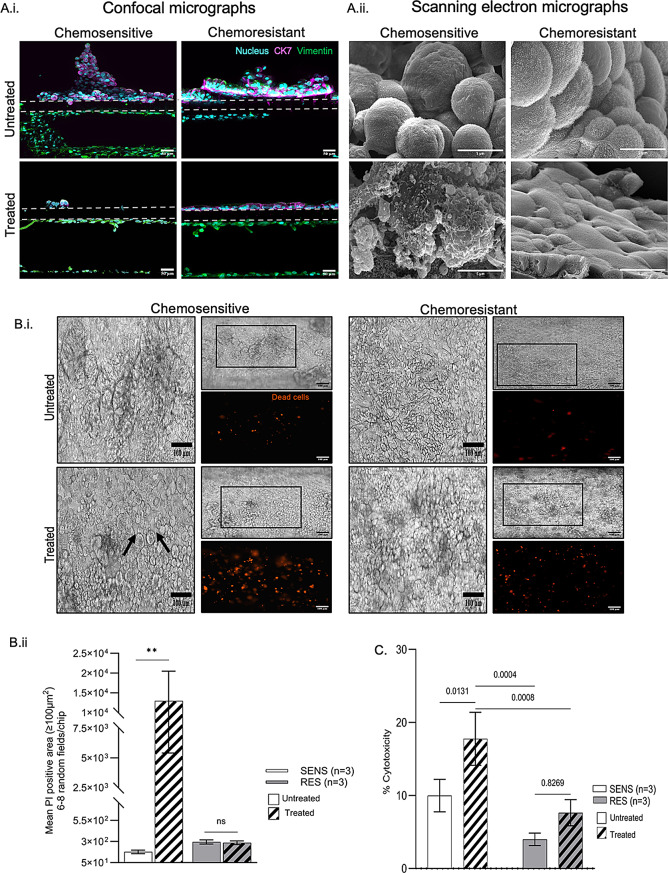
Methods: Adapting a patient-centered approach, we leveraged a microfluidic cell culture technology platform (Emulate), organoids derived from treatment-naive patient tumors or adjacent normal tissues, and patient-matched cancer-associated or normal fibroblasts respectively, to develop a novel, physiologically relevant, high-fidelity preclinical esophagus-on-a-chip model. H&E, immunofluorescence staining, live/dead assay, LDH assay, and ELISA-based detection of tumor biomarkers were used to assess treatment responses.

Results: Each patient-specific stroma-inclusive microfluidic esophageal adenocarcinoma on-a-chip (EAC chip) faithfully recreates the tumor-stroma interface while preserving the full diversity of two cell types (epithelia and fibroblasts), genetic landscapes and histological architecture of the source tumors. EAC chips also accurately predict the response to neoadjuvant chemotherapy (NACT) within a clinically useful timeframe (approx. 12 days). A docetaxel-based triplet chemotherapy regimen matched with the treatment of the source patient was successfully perfused through the interstitial space within this model. Therefore, EAC chips more accurately recapitulate inpatient pathological and objective responses than the corresponding static 3D-organoid-only cultures.

Conclusions: Overall, this model is an effective tool for predicting patients' responses to chemotherapy and testing tumor- or stroma-targeted alternative therapies. Moreover, these high-fidelity, low-throughput EAC chips effectively complement high-throughput PDO culture-based drug testing and provide improved insights into drug efficacy before human studies.

Keywords: Cancer-associated fibroblasts; Esophageal adenocarcinoma; Organ-on-a-chip; Organoids.


Conflict of interest statement

Declarations. Ethics approval and consent to participate: Patient data were used in this study, with informed consent obtained from each patient prior to the diagnostic biopsy. The study was approved by the Research Ethics Board of the McGill University Health Centre (protocols 2007 − 856 and 2021–7681). Consent for publication: The study includes data from individuals who provided consent for their information without identity to be used exclusively for research purposes. Conflict of interest: Dr. Donald E. Ingber is a founder, board member, and chairs the SAB of Emulate Inc., and holds equity. The remaining authors disclose no conflicts.

Figures

Fig. 1
Fig. 1
Patient treatment timeline. Timelines of diagnosis, treatment, response to treatment, and PDO generation in representative (A) chemosensitive (SEN) and (B) chemoresistant (RES) patients
Fig. 2
Fig. 2
Static PDO cultures maintained without associated fibroblasts display higher throughput but lower fidelity. (A) (i) H&E-stained sections of representative chemonaïve primary tissues and corresponding PDOs. (ii) Representative EVOS images of mature EAC tissue-derived PDOs stained with DAPI (cyan) and Ki67 (magenta). 20X objective. Scale bars, 50 mm. (iii) PDOs and CAFs were characterized using relevant epithelial and fibroblasts lineage-specific biomarkers. (B) Response to treatment measured via the CellTiter-Glo® assay. (i) 3D-PDOs were treated with triplet chemotherapy for 72 h, and dose‒response curves are displayed. Differences in subcytotoxic IC50 doses (determined via GraphPad) were not significant between chemosensitive (green; n = 3) and chemoresistant (red n = 3) PDOs. (ii) Fragmented PDOs seeded on ECM-coated wells and exposed to either Cmax or subcytotoxic dose (< Cmax) concentrations of FOT (ST2) on day 8. Viability was determined on day 12. The representative bar graphs (means ± SEMs) show no significant differences in response between the groups
Fig. 3
Fig. 3
Establishment and characterization of esophageal organ-on-chip systems. PDO-derived normal or EAC epithelial cells were cultured in the upper “epithelial” channel, along with patient- and tissue-matched fibroblasts in the lower “stromal” channel, for 12 days under continuous flow. (A) Brightfield (left) and immunofluorescence (right) images of vertical sections show cell patterns as well as interfaces between the epithelial layer and fibroblasts. The gray dashed lines indicate the boundaries of the porous PDMS membrane. (B) Horizontal brightfield images of chip microtissues showing a characteristic squamous epithelium layer (normal esophagus-on-chip) or irregular adenocarcinoma-like glandular formations (EAC chip). (C) SEM images of representative vertical cross-sections of epithelial surfaces of the EAC chip
Fig. 4
Fig. 4
EAC chips recapitulate the histology and genetic features of the source patient tissue. (A) Representative brightfield image of a horizontal section of the EAC-Chip derived from patient MGE-1023. (B) Representative H&E-stained image of an OCT®-embedded cryosection (30 μm) of on-chip EAC epithelium of MGE-1023 (right) demonstrating histological similarity to a matched primary tumor section (5 μm, left). (C) Genetic materials from EAC patient (MGE-1023) on-a-chip recapitulates the genetic landscapes of primary tissues despite undergoing in vitro culture for several generations
Fig. 5
Fig. 5
Evaluation of clinical mimicry in EAC chips. EAC chips were subjected to 1 cycle (24 h) of FOT (Supplementary Table 2) at clinically relevant concentrations on day 8 via the stromal channel. The chips were maintained for another 72 h without chemotherapy. (A) (i) Representative confocal image showing near-complete eradication of epithelial cells (CK7+) after 1 cycle of chemotherapy in the chemosensitive sample, whereas the epithelial layer remained intact in the chemoresistant sample. (ii) Representative SEM images demonstrating the disrupted surface of epithelial cell membranes in treated chemosensitive but not chemoresistant EAC-Chips. (B) (i) Representative live brightfield and fluorescence micrographs at 72 h posttreatment depict the different sensitivities of chemosensitive and chemoresistant samples. The black arrows indicate intercellular spaces affected by chemotherapy. (ii) The sizes of the dead (PI-positive; average cell area ≥ 100 µm2) regions in 6–8 random fields of intact chips were measured, and the sizes (mean ± SEM) are displayed as a bar graph, which shows larger affected area and additional dead cells in chemosensitive versus chemoresistant samples. p value **< 0.01, ns: not significant; Mann‒Whitney U‒Wilcoxon test. (C) EAC-chip effluents were collected on day 9 from the epithelial channel to determine LDH levels. The data revealed significantly greater LDH release in the chemosensitive samples than in the chemoresistant samples, indicating greater chemotherapy-mediated cytotoxicity. The data are presented as the mean percentage of cytotoxicity ± SEM. p values < 0.05; 2-way ANOVA
Fig. 6
Fig. 6
The FOT regimen as reconstituted on EAC chips is highly effective with respect to the clinical response (RECIST v1.1). The merged comparative bar graph depicts matched inpatient objective responses to NACT (4 cycles) and in-chip NACT responses (1 cycle). All SENS patients had a > 50% reduction in tumor burden from baseline CT according to RECIST 1.1. Chip effluents were collected on day 11 (48 h after chemotherapy) for the CK19 fragment assay. The alterations in soluble CK19 fragment concentrations within EAC chip effluents before and after triplet chemotherapy are expressed as the mean percentage reduction in CK19 concentration ± SEM. This reduction is calculated separately by comparing the treated EAC-chips to their untreated counterparts

References

    1. Morgan E, Soerjomataram I, Rumgay H, Coleman HG, Thrift AP, Vignat J, Laversanne M, Ferlay J, Arnold M. The global landscape of esophageal squamous cell carcinoma and esophageal adenocarcinoma incidence and mortality in 2020 and projections to 2040: new estimates from GLOBOCAN 2020. Gastroenterology. 2022;163(3):649–58. 10.1053/j.gastro.2022.05.054. - PubMed
    1. Ajani JA, D’Amico TA, Bentrem DJ, Cooke D, Corvera C, Das P, Enzinger PC, Enzler T, Farjah F, Gerdes H, et al. Esophageal and esophagogastric junction cancers, version 2.2023, NCCN clinical practice guidelines in oncology. J Natl Compr Canc Netw. 2023;21(4):393–422. 10.6004/jnccn.2023.0019. - PubMed
    1. Derouet MF, Allen J, Wilson GW, Ng C, Radulovich N, Kalimuthu S, Tsao MS, Darling GE, Yeung JC. Towards personalized induction therapy for esophageal adenocarcinoma: organoids derived from endoscopic biopsy recapitulate the pre-treatment tumor. Sci Rep. 2020;10(1):14514. 10.1038/s41598-020-71589-4. - PMC - PubMed
    1. Karakasheva TA, Gabre JT, Sachdeva UM, Cruz-Acuna R, Lin EW, DeMarshall M, Falk GW, Ginsberg GG, Yang Z, Kim MM, et al. Patient-derived organoids as a platform for modeling a patient’s response to chemoradiotherapy in esophageal cancer. Sci Rep. 2021;11(1):21304. 10.1038/s41598-021-00706-8. - PMC - PubMed
    1. Liu H, Wang X. Esophageal organoids: applications and future prospects. J Mol Med (Berl). 2023;101(8):931–45. 10.1007/s00109-023-02340-5. - PubMed

Substances

Supplementary concepts