. 2025 Jun;34(6):e70174.
doi: 10.1002/pro.70174.

Defining short linear motif binding determinants by phage display-based deep mutational scanning

Affiliations

Defining short linear motif binding determinants by phage display-based deep mutational scanning

Caroline Benz et al. Protein Sci. 2025 Jun.

Abstract

Deep mutational scanning (DMS) has emerged as a powerful approach for evaluating the effects of mutations on binding or function. Here, we developed a DMS by phage display protocol to define the specificity determinants of short linear motifs (SLiMs) binding to peptide-binding domains. We first designed a benchmarking DMS library to evaluate the performance of the approach on well-known ligands for 11 different peptide-binding domains, including the talin-1 PTB domain, the G3BP1 NTF2 domain, and the MDM2 SWIB domain. Comparison with a set of reference motifs from the eukaryotic linear motif (ELM) database confirmed that the DMS by phage display analysis correctly identifies known motif binding determinants and provides novel insights into specificity determinants, including defining a non-canonical talin-1 PTB binding motif with a putative extended conformation. A second DMS library was designed, aiming to provide information on the binding determinants for 19 SLiM-based interactions between human and SARS-CoV-2 proteins. The analysis confirmed the affinity determining residues of viral peptides binding to host proteins and refined the consensus motifs in human peptides binding to five domains from SARS-CoV-2 proteins, including the non-structural protein (NSP) 9. The DMS analysis further pinpointed mutations that increased the affinity of ligands for NSP3 and NSP9. An affinity-improved cell-permeable NSP9-binding peptide was found to exert stronger antiviral effects than the wild-type peptide. Our study demonstrates that DMS by phage display can efficiently be multiplexed and applied to refine binding determinants and shows how the results can guide peptide-engineering efforts.

Keywords: NSP9; SARS‐CoV‐2; deep mutational scanning; peptide‐phage display; short linear motif.


Conflict of interest statement

The authors declare no conflicts of interest.

Figures

FIGURE 1
FIGURE 1
Overview of the benchmarking DMS analysis. (a) Bait protein domains selected for developing the DMS by phage protocol. (b) Schematic of the design of the DMS‐BM library. Overlapping parental peptides were selected based on data reported in the ELM database or in the ProP‐PD portal and used to generate the DMS phage library. (c) Coverage of the DMS‐BM phage display library. The read count distribution of all peptides found by input library sequencing is shown together with the percentage of wt/mutant pairs confirmed by NGS analysis. For 2% of the pairs, the mutant peptides were missing. (d) Schematic of the selections against the DMS‐BM library. The binding enriched phage pools were analyzed by NGS. (e) Receiver operating characteristic (ROC) analysis benchmarking the DMS by phage display analysis using Position‐Specific Scoring Matrices (PSSMs) created from the DMS‐BM selections, compared to PSSMs generated from peptide instances curated in the ELM database. Two scoring methods are compared: ranks and p‐values. Ranks refer to the ordered comparison scores from PSSM‐PSSM matching, where the rank of the expected ELM class relative to other classes is used for ROC analysis. p‐values correspond to the statistical significance of each PSSM‐PSSM comparison, quantifying the likelihood of observing the match between the expected ELM class and the DMS‐BM‐derived PSSMs by chance. Both methods assess the specificity and sensitivity of the predicted matches. (f) Comparison of the similarities of PSSMs generated based on results from overlapping peptides, from different peptides designed for the same bait, and from PSSMs for unrelated baits. **** indicates p < 0.0001.
FIGURE 2
FIGURE 2
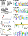
Examples of PSSMs generated by selections against the DMS‐BM library together with validation of an extended TLN1 PTB binding motif in TPTE2. (a‐h) Representative examples of PSSMs generated for MDM2 (a, b), PP2A B56 (c, d), G3BP1 (e, f), and TLN1 (g, h). (i) Heatmap representation of the PSSMs generated for the TPTE2 peptide binding to TLN1 PTB. The “DMS score” indicates the residue frequency from the PSSM. (j) Fold‐change of affinities of TLN1 PTB‐binding TPTE2 peptides upon mutation as determined using fluorescence polarization‐based affinity measurements. (k) AlpaFold3 model of the TLN1 PTB–TPTE2 complex overlayed with the previously solved NMR structure of TLN1 PTB in complex with PIP5K1C (PDB ID 2G35; peptide in magenta). The TPTE2 peptide is colored according to the pLDDT score, which shows the high confidence of the model (dark blue very high confidence pLDDT >90; light blue pLDDT 90 > pLDDT >70).
FIGURE 3
FIGURE 3
DMS‐CoV library quality and results for selections against human bait proteins. (a) Bait proteins selected for the DMS‐CoV analysis. (b) DMS‐CoV phage library design parameters, coverage, and counts distribution of all unique peptides. (c) DMS‐CoV library coverage on the wt/mutant peptides pair level. For 4% of the wt‐mutant peptide pairs, the mutant peptides were not found, and for 2.5% of the pairs, the wt peptides were also lacking, resulting in 2% of the pairs being absent from the physical phage library (top). The read count ratios for each wt/mutant peptide pair were evenly distributed, with the majority of the pairs having ratios of 13–1 or lower (bottom). (d) Completeness of DMS‐CoV selection results as compared to the DMS‐BM results. A low completeness score (y‐axis) indicates that sequencing data is missing for many mutations and amino acid positions for a given parent peptide (instance, x‐axis). (e–g, i) Representative PSSMs generated for viral peptides binding to the human bait protein domains EZR FERM (e), AP2 M1 (f), and G3BP1 NTF2 (g, i). (h) Fold‐change of affinities of G3BP1 NTF2‐binding N peptides upon mutation.
FIGURE 4
FIGURE 4
DMS analysis of peptides binding to the ADRP, UBl1, and SUD‐M domains of NSP3 PSSM (a–c) and heatmap (d–f) representations of the DMS data for the MBOAT1 peptide binding to NSP3 ADRP (a, d), the NCOA2 peptide binding to NSP3 UBl1 (b, e), and the PRDM14 peptide binding to NSP3 SUD‐M (c, f). (g–i) Fold‐change of affinities upon mutation of the respective wild‐type peptide binding to NSP3 ADRP (g), UBl1 (h), and SUD‐M (i).
FIGURE 5
FIGURE 5
DMS analysis of NSP9‐binding peptides guides the engineering of more potent antiviral inhibitors. (a) PSSM representation of the DMS results of the Nsp9 binding AXIN1 peptide. (b, c) PSSM and heat map representation of the DMS results of the NSP9 binding NOTCH4 peptide. (d) Fold‐change of affinities upon point mutation of the NOTCH4 peptide. (e) FP‐based affinity determinations of wt, single (A1603V and P1613V) and double mutants of the NOTCH4 peptide binding to NSP9 (A1603V/P1613V). (f) AlphaFold3 model of the complex of NSP9 and the NOTCH4 A1603/P1613V peptide. Peptide coloring is according to the pLDDT score (deep blue = high confidence). (g) Evaluation of the antiviral effect of cell‐permeable Tat‐tagged variants of the NOTCH4 peptides in VeroE6 cells. VeroE6 cells were infected with SARS‐CoV‐2 (multiplicity of infection: 0.5) and 8 h post‐infection viral RNA was quantified using qPCR. Viral RNA was normalized to the RNA levels in mock‐treated cells and presented as % RNA of control. Data are cumulative of two independent experiments done in triplicates (N = 6). (h) Unaffected cell viability upon treatment with the Tat‐tagged peptides. Cellular viability after peptide treatment was measured using Celltiter Glo. Data are cumulative of two independent experiments done in triplicates (N = 6).

References

    1. Abramson J, Adler J, Dunger J, Evans R, Green T, Pritzel A, et al. Accurate structure prediction of biomolecular interactions with AlphaFold 3. Nature. 2024; 630(8016):49‐350 - PMC - PubMed
    1. Baker DL, Wang B, Wilkinson‐White LE, El‐Kamand S, Allport TA, Ataide SF, et al. A biochemical and biophysical analysis of the interaction of nsp9 with nsp12 from SARS‐CoV‐2‐implications for future drug discovery efforts. Proteins. 2024;92(11):1308–1317. - PMC - PubMed
    1. Bandyopadhyay S, Bhaduri S, Ord M, Davey NE, Loog M, Pryciak PM. Comprehensive analysis of G1 cyclin docking motif sequences that control CDK regulatory potency in vivo. Curr Biol. 2020;30(22):4454–4466. - PMC - PubMed
    1. Benz C, Ali M, Krystkowiak I, Simonetti L, Sayadi A, Mihalic F, et al. Proteome‐scale mapping of binding sites in the unstructured regions of the human proteome. Mol Syst Biol. 2022;18(1):e10584. - PMC - PubMed
    1. Biswal M, Lu J, Song J. SARS‐CoV‐2 nucleocapsid protein targets a conserved surface groove of the NTF2‐like domain of G3BP1. J Mol Biol. 2022;434(9):167516. - PMC - PubMed