. 2025 May 24;74(7):220.
doi: 10.1007/s00262-025-04047-7.

Exosomal PD-L1 derived from hypoxia nasopharyngeal carcinoma cell exacerbates CD8+ T cell suppression by promoting PD-L1 upregulation in macrophages

Affiliations

Exosomal PD-L1 derived from hypoxia nasopharyngeal carcinoma cell exacerbates CD8+ T cell suppression by promoting PD-L1 upregulation in macrophages

Xiaofei Yuan et al. Cancer Immunol Immunother. .

Abstract

Immunotherapy targeting the programmed death ligand-1/programmed cell death protein-1 (PD-L1/PD-1) pathway exhibits limited effectiveness in individuals with recurrent and metastatic nasopharyngeal carcinoma (NPC). Recent studies have noted that hypoxia within the tumor microenvironment (TME) triggers intricate interplay, termed "hypoxia-induced exosome-mediated communication", between cancer cells and various immune cells. However, the role of hypoxia in modulating the immunosuppressive environment and its implications on the efficacy of immunotherapy in NPC remains poorly understood. In this study, we found hypoxia inducible factor-1 (HIF-1α) was positively associated with increased PD-L1 levels and decreased CD8+ T cell infiltration, and correlated with a poor prognosis. Mechanistically, we demonstrated that hypoxia regulated the expression of PD-L1 in NPC cells and their exosomes by activating the binding of HIF-1α to the PD-L1 promoter. Meanwhile, using in vitro approaches, we found that macrophages could upregulate their PD-L1 expression through the phagocytosis of exosomal PD-L1 derived from NPC cells. Furthermore, we confirmed that PD-L1+ macrophages could induce CD8+ T cell exhaustion and reduce their proliferation. In conclusion, our study revealed that hypoxia (via HIF-1α) upregulated the expression of PD-L1 in exosomes derived from NPC cells, while macrophages induce the suppression of CD8+ T cells by phagocytosis of exosomal PD-L1. Targeting the PD-L1+ macrophages could potentially serve as a promising approach to augment the effectiveness of immune checkpoint blockade in NPC.

Keywords: Hypoxia; Immune escape; Macrophage; Nasopharyngeal carcinoma; PD-L1.


Conflict of interest statement

Declarations. Conflict of interest: The authors declare that there are no conflicts of interest regarding the publication of this paper. Ethical approval: This study was approved by the Ethics Committee of Nanfang Hospital of Southern Medical University (Approval no.: NFEC-2017-165). All participants provided written informed consent for the use of their samples and data in research. The study adhered to the Declaration of Helsinki and ensured participant confidentiality.

Figures
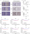
Fig. 1
Fig. 1
HIF-1α expression was positively correlated with elevated PD-L1 expression and reduced CD8 expression, and high HIF-1α or CD8 expression was associated with a negative prognosis. a Representative pictures of PD-L1 and CD8 expression in NPC patients in HIF-1α-low and high expression groups. Scale bars represented 50 or 100um. b Pearson correlation analysis of the association between HIF-1α and PD-L1 (r = 0.6799, p < 0.001). c Pearson correlation analysis of the association between HIF-1α and CD8 (r = −0.4143, p < 0.001). d Pearson correlation analysis of the association between PD-L1 and CD8 (r = −0.5105, p < 0.001). e Kaplan–Meier curves for OS between high HIF-1α, PD-L1 or CD8 groups and low HIF-1α, PD-L1 or CD8 groups. f Kaplan–Meier curves for PFS between high HIF-1α, PD-L1 or CD8 groups and low HIF-1α, PD-L1 or CD8 groups
Fig. 2
Fig. 2
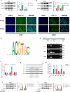
Hypoxia upregulated PD-L1 expression in NPC cells through HIF-1α. a, b Western blot analysis of HIF-1α and PD-L1 within normoxia condition or different hypoxia conditions in HK-1 and HNE-1 cells. c IF staining localization of HIF-1α expression in within normoxia condition or hypoxia condition in HK-1 and HNE-1 cells. d The binding motif of HIF-1α from JASPAR database. e The CHIP-PCR assay was used to assess the binding of PD-L1 promoter region. f Anti-HIF-1α-pulled down chromatins were analyzed by qRT-PCR. g A diagram showing the relationship of full-length and mutant PD-L1 promoters. h Dual-luciferase reporter gene assay was performed to indicate the interaction between HIF-1α and PD-L1. i, j Western blot analysis of HIF-1α and PD-L1 within normoxia condition or hypoxia condition after silencing HIF-1α in HK-1 and HNE-1 cells. Notes: *p < 0.05, **p < 0.01, ***p < 0.001; ns, not significant
Fig. 3
Fig. 3
Hypoxia NPC cells upregulate the expression of PD-L1 in macrophages. a IHC staining of PD-L1+ NPC cells and PD-L1+ immune cells. b Western blot analysis of PD-L1 in macrophages co-cultured with differently pretreated HK-1 and HNE-1 cells (normoxia and hypoxia) for 48 h. c The mIF staining of PD-L1+ macrophages and CD8+ T cells was performed in NPC tissue samples. d Pearson correlation analysis of the association between the levels of PD-L1+ macrophages and CD8+ T cells (r = −0.7080, p < 0.001). e, f Western blot analysis of PD-L1 in macrophages co-cultured with differently pretreated HK-1 and HNE-1 cells (normoxia, normoxia/shHIF-1α, hypoxia and hypoxia/shHIF-1α) for 48 h. g, h Flow cytometry was performed to detect the expression of HLA-DR and CD163 in in macrophages co-cultured with differently pretreated HK-1 and HNE-1 cells (normoxia, normoxia/shHIF-1α, hypoxia and hypoxia/shHIF-1α). Notes: *p < 0.05, **p < 0.01, ***p < 0.001; ns, not significant
Fig. 4
Fig. 4
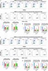
PD-L1+ macrophages induced by hypoxic NPC cells could inhibit CD8+ T cells. a Flow cytometry was performed to detect the levels of CD8+ T cells and PD-1+ CD8+ T cells in PBMCs co-cultured with differently pretreated macrophages (macrophages were co-cultured with differently pretreated HK-1 cells). b IL-2, IFN-γ and Granzyme B levels were detected in the culture supernatants of human peripheral CD8+ T cells co-cultured with macrophages that underwent different pretreatments (macrophages were co-cultured with differently pretreated HK-1 cells) by ELISA. c Flow cytometry was performed to detect the levels of CD8+ T cells and PD-1+ CD8+ T cells in PBMCs co-cultured with differently pretreated macrophages (macrophages were co-cultured with differently pretreated HNE-1 cells). d IL-2, IFN-γ and Granzyme B levels were detected in the culture supernatants of human peripheral CD8+ T cells co-cultured with macrophages that underwent different pretreatments (macrophages were co-cultured with differently pretreated HNE-1 cells) by ELISA. Notes: *p < 0.05, **p < 0.01, ***p < 0.001; ns, not significant
Fig. 5
Fig. 5
Hypoxia increases exosomal PD-L1 levels in NPC cells, macrophages increase their PD-L1 expression by phagocytizing PD-L1 in exosomes. a The PD-L1 mRNA expression of macrophages co-cultured with differently pretreated HK-1 and HNE-1 cells (normoxia and hypoxia) for 48 h by qRT-PCR. b Transmission electron micrograph of HK-1 or HNE-1 cells-derived exosomes. c Exosomes released by HK-1 cells or HNE-1 cells were detected by nanosight particle tracking analysis or ZETA view analysis. d Exosome markers CD9, TSG101 and Calnexin proteins were detected by western blot. e IF detection of macrophages phagocytosing exosomal PD-L1(exosomes: green fluorescent; PD-L1: red fluorescent). f, g Western blot analysis of PD-L1 in exosomes derived from differently pretreated HK-1 and HNE-1 cells (normoxia, normoxia/shHIF-1α, hypoxia and hypoxia/shHIF-1α). h, i Western blot analysis of PD-L1 in macrophages co-cultured with exosomes derived from differently pretreated HK-1 and HNE-1 cells (normoxia, normoxia/shHIF-1α, hypoxia and hypoxia/shHIF-1α) for 48 h. j, k Flow cytometry was performed to detect the expression of HLA-DR and CD163 in in macrophages co-cultured with exosomes derived from differently pretreated HK-1 and HNE-1 cells (normoxia, normoxia/shHIF-1α, hypoxia and hypoxia/shHIF-1α). Notes: *p < 0.05, **p < 0.01, ***p < 0.001; ns, not significant
Fig. 6
Fig. 6
PD-L1+ macrophages induced by exosomal PD-L1 derived from hypoxic NPC cells could inhibit CD8+ T cells. a Flow cytometry was performed to detect the levels of CD8+ T cells and PD-1+ CD8+ T cells in PBMCs co-cultured with differently pretreated macrophages (macrophages were co-cultured with exosomes derived from differently pretreated HK-1 cells). b IL-2, IFN-γ and Granzyme B levels were detected in the culture supernatants of human peripheral CD8+ T cells co-cultured with macrophages that underwent different pretreatments (macrophages were co-cultured with exosomes derived from differently pretreated HK-1 cells) by ELISA. c Flow cytometry was performed to detect the levels of CD8+ T cells and PD-1+ CD8+ T cells in PBMCs co-cultured with differently pretreated macrophages (macrophages were co-cultured with exosomes derived from differently pretreated HNE-1 cells). d IL-2, IFN-γ and Granzyme B levels were detected in the culture supernatants of human peripheral CD8+ T cells co-cultured with macrophages that underwent different pretreatments (macrophages were co-cultured with exosomes derived from differently pretreated HNE-1 cells) by ELISA. Notes: *p < 0.05, **p < 0.01, ***p < 0.001; ns, not significant
Fig. 7
Fig. 7
The schematic diagram of this article. a Immune response process of NPC. b Hypoxia (via HIF-1α) upregulated the expression of PD-L1 in exosomes derived from NPC cells, while macrophages induce the suppression of CD8+ T cells by phagocytosis of exosomal PD-L1

Similar articles

Cited by

References

    1. Wong K, Hui EP, Lo KW et al (2021) Nasopharyngeal carcinoma: an evolving paradigm. Nat Rev Clin Oncol 18(11):679–695 - PubMed
    1. Chen YP, Chan A, Le QT et al (2019) Nasopharyngeal carcinoma. Lancet 394(10192):64–80 - PubMed
    1. Robert C (2020) A decade of immune-checkpoint inhibitors in cancer therapy. Nat Commun 11(1):3801 - PMC - PubMed
    1. Li W, Duan X, Chen X et al (2022) Immunotherapeutic approaches in EBV-associated nasopharyngeal carcinoma. Front Immunol 13:1079515 - PMC - PubMed
    1. Fang W, Yang Y, Ma Y et al (2018) Camrelizumab (SHR-1210) alone or in combination with gemcitabine plus cisplatin for nasopharyngeal carcinoma: results from two single-arm, phase 1 trials. Lancet Oncol 19(10):1338–1350 - PubMed

MeSH terms

LinkOut - more resources