. 2025 May 24;25(1):107.
doi: 10.1007/s10142-025-01617-x.

N4-acetylcytidine modification of LncRNA GFOD1-AS1 promotes high glucose-induced dysfunction in human dermal microvascular endothelial cells through stabilization of DNMT1 protein

Affiliations

N4-acetylcytidine modification of LncRNA GFOD1-AS1 promotes high glucose-induced dysfunction in human dermal microvascular endothelial cells through stabilization of DNMT1 protein

Jingjing Yuan et al. Funct Integr Genomics. .

Abstract

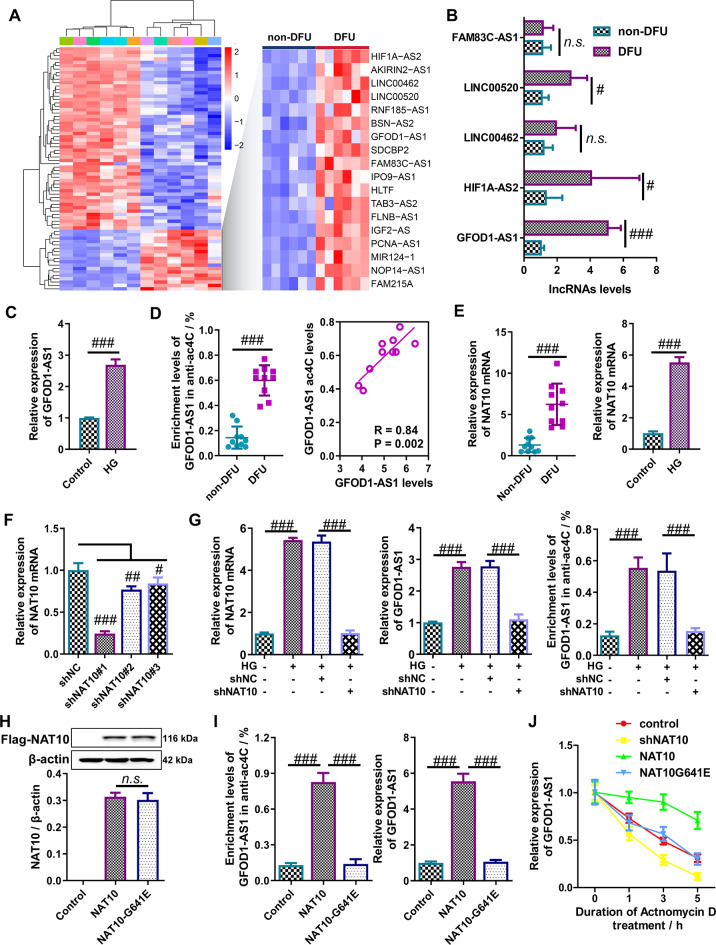
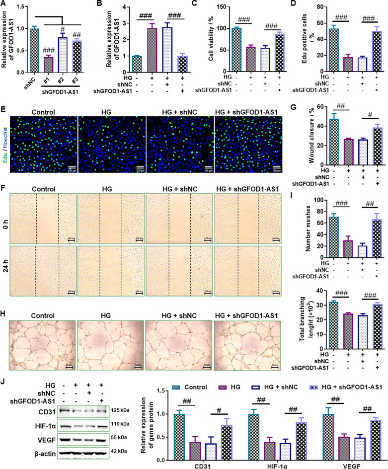
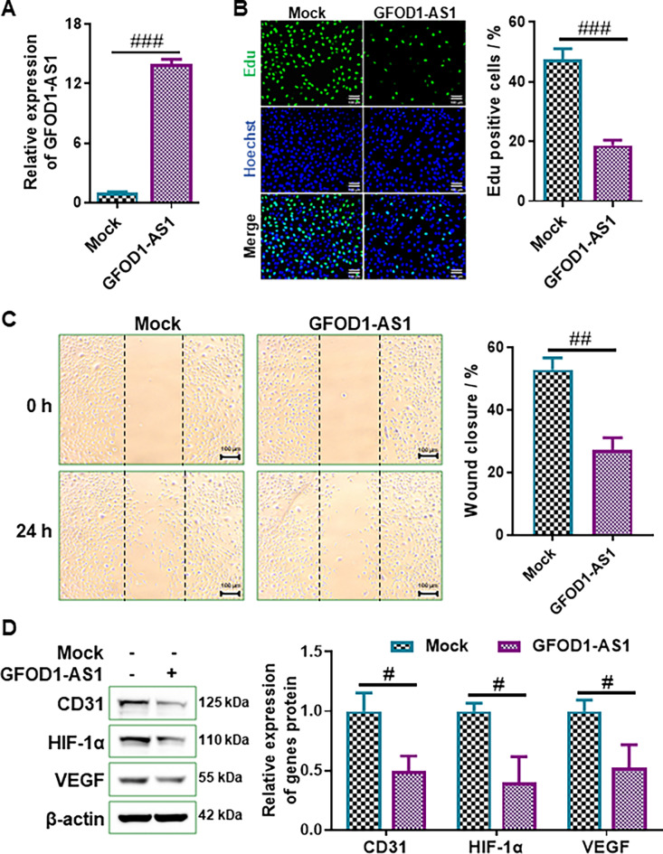
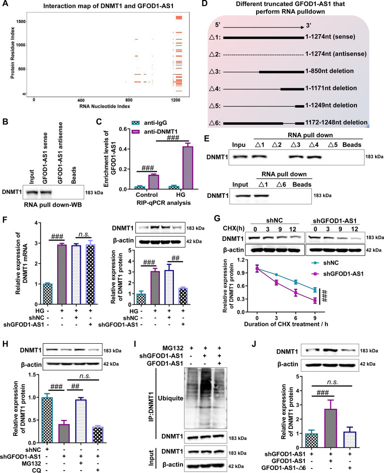
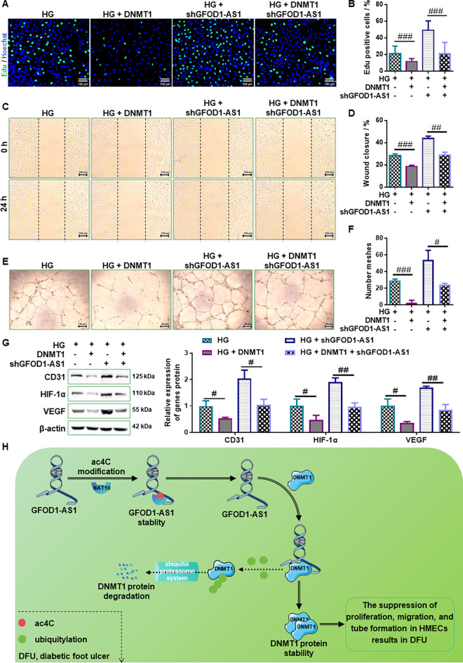
Emerging evidence supports that angiogenesis is essential for the wound healing of diabetic foot ulcer (DFU), and high glucose (HG)-induced dysfunction of human dermal microvascular endothelial cells is a key factor that hinders angiogenesis. However, the underlying mechanisms by which HG leads to the dysfunction of human dermal microvascular endothelial cells has not been fully elucidated. In the present investigation, we discovered a significant upregulation of the long non-coding RNA GFOD1-AS1(GFOD1-AS1) in the ulcer margin samples of patients with DFU and the HG-induced dysfunction model of human dermal microvascular endothelial cells, attributing its dysregulation to the stabilizing effect of NAT10-mediated ac4C modification, as corroborated by an integrated approach of data mining and experimental validation. Subsequently, a series of in vitro functional analyses showed that ectopic expression of GFOD1-AS1 promoted impaired function of human dermal microvascular endothelial cells. In contrast, knockdown of GFOD1-AS1 significantly alleviated the HG-induced functional impairment in human dermal microvascular endothelial cells, as indicated by the enhanced cell proliferation, migration, and tube formation. Mechanistically, GFOD1-AS1 directly interacts with DNA methyltransferase DNMT1 to block its ubiquitin-proteasome degradation, thereby enhancing the protein stability of DNMT1.This stability elevates DNMT1 protein expression, ultimately inducing HG-induced dysfunction in human dermal microvascular endothelial cells. In summary, our results reveal that GFOD1-AS1 serves as a potential therapeutic target for DFU, and highlight the critical role of the NAT10/GFOD1-AS1/DNMT1 axis in the dysfunction of human dermal microvascular endothelial cells in DFU.

Keywords: DNMT1; Diabetic foot ulcer; GFOD1-AS1; Human dermal microvascular endothelial cells; N4-acetylcytidine modification.


Conflict of interest statement

Declarations. Competing interests: The authors declare no competing interests.

Figures

Fig. 1
Fig. 1
NAT10-mediated ac4C modification increases GFOD1-AS1 stability in diabetic foot ulcer (DFU). (A) Analysis of the GSE80178 dataset using a heatmap disclosed distinct long non-coding RNA (lncRNA) expression patterns, with significant differential expression (|logFC| > 1.0 and P < 0.01) observed in ulcer margin samples from six individuals with DFU and normal skin samples from six non-DFU patients. Notably, 18 lncRNAs were markedly up-regulated specifically in ulcer margin samples from patients with DFU. (B) Reverse transcription quantitative polymerase chain reaction (RT-qPCR) was employed to quantify the expression levels of long non-coding RNAs HIF1A-AS2, LINC00520, LINC00462, GFOD1-AS1, and FAM83C-AS1 in ulcer margin samples from ten individuals with DFU and wound margin normal skin samples from ten non-DFU patients. (C) RT-qPCR was utilized to assess the expression levels of GFOD1-AS1 in human dermal microvascular endothelial cells (HMEC-1 cells) under conditions of high glucose (HG) treatment and normal glucose control. (D) The enrichment of acetylation at lysine 4 (ac4C) modification on GFOD1-AS1 was determined using anti-acetyl-lysine RNA immunoprecipitation (acRIP) in ulcer margin samples from ten individuals with DFU and wound margin normal skin samples from ten non-DFU patients, and Pearson correlation coefficient analysis revealed a significant positive correlation between the transcript levels of GFOD1-AS1 and its ac4C modification levels in ulcer margin samples from patients with DFU. (E) RT-qPCR was employed to measure NAT10 expression levels in ulcer margin samples from ten individuals with DFU and wound margin normal skin samples from ten non-DFU patients, as well as HMEC-1 cells under conditions of HG treatment and normal glucose control. (F) RT-qPCR was utilized to assess GFOD1-AS1 expression levels following transfection of HMEC-1 cells with shNC or shNAT10. (G) acRIP-qPCR and RT-qPCR were employed to evaluate the enrichment of GFOD1-AS1 with the ac4C antibody and the expression levels of GFOD1-AS1 and NAT10 in HMEC-1 cells under HG conditions. (H) Western blot analysis revealed no significant difference in NAT10 protein expression levels between HMEC-1 cells overexpressing NAT10 and those overexpressing the NAT10-G641E. (I) acRIP-qPCR and RT-qPCR were used to assess the enrichment of GFOD1-AS1 with the ac4C antibody and to determine GFOD1-AS1 expression levels in HMEC-1 cells overexpressing either NAT10 or the NAT10-G641E. (J) RNA stability assays were conducted to evaluate the impact of NAT10 on the degradation of GFOD1-AS1. DFU, diabetic foot ulcers; HG, high glucose; shNC, negative control short hairpin RNA vector; shNAT10, NAT10 short hairpin RNA vector; NAT10, NAT10 overexpression vector; NAT10G641E, NAT10G641E overexpression vector. N = 3, n.s.p > 0.05, #p < 0.05, ##p < 0.01, and ###p < 0.001
Fig. 2
Fig. 2
Knockdown of GFOD1-AS1 enhances the proliferation, migration, and tube formation of human dermal microvascular endothelial cells under high-glucose. (A) RT-qPCR was used to determine the expression levels of GFOD1-AS1 in HMEC-1 cells following transfection with three different shGFOD1-AS1 or shNC, thereby assessing the efficiency of transfection. (B) RT-qPCR was conducted to measure GFOD1-AS1 expression levels in the specified HMEC-1 cells. (C-E) The CCK-8 and EdU assays were used to measure the proliferation ability of the indicated HMEC-1 cells. Scale bar = 100 μm. (F-G) The wound healing assay was applied to detect the migration of the indicated HMEC-1 cells. Scale bar = 100 μm. (H-I) The tube formation assay was utilized to evaluate the angiogenic tube formation capacity of the specified HMEC-1 cells. Scale bar = 50 μm. (J) Western blot analysis was conducted to assess the protein expression levels of angiogenic markers, including VEGF, HIF-1α, and CD31, in the indicated HMEC-1 cells. HG, high glucose; shNC, negative control short hairpin RNA vector; shGFOD1-AS1, GFOD1-AS1 short hairpin RNA vector. N = 3 ~ 5, #p < 0.05, ##p < 0.01, and ###p < 0.001
Fig. 3
Fig. 3
Elevated levels of GFOD1-AS1 suppress the proliferation, migration, and tube formation capabilities of human dermal microvascular endothelial cells. (A) RT-qPCR was used to determine the expression levels of GFOD1-AS1 in HMEC-1 cells following transfection with GFOD1-AS1 or mock, thereby assessing the efficiency of transfection. (B) The EdU assay was used to measure the proliferation ability of HMEC-1 cells with or without GFOD1-AS1 overexpression. Scale bar = 100 μm. (C) The wound healing assay was employed to assess the migratory capability of HMEC-1 cells with or without GFOD1-AS1 overexpression. Scale bar = 100 μm. (D) Western blot analysis was conducted to assess the protein expression levels of angiogenic markers (VEGF, HIF-1α, and CD31) in HMEC-1 cells with or without GFOD1-AS1 overexpression. Mock, negative control vector; GFOD1-AS1, GFOD1-AS1 expressed vector. N = 3, #p < 0.05, ##p < 0.01, and ###p < 0.001
Fig. 4
Fig. 4
GFOD1-AS1 interacts with DNMT1 to block the degradation of DNMT1 mediated by ubiquitin-proteasome pathway and up-regulate the expression of DNMT1. (A) The CatRAPID online platform was utilized to predict interactions between DNMT1 and GFOD1-AS1. (B) Western blot analysis confirmed the enrichment of DNMT1 in the fractions specifically pulled down by GFOD1-AS. (C) RNA immunoprecipitation (RIP) assays were performed on HMIC-1 cells from the HG and control groups using anti-IgG antibody (negative control) and anti-DNMT1 antibody, and the enrichment of GFOD1-AS1 in the precipitates was quantified by RT-qPCR. RIP-qPCR results confirmed that the DNMT1-directed antibody was capable of precipitating GFOD1-AS1. (D) The schematic diagram illustrates the full-length, antisense, and different truncated fragments of GFOD1-AS1. (E) RNA pull down-western blot analysis revealed that the truncated fragment #6 of GFOD1-AS1 was indispensable for the interaction with DNMT1. (F) RT-qPCR and Western blot analysis were employed to determine the relative mRNA and protein levels of DNMT1 in indicated HMEC-1 cells. (G) HMEC-1 cells transfected with shGFOD1-AS1 or shNC were treated with 50 µg/mL cycloheximide (CHX) for specified time periods, after which DNMT1 protein levels were assessed using Western blot analysis. (H) HMEC-1 cells transfected with shGFOD1-AS1 or shNC were treated with 10 µM MG132 or 15 µM chloroquine (CQ) for specified time periods, and then DNMT1 protein levels was detected by Western blot analysis. (I) HMEC-1 cells transfected with shGFOD1-AS1 or overexpressing GFOD1-AS1 were treated with 10 µM MG132 for 10 h, and then the ubiquitination level of DNMT1 was evaluated using co-immunoprecipitation (Co-IP) followed by Western blot analysis. (J) Western blot analysis was performed to detect the expression of DNMT1 protein in HMEC-1 cells after knockdown of GFOD1-AS1, followed by re-expression of GFOD1-AS1 or GFOD1-AS1-△6. HG, high glucose; DNMT1, DNMT1 overexpression vector; shGFOD1-AS1, GFOD1-AS1 short hairpin RNA vector; GFOD1-AS1, GFOD1-AS1overexpression vector; △1, Full-length vector of GFOD1-AS1; △2, Antisense vector of GFOD1-AS1; △3, A truncated GFOD1-AS1 containing 851-1274nt; △4, A truncated GFOD1-AS1 containing 1172-1274nt; △5, A truncated GFOD1-AS1 containing 1248-1274nt; △6, A truncated GFOD1-AS1 deletion of 1172-1248nt (GFOD1-AS1-△6). N = 3, n.s.p > 0.05, ##p < 0.01, and ###p < 0.001
Fig. 5
Fig. 5
GFOD1-AS1 knockdown mitigates high-glucose vascular endothelial cell injury by suppressing DNMT1 expression, and the detailed comparisons between high glucose and control groups provided in Figs. 1 and 2. (A and B) The EdU assay was used to measure the proliferation ability in the indicated HMEC-1 cells. Scale bar = 100 μm. (C and D) The wound healing assay was applied to detect the migration in the indicated HMEC-1 cells. Scale bar = 100 μm. (E and F) The tube formation assay was utilized to evaluate the angiogenic tube formation capacity in the indicated HMEC-1 cells. Scale bar = 50 μm. (G) Western blot analysis was conducted to assess the protein expression levels of angiogenic markers (VEGF, HIF-1α, and CD31) in the indicated HVEC cells. HG, high glucose; DNMT1, DNMT1 overexpression vector; shGFOD1-AS1, GFOD1-AS1 short hairpin RNA vector. (H) Schematic diagram of the mechanism by which GFOD1-AS1 promotes the dysfunction in microvascular endothelial cells of diabetic foot ulcers. N = 3, #p < 0.05, ##p < 0.01, and ###p < 0.001

Similar articles

References

    1. Arango D, Sturgill D, Alhusaini N, Dillman AA, Sweet TJ, Hanson G, Hosogane M, Sinclair WR, Nanan KK, Mandler MD, Fox SD, Zengeya TT, Andresson T, Meier JL, Coller J, Oberdoerffer S (2018) Acetylation of cytidine in mRNA promotes translation efficiency. Cell 175:1872–1886e24 - PMC - PubMed
    1. Architha T, Juanitaa GR, Vijayalalitha R, Jayasuriya R, Athira G, Balamurugan R, Ganesan K, Ramkumar KM (2024) LncRNA NEAT1/miR-146a-5p Axis restores normal angiogenesis in diabetic foot ulcers by targeting MafG. Cells.;13 - PMC - PubMed
    1. Armstrong DG, Tan TW, Boulton AJM, Bus SA (2023) Diabetic foot ulcers: A review. JAMA 330:62–75 - PMC - PubMed
    1. Bian EB, Xiong ZG, Li J (2019) New advances of LncRNAs in liver fibrosis, with specific focus on lncRNA-miRNA interactions. J Cell Physiol 234:2194–2203 - PubMed
    1. Chen L, Shen S, Wang S (2023) LncRNA SNHG16 knockdown promotes diabetic foot ulcer wound healing via sponging MiR-31-5p. Tohoku J Exp Med - PubMed

MeSH terms

LinkOut - more resources