. 2025 Aug:66:101182.
doi: 10.1016/j.neo.2025.101182. Epub 2025 May 23.

Escitalopram facilitates tumor growth and metastasis in rodents: Is it safe?

Affiliations

Escitalopram facilitates tumor growth and metastasis in rodents: Is it safe?

Yosi Azan et al. Neoplasia. 2025 Aug.

Abstract

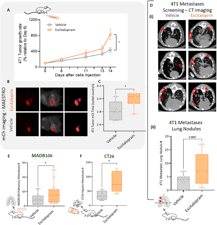
Cancer patients are often treated perioperatively with serotonin reuptake inhibitors (SSRIs) to counteract anxiety and depression. Recent studies suggest that long-term cancer outcomes may also be affected by SSRI use in an agent-dependent manner. Importantly, the perioperative use of SSRIs is prevalent clinically, but has rarely been studied empirically. Herein, we studied escitalopram, a commonly prescribed SSRI in cancer patients, in vitro, and in vivo in the context of surgery and/or cancer progression in immune-competent rodents, employing the Panc02 (pancreatic), MADB106, 4T1, EO771 (mammary), and CT26 (colon) syngeneic tumor models, assessing primary tumor growth and metastasis. Escitalopram (10-15mg/kg/day, 14-30 days) was administered along tumor and/or metastatic progression, via intraperitoneal injections, Alzet osmotic pumps, or drinking water. In vitro, escitalopram affected proliferation rates in a cell-line-, dose-, and exposure duration- dependent manner, mostly increasing or not affecting proliferation. In contrast, in vivo escitalopram consistently increased primary tumor growth, and experimental and spontaneous metastasis in all models tested. In pancreatic tumor-bearing mice, escitalopram increased tumor growth in two different studies by ∼1.5-fold, as indicated by bioluminescence imaging. In the mammary primary tumor models, escitalopram increased 4T1 and EO771 growth by 1.4 to 2.2-fold. Last, escitalopram increased experimental MADB106 lung metastasis and CT26 liver metastasis, as well as spontaneous post-excision 4T1 lung metastasis by 1.6 to 2.3-fold. Taken together, although additional research is needed to elucidate mediating in vivo mechanisms, and to assess clinical oncological risks of escitalopram, these findings raise concerns regarding the prevalent perioperative use of escitalopram in cancer patients.

Keywords: Cancer; Escitalopram; Metastasis; Periopearative period; Rodents.


Conflict of interest statement

Declaration of competing interest The authors declare the following financial interests/personal relationships which may be considered as potential competing interests: Shamgar Ben-eliyahu reports financial support was provided by Israel Science Foundation. If there are other authors, they declare that they have no known competing financial interests or personal relationships that could have appeared to influence the work reported in this paper.

Figures

Fig. 1
Fig. 1
In vitro effects of escitalopram on tumor cell proliferation. In vitro effect of escitalopram on tumor cell proliferation. (A) 4T1, (B) Panc02, (C) MADB106, (D) CT26, and (E) EO771 cells were co-incubated with escitalopram at 10-fold increasing concentrations from 5 × 10⁻⁹ to 5 × 10⁻⁵ M along 1, 2, or 3 days. Proliferation was assessed using the XTT cell assay. Results show inconsistent effects across different cell lines, with CT26 and MADB106 cells showing increased proliferation at clinically relevant plasma concentrations (5 × 10⁻⁸ to 5 × 10⁻⁷ M) [43,44,47], while Panc02 and 4T1 cells exhibited decreased proliferation only at the highest concentration. EO771 showed increased proliferation when exposed to escitalopram on day 3, however this effect was only marginally significant (p = 0.096). Data are presented as a percentage from control levels (mean ± SEM), with values higher or lower than 100 indicating increased or decreased proliferation, respectively. Significant differences from control are marked as follows: *p < 0.05, **p < 0.01, ***p < 0.001, ****p < 0.0001.
Fig. 2
Fig. 2
Deleterious in-vivo effects of Escitalopram on tumor progression across pancreatic (Panc02) and mammary cancer (4T1 and EO771) models. (A) Experiment 1, Panc02 tumor growth rate relative to day 7, based on BLI, in mice treated with daily i.p. escitalopram or saline injections (n = 10, 11). Tumor growth significantly increased over time compared to controls (∼1.5-fold) (F(1,26)=5.885, p = 0.0225). (B) Experiment 2, Panc02 tumor growth rate (BLI) in animals receiving continuous escitalopram or saline via ALZET osmotic pumps (n = 10, 11), relative to day 7. Escitalopram infusion resulted in significantly increased tumor growth (F(1,19)=4.432, p = 0.0485). By day 30, tumors in the escitalopram group had increased 22 % from baseline, whereas control tumors exhibited a 35 % reduction. (C) Representative BLI images of Panc02 tumors over time. (D-E) 4T1 mammary cancer tumor volume measured over time (n = 14 per group). Escitalopram significantly increased tumor growth by 1.6-fold compared to controls (F(1,26)=7.631, p = 0.0104) (D). Ex-vivo, tumor weight on day 14 was marginally heavier in the escitalopram group, with a 1.4-fold increase (t(26)=1.728, p = 0.0958) (E). (F-G) In the EO771 mammary cancer model, tumor volume measured over time increased by 2.2-fold in escitalopram-treated mice compared to vehicle-treated mice (n = 15, 12, F(1,28)=11.01, p = 0.0025) (F). Ex-vivo, tumor weight on day 25 increase by 1.75-fold in the escitalopram group (n = 11,15, t(24)=2.862, p = 0.0086) (G). Graphs represent mean ± SEM, boxes represent the 2nd and 3rd quartiles, and whiskers show Min and Max values. Significant differences are marked as follows: *p < 0.05, **p < 0.01.
Fig. 3
Fig. 3
Experimental design to assess the effects of escitalopram on 4T1 mammary tumor growth and metastasis in mice. 14 Female BALB/c mice received escitalopram (15 mg/kg/day, i.p.) from day 23 to 14. 4T1 cells were inoculated on day 0. From day 0 to day 14, tumors were measured by caliper. Drug treatment lasted until day 14, when primary tumors were excised after in-vivo mCherry flourescence imaging. Lung metastases were assessed on day 29 using MicroCT.
Fig. 4
Fig. 4
Escitalopram enhances tumor growth and metastasis in mammary (4T1 and MADB106) and colorectal (CT26) tumor models. (A-D) 4T1 mammary tumor growth and metastasis rate in BALB/c mice (n = 7 per group). Escitalopram via osmotic pumps significantly increased tumor growth relative to vehicle (F(1,12)=5.176, p = 0.042), and showed significant interaction with time (F(4,48)=40, p < 0.0001) (A). By day 14, escitalopram-treated tumors grew twice as much as controls (800 % vs. 400 % relative to day 6). Representative in vivo mCherry fluorescence images of primary tumors on day 14 (B), and quantification of fluorescence intensity, showing significantly higher signal in the escitalopram group (t(12) = 2.405, p = 0.0332) (C). Representative micro-CT images of lungs from vehicle and escitalopram treated mice (D); (i) screening CT, (ii) escitalopram treated mice showed greater number of lung metastatic lesions, approaching significance (t(11) = 1.861, p = 0.0897), with twice as many nodules. (E) In the MADB106 lung metastasis model, escitalopram-treated rats (n = 14, 15) exhibited a 1.6-fold increase in pulmonary metastases number relative to controls (U = 58.5, p = 0.0367). (F) In the CT26 hepatic metastasis model, escitalopram-treated BALB/c mice (n = 6 per group) showed a 2.3-fold increase in hepatic metastases compared to controls (t(10)=2.789, p = 0.0192). Graphs represent mean ± SEM, boxes represent the 2nd, and 3rd quartiles, and whiskers show Min and Max values. Significant differences are marked as follows: *p < 0.05, **p < 0.01.

References

    1. Zou J., Zhu Y. Antidepressant use pattern and disparities among cancer patients in the United States. Front. Public health. 2022;10 doi: 10.3389/fpubh.2022.1000000. - DOI - PMC - PubMed
    1. Sangkuhl K., Klein T.E., Altman R.B. Selective serotonin reuptake inhibitors pathway. Pharmacogenetics Genom. 2009;19(11):907–909. doi: 10.1097/FPC.0b013e32833132cb. - DOI - PMC - PubMed
    1. Owens M.J., Knight D.L., Nemeroff C.B. Second-generation SSRIs: human monoamine transporter binding profile of escitalopram and R-fluoxetine. Biol. Psychiatry. 2001;50(5):345–350. doi: 10.1016/s0006-3223(01)01145-3. - DOI - PubMed
    1. Vaswani M., Linda F.K., Ramesh S. Role of selective serotonin reuptake inhibitors in psychiatric disorders: a comprehensive review. Prog. Neuro-Psychopharmacol. Biol. Psychiatry. 2003;27(1):85–102. doi: 10.1016/s0278-5846(02)00338-x. - DOI - PubMed
    1. Mandrioli R., Mercolini L., Saracino M.A., Raggi M.A. Selective serotonin reuptake inhibitors (SSRIs): therapeutic drug monitoring and pharmacological interactions. Curr. Med. Chem. 2012;19:1846–1863. doi: 10.2174/092986712800099749. - DOI - PubMed

Publication types

Substances