. 2025 May 24;16(1):4846.
doi: 10.1038/s41467-025-60174-w.

Alveolar epithelial and vascular CXCR2 mediates transcytosis of CXCL1 in inflamed lungs

Affiliations

Alveolar epithelial and vascular CXCR2 mediates transcytosis of CXCL1 in inflamed lungs

Katharina Thomas et al. Nat Commun. .

Abstract

Pulmonary infections are characterized by neutrophil recruitment into the lung driven by chemokine ligands of CXCR2, which is expressed on neutrophils, but also present in non-hematopoietic lung cells, in which its role remains unclear. We hypothesize that CXCR2 in epithelial and endothelial cells contributes to neutrophil recruitment into the lung by modifying the availability of its cognate chemokines in lung alveoli. Using conditional endothelial and epithelial CXCR2 knockout mice, we demonstrate that selective CXCR2 deletion in either compartment impairs neutrophil recruitment into the lung during bacterial pneumonia and reduces bacterial clearance. We show that CXCR2 ablation in epithelial and endothelial cells compromises respective trans-epithelial and trans-endothelial transcytosis of alveolar CXCL1. Mechanistically, CXCR2-mediated CXCL1 endothelial and epithelial cell transcytosis requires the function of Bruton's tyrosine kinase in these cells. In conclusion, CXCR2 plays an important role in alveolar epithelial and endothelial cells, where it mediates cognate chemokine transcytosis, thus actively supporting their activities in neutrophil recruitment to the infected lungs.


Conflict of interest statement

Competing interests: The authors declare no competing interests.

Figures
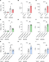
Fig. 1
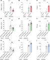
Fig. 1. Haematopoietic and non-haematopoietic CXCR2 is required for host defense during bacterial pneumonia.
CXCR2 KO mice (red = CXCR2−/−, green = CXCR2fl/flCdh5Cre+, blue = CXCR2fl/flShhCre+) and appropriate WT/Cre- control (gray) mice were intratracheally administered with sterile saline or with viable K. pneumoniae. Neutrophil recruitment into the lung (A, D, G), as well as CFUs in the lung (B, E, H) and spleen (C, F, I) were assessed 24 h after infection. Data are mean ± SD, n = 5–10 biologically independent mice pre group, age 8–16 weeks, equal gender distribution, one-way ANOVA followed by Bonferroni correction, *p < 0.05, **p < 0.01.
Fig. 2
Fig. 2. Endothelial and epithelial CXCR2 are required for host defense during K. pneumoniae-induced pneumonia.
CXCR2 KO mice (red=CXCR2−/−, green=CXCR2fl/flCdh5Cre+, blue=CXCR2fl/flShhCre+) and appropriate WT/Cre- control (gray) mice were intratracheally administered with sterile saline or with viable K. pneumoniae. Neutrophil recruitment into BAL (A, D, G), as well as CFUs in the BAL (B, E, H) and blood (C, F, I) were assessed 24 h after infection. Data are mean ± SD, n = 5–10 biologically independent mice pre group, age 8–16 weeks, equal gender distribution, one-way ANOVA followed by Bonferroni correction, *p < 0.05, **p < 0.01.
Fig. 3
Fig. 3. Loss of endothelial and epithelial CXCR2 leads to reduced survival during K. pneumoniae-induced pneumonia.
CXCR2 KO mice (red=CXCR2−/−, green=CXCR2fl/flCdh5Cre+, blue=CXCR2fl/flShhCre+) and appropriate WT/Cre- control (gray) mice were intratracheally administered with a lethal CFU dose of viable K. pneumoniae. AC Survival was analyzed 24 h. Data are mean ± SD, n = 5–8 biologically independent mice pre group, age 8–16 weeks, equal gender distribution, Kaplan-Meier analysis, *p < 0.05.
Fig. 4
Fig. 4. Neutrophil recruitment into the alveoli is diminished in CXCR2 deficient mice upon CXCL1 but not fMLP treatment.
Intravital microscopy of the lung was performed in CXCR2 KO mice (red = CXCR2−/−, green = CXCR2fl/flCdh5Cre+, blue = CXCR2fl/flShhCre+) and appropriate WT/Cre- controls (gray) 4 h after intratracheal injection of CXCL1 or fMLP. Neutrophils were visualized in the alveoli by intravenous administration of an AF488-coupled Gr-1 antibody before starting microscopy (A, F, K). Neutrophil numbers were determined per field of view after CXCL1 (B, G, L) or fMLP (C, H, M) treatment. BALF was collected, and neutrophil infiltration into the alveoli was assessed by flow cytometry after CXCL1 (D, I, N) or fMLP (E, J, O) stimulation. Data are mean ± SD, scale bars equal 20 µm, n = 3–4 biologically independent mice pre group, age 8–16 weeks, equal gender distribution, t-test, *p < 0.05, **p < 0.01.
Fig. 5
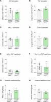
Fig. 5. Neutrophil recruitment in cremaster and kidney tissue is not altered in CXCR2fl/flCdh5Cre+ mice.
CXCR2fl/flCdh5Cre+ and Cre mice were analyzed for neutrophil recruitment after acute inflammation of the cremaster muscle (AF). Adherent and emigrated cells were analyzed after TNF stimulation (A, B) and CXCL1 superfusion of the cremaster muscle (C, D) or before and after fMLP superfusion (E, F). Neutrophil recruitment into the kidney (G) and plasma creatinine (H) were evaluated in CXCR2fl/flCdh5Cre+ and Cre control mice after ischemia reperfusion injury (IRI). Data are mean ± SD, n = 3–4 biologically independent mice pre group, age 8–16 weeks, only male mice for cremaster IMV, all other equal gender distribution, t-test.
Fig. 6
Fig. 6. Loss of endothelial and epithelial CXCR2 decreases transmigration of neutrophils through an endothelial-epithelial bilayer in vitro.
Primary murine lung microvascular endothelial cells (MLMVEC) and alveolar epithelial cells (AEC) were cultured on different sides of a Transwell™ insert. Transmigration of WT bone marrow-derived neutrophils towards a CXCL1 gradient was analyzed in case of different WT and CXCR2−/− layer combinations (A). Confluency was assured via Phalloidin staining of (B) AECs and (C) MLMVECs on Transwell™ membranes. D To further confirm the development of confluent and tight cell layers a Blue Dextran solution was transferred into the upper Transwell™ chamber and allowed to distribute for 3 h at 37 °C. The amount of Blue Dextran in the lower chamber was assessed in a plate reader. E Reliable transmigration of neutrophils was evaluated by determining transmigration efficiency of WT and CXCR2−/− neutrophils towards a CXCL1 gradient through either the Transwell membrane (w/o layer) or a bilayer consisting of WT AECs and MLMVECs (WT/WT bilayer). Transwells were equipped with WT/WT control layer or with CXCR2 deficient epithelial and endothelial layers, epithelial WT and endothelial KO layers or epithelial KO but endothelium WT layers (F). The percent of transmigrated neutrophils was assessed by the help of a Sysmex haemocytometer. Data are mean ± SD, n = 3–7 biologically independent samples, one-way ANOVA followed by Bonferroni correction, *p < 0.05.
Fig. 7
Fig. 7. Transmission electron microscopy of murine lung sections reveals evidence for a CXCR2-mediated transcytosis of CXCL1.
Colocalization of CXCR2 (blue), CXCL1 (red), and clathrin (green) was analyzed in ultrathin cross-sectioned lung tissue from K. pneumoniae infected CXCR2 KO and ctrl mice by transmission electron microscopy (TEM). Immunological staining was performed using gold particles of different sizes (6, 12, and 18 nm) coupled with antibodies against CXCR-2, CXCL-1, and clathrin. Lung sections from WT and CXCR2 full knockout mice (A) as well as conditional KO mice and corresponding Cre- controls (B, C). Abbreviations: ery erythrocyte, ca capillary, en endothelium, ep epithelium, al alveolar space. Scale bar: 500 nm (A), 200 nm (B, C). D Schematic cartoon of the morphological structures observable in (A). Numbers of CXCL1-coupled gold particles on (E) lung epithelial (EP) and (F) lung endothelial cells (EC) (data are mean ± SD analysis of n = 9 representative images per group), one-way ANOVA followed by Bonferroni correction, *p < 0.05.
Fig. 8
Fig. 8. Inhibition of clathrin-mediated transcytosis reduces CXCR2-mediated transcytosis of CXCL1.
A Primary murine lung microvascular endothelial cells (MLMVEC) and alveolar epithelial cells (AEC) were cultured on different sides of a Transwell™ insert. Transmigration of WT bone marrow-derived neutrophils towards a CXCL1 gradient was analyzed under control conditions (solvent DMSO) and after incubation of the epithelium and endothelium with the inhibitor Pitstop-2 (10 µM), MBCD (1 µM), and Dyngo 4a (30 µM) for 30 min. B CXCL1 concentrations in the upper well after 30 min (data are mean ± SD, n = 6 biologically independent samples), one-way ANOVA followed by Bonferroni correction, *p < 0.05.
Fig. 9
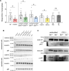
Fig. 9. Loss of endothelial and epithelial CXCR2 decreases transcytosis of CXCL1.
A CXCR2 KO mice (red=CXCR2−/−, green=CXCR2fl/flCdh5Cre+, blue=CXCR2fl/flShhCre+), appropriate WT/Cre- control (gray) mice, lethally irradiated WT or Btk−/− recipient mice reconstituted with isolated WT donor bone marrow, and WT mice pretreated with 4 µg Ptx/mouse i.v. or PBS as a control were injected intratracheally with 5 µg streptavidin-coupled CXCL1. After 4 h, blood samples were obtained and the levels of transcytosed streptavidin-coupled CXCL1 were analyzed by an ELISA using a CXCL1 capture antibody and a biotin-HRP conjugated detection antibody. Data are mean ± SD, n = 4–19 biologically independent mice pre group, age 8–16 weeks, equal gender distribution, one-way ANOVA followed by Bonferroni correction, *p < 0.05. B Protein expression of clathrin (MW 190 kDa), dynamin (MW 100 kDa), caveolin (MW 24 kDa) and total p38 (MW 38 kDa) as protein loading control was analyzed by western blotting of pulmonary epithelial and endothelia cells isolated form the indicated mouse strains (exemplary images from n = 3 independent biological experiments). C Akt and p38 phosphorylation in alveolar epithelial cells isolated from Btk+/+ and Btk−/− mice (exemplary images from n = 3 independent biological experiments), one-way ANOVA followed by Bonferroni correction, *p < 0.05.
Fig. 10
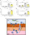
Fig. 10. Loss of non-hematopoietic Btk decreases transcytosis of CXCL1.
WT and Btk−/− mice were leathally irradiated and bone marrow reconstitution was performed by i.v. injection of isolated WT or Btk−/− bone marrow cells. 6 weeks after irradiation, bone marrow chimeric mice (WT donor -> WT recipient and WT donor -> Btk KO recipient) mice were intratracheally administered with sterile saline or with viable K. pneumoniae. Neutrophil recruitment into the BAL (A) and lung (B) as well as CFUs in the BAL (C) and lung (D) were assessed 24 h after infection. E Graphical abstract visualizing the main findings of this study. Data are mean ± SD, n = 3 biologically independent mice pre group, age 8–16 weeks, random gender distribution, one-way ANOVA followed by Bonferroni correction, *p < 0.05, **p < 0.01, ***p < 0.001.

References

    1. Mizgerd, J. P. Lung infection–a public health priority. PLoS Med.3, e76 (2006). - PMC - PubMed
    1. Matthay, M. A., Ware, L. B. & Zimmerman, G. A. The acute respiratory distress syndrome. J. Clin. Investig.122, 2731–2740 (2012). - PMC - PubMed
    1. Galeas-Pena, M., McLaughlin, N. & Pociask, D. The role of the innate immune system on pulmonary infections. Biol. Chem.400, 443–456 (2019). - PMC - PubMed
    1. Rossaint, J. & Zarbock, A. Tissue-specific neutrophil recruitment into the lung, liver, and kidney. J. Innate Immun.5, 348–357 (2013). - PMC - PubMed
    1. Pober, J. S. & Sessa, W. C. Evolving functions of endothelial cells in inflammation. Nat. Rev. Immunol.7, 803–815 (2007). - PubMed

MeSH terms

LinkOut - more resources