

GTV delineating for patients with postoperative glioma based on enhanced T2-FLAIR sequence instead of enhanced T1-TFE sequence: a feasibility study
- PMID: 40413710
- PMCID: PMC12104131
- DOI: 10.1007/s12672-025-02697-8
GTV delineating for patients with postoperative glioma based on enhanced T2-FLAIR sequence instead of enhanced T1-TFE sequence: a feasibility study
Abstract
Objective: To investigate the comparison of MRI Enhanced T2-Fluid Attenuated Inversion Recovery(T2-FLAIR+C) sequence and Enhanced T1-Turbo Field Echo(T1-TFE+C) sequence in delineating Gross Tumor Volume (GTV) of postoperative glioma.
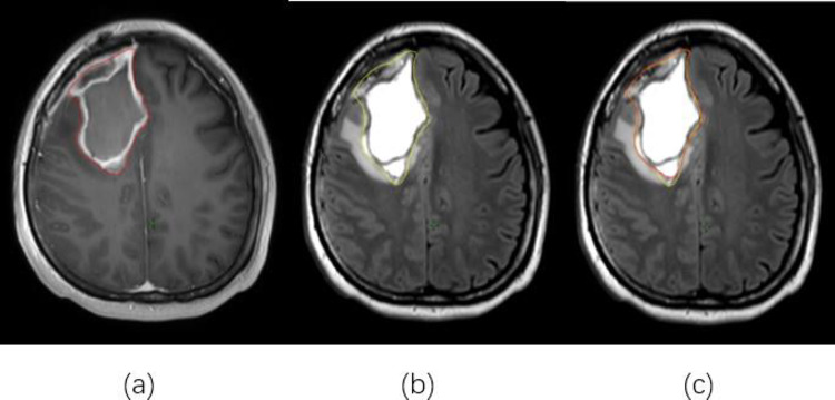
Method: Twenty patients with postoperative glioma underwent MRI simulation(MRI-sim) were enrolled. The T1-TFE+C sequence and T2-FLAIR+C sequence were separately registered with CT simulation(CT-sim). GTV was delineated by the same physician based on CT/T1 and CT/T2, respectively. Subsequently, the number, volume and overlapping ratio(OR) of GTV between the two groups were quantified and analyzed statistically. The signal intensity(SI) of the tumor area, normal gray matter and white matter (background of normal brain tissue)were measured on T1-TFE+C and T2-FLAIR+C sequences, respectively. The contrast ratio(CR) of the tumor in the two sequences were calculated and statistically analyzed.
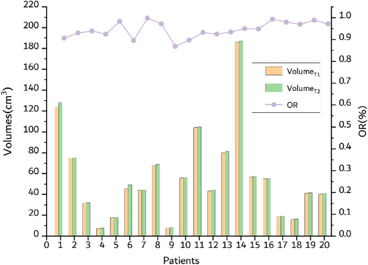
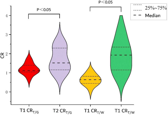
Results: The volumes of GTV delineated based on CT/T1 and CT/T2 were (55.89 ± 30.20) cm3 and (56.75 ± 30.52) cm3, respectively. There was no statistically significance between the two groups of GTV volumes (P > 0.05). The maximum OR, minimum OR and average OR of GTV volumes between the two groups were 99.77%, 86.90%, and 94.51%, respectively. The CR of tumor/white matter and tumor/gray matter in T2-FLAIR+C were significantly higher than those in the T1-TFE+C sequence (P < 0.05).
Conclusion: The volume of GTV delineated by T2-FLAIR+C was slightly larger compared to that by T1-TFE+C, and T2-FLAIR+C could provide a more comprehensive range of GTV delineation. CR was statistically significant between the two groups (P < 0.05), and T2-FLAIR+C demonstrated the ability to accurately depict changes in tumor boundaries and surrounding edema with a higher tumor enhancement signal. Therefore, GTV delineation of gliomas based on T2-FLAIR+C may offer certain advantages and could potentially serve as a complete replacement for T1-TFE+C in future clinical applications.
Keywords: GTV; MRI-sim; Postoperative glioma; T1-TFE+C; T2-FLAIR+C.
© 2025. The Author(s).
Conflict of interest statement
Declarations. Conflict of interests: The authors declare no competing interests. Consent for publication: All authors were consent for publication. Ethical Approval: We state that the study was approved by the institutional review board of National Cancer Center/National Clinical Research Center for Cancer/Cancer Hospital & Shenzhen Hospital and informed consent to participate was waived by the Institu- tional Review Board(NO. SZCHY2023069). We confirm that all methods were carried out in accordance with relevant guidelines and regulations.
Figures
References
-
- Han Q, Lu Y, Wang D, Li X, Ruan Z, Mei N, Ji X, Geng D, Yin B. Glioblastomas with and without peritumoral fluid-attenuated inversion recovery (flair) hyperintensity present morphological and microstructural differences on conventional mr images. Eur Radiol. 2023;33(12):9139–51. 10.1007/s00330-023-09924-2. - DOI - PubMed
-
- Louis DN, Perry A, Wesseling P, Brat DJ, Cree IA, Figarella-Branger D, Hawkins C, Ng HK, Pfister SM, Reifenberger G, Soffietti R, Deimling A, Ellison DW. The 2021 WHO classification of tumors of the central nervous system: a summary. Neuro Oncol. 2021;23(8):1231–51. 10.1093/neuonc/noab106. - DOI - PMC - PubMed
Grants and funding
LinkOut - more resources
Full Text Sources