. 2025 May 25;23(1):240.
doi: 10.1186/s12964-025-02249-1.

Conjugated bile acids promote metabolic dysfunction-associated steatotic liver disease through inducing nuclear translocation of sphingosine-1-phosphate receptor 2 to disrupt peroxisome proliferator-activated receptor alpha

Affiliations

Conjugated bile acids promote metabolic dysfunction-associated steatotic liver disease through inducing nuclear translocation of sphingosine-1-phosphate receptor 2 to disrupt peroxisome proliferator-activated receptor alpha

Rong-Rong Miao et al. Cell Commun Signal. .

Abstract

Background: Conjugated bile acids (CBAs) induced metabolic dysfunction-associated steatotic liver disease (MASLD) through activating sphingosine-1-phosphate receptor 2 (S1PR2). However, the precise mechanisms have not been fully understood.

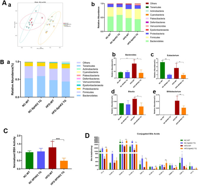
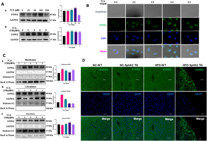
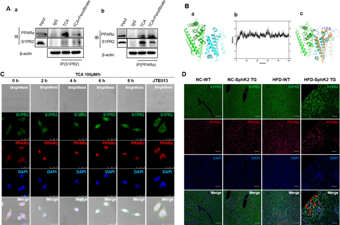
Methods: We established a link between CBAs and MASLD in IBD patients with high SphK2 and Villin-SphK2TG mice. Villin-SphK2TG mice were fed high-fat diet (HFD) for inducing MASLD. Gut microbiota composition was analyzed by 16 S rDNA Amplicon sequencing assay. We performed the UPLC/TQMS based targeted metabolomics assay to analyze the compositions of BAs in liver, serum and feces. In vitro assays, hepatocytes transfected with N-terminal truncated-S1PR2 analyzed the dynamics of S1PR2 stimulated by CBAs. PPARα function was assayed by analyzing the DNA-protein interactions by using Electrophoretic mobility shift assay (EMSA).

Results: The IBD patients with high colonocyte SphK2 conferred the development of MASLD. Feeding high-fat diet, Villin-SphK2TG mice developed MASLD more severely than WT mice. Analysis of gut microbiota showed that colonic SphK2 shaped microbiota by reducing the BSH-producing bacteria, thus leading to the accumulation of CBAs in liver via the gut-liver axis. CBAs induced nuclear translocation of S1PR2 through cleavage the N-terminal sequences of Ala-Ser-Ala-Phe-Iso in hepatocytes. Cleaved S1PR2 (S1PR2') was thus translocated into the nucleus to bind with PPARα, thereby interdicting the function of PPARα in regulating the genes involved in lipid catabolism. S1PR2 antagonist JTE-013 blocked the CBAs-induced nuclear translocation of S1PR2 and S1PR2 is thus identified as a potential therapeutic target for MASLD treatment.

Conclusion: CBAs promoted MASLD through inducing S1PR2 translocation into the nucleus, where it bound PPARα to interdict the function of PPARα in regulating genes involved in lipid catabolism.

Keywords: Conjugated bile acid (CBAs); MASLD; Nuclear translocation of S1PR2; PPARα; S1PR2.


Conflict of interest statement

Declarations. Ethical approval: Ethics Committee of Binzhou Medical College Hospital approved the use of human hepatocellular carcinoma samples (No.2020-012). Animal Welfare Committee of Capital Medical University approved the protocols of animal studies (AEEI-2019–043). Consent for publication: Yes. Competing interests: The authors declare no competing interests.

Figures

Fig. 1
Fig. 1
Villin-SphK2TG mice fed HFD developed more severely of MASLD than their WT littermates. (A) The levels of SphK2 in the IECs of SphK2TG mice and WT littermates. 1, 2, 3 represented three mice. (B) Body weight gains of mice during HFD-induced MASLD. (C) Representative images of the liver, Oil Red and hematoxylin and eosin (H&E) staining of the liver slides. (D) Quantification of lipid droplets in the livers of SphK2TG and WT mice. (E) Sirius red staining identified collagen I (red) in the livers, showing strong accumulation of collagen I in HFD-SphK2TG mice and a moderate level of collagen I in HFD-WT mice (red arrow). Feeding standard diet did not induce collagen I accumulation in both WT and SphK2TG.. n = 6. *p < 0.05, **p < 0.01, ***p < 0.001, ****p < 0.0001 between SphK2TG mice and WT mice
Fig. 2
Fig. 2
High IECs SphK2 shaped gut microbiota by reducing the BSH-producing microbiota, thereby accumulating hepatic CBAs through impairing the gut-to-liver axis in SphK2TG mice. A. a. Principal Coordinates Analysis (PCoA) ordination plot on unweighted UniFrac distance matrix; b. The compositions of gut microbiota at the phylum level. B. a. The abundance of the BSH-enriched microbes in colonic contents at the genus level; b, c, d, e. The abundance of BSH-enriched microbes, shch as Bacteroides, Eubacterium, Blautia and Bifidobacterium.n = 5. *p < 0.05, **p < 0.01, and ***P < 0.001. (C) The levels of BSH activity. n = 5. ***p < 0.001. (D) Statistical graph of CBAs in the colonic contents. *p < 0.05, **p < 0.01
Fig. 3
Fig. 3
The levels of S1PR2 in the subcellular organelles and the dynamics of S1PR2 induced by TCA in hepatocytes. (A) The TCA-activated S1PR2 levels in hepatocytes. (B) Immunofluorescence staining showed nuclear translocation of S1PR2 in NCTC1469 cells. (C) Western blot analysis showed the nuclear translocation of S1PR2 in NCTC1469 cells. n = 3. ***p < 0.001, ****p < 0.0001 vs. 0 h. (D) Immunofluorescence analysis identified nuclear S1PR2 in hepatocytes of HFD-SphK2TG mice
Fig. 4
Fig. 4
Responded to TCA, the plasma-membrane S1PR2 was translocated into the nucleus through cleaving the N-terminal Ala-Ser-Ala-Phe-Iso sequence in hepatocytes. A. Two bands of S1PR2 in plasma-membrane and cytoplasm, and one band in nucleus. AML12 cells were exposed to TCA for 6 h. The plasma-membrane S1PR2 (Mr = 40 kDa) was gradually attenuated, while cytoplasm S1PR2 (Mr = 40 kDa and Mr = 36 kDa) was parallel increased and S1PR2 (Mr = 36 kDa) was increased in nucleus. B. Trajectory of S1PR2 in AML12 cells. Cells were transfected with the N-terminal RFP-tagged full-length S1PR2. Natural S1PR2 (green) and N-terminal RFP-tagged full-length S1PR2 (red) were both localized in plasma-membrane. Nuclei had no S1PR2 in the intact cells (green- and red-stained S1PR2, first line). Exposed to TCA, plasma membrane S1PR2 were internalized into cytoplasm (second line), and then translocated into nuclei (green). But, nuclear S1PR2 did not include the N-terminal RFP-tagged full-length S1PR2 (no red-stained S1PR2). The N-terminal RFP-tagged full-length S1PR2 remained in plasma membrane. C. Immunofluorescence analyzed the mechanism of the constitutively shortened S1PR2 induced by TCA. Truncated S1PR2 (S1PR2’), lacking the first 37 amino acids, were created with RFP-tag at C-terminal and exhibited red fluorescence. Natural S1PR2 was identified in membrane (green-stained), and the S1PR2’ was identified in cytoplasm (red-stained). When exposed to TCA, only the full-length S1PR2 from the cell membrane can translocate into the nucleus following N-terminal truncation. In contrast, the overexpressed truncated form of S1PR2 (S1PR2’) is unable to respond to TCA stimulation and translocate into the nucleus, as it remains localized to the cell cytoplasm
Fig. 5
Fig. 5
The N-terminal truncated-S1PR2 (S1PR2’) bound with PPARα to form the S1PR2/PPARα complex in the nucleus of hepatocytes. A. Co-IP analysis showed the S1PR2’/PPARα complex in the TCA-treated hepatocytes. Fenofibrate reversed the TCA-induced S1PR2’/PPARα complex. B. Molecular docking and MD simulations analyzed complex of the S1PR2’ with PPARα. a: The helix region of PPARα (green) form close contacts with S1PR2’ (cyan) through hydrophobic and electrostatic interactions at contacting interface. b: MD analyzed RMSD for the backbone of the S1PR2’/PPARα complex. The RMSD values for the S1PR2’/PPARα complex reached about 3.5Å in 10ns and maintained constant in the following 90ns. c: The superimposition of the intact S1PR2 (orange) with S1PR2’ (blue). C. Immunofluorescence analyzed the S1PR2’/PPARα complex in hepatocytes. D. Immunofluorescence analyzed the S1PR2’/PPARα complex in hepatocytes of HFD-SphK2TG mice (fourth column, arrow point)
Fig. 6
Fig. 6
The forming of S1PR2-PPARα complex interdicted the PPARα’s function in regulating the genes involved in lipid catabolism. A. The level of FGF21 in primary hepatocytes of Villin-SphK2TG mice. B. The level FGF21 in livers of four groups of mice. HFD-SphK2 TG mice had the lowest level of FGF21. C. EMSA analyzed the PPARα’s ability in binding the genes, which contain the functional PPRE in hepatocytes. D. The qRT-PCR assay analyzed the levels of target genes of PPARα in AML12 and NCTC1469 exposed to TCA (100 µM). E. The qRT-PCR assay analyzed the levels of target genes of PPARα in livers of SphK2TG mice and control mice. F. Western blotting analysis showed the levels of FASN, CPT1A, CD36, ACOX1 in hepatocytes of WT mice and SphK2TG mice fed normal diet or HFD diet. n = 3. *p < 0.05, **p < 0.01, and ***P < 0.001

References

    1. Kumar V, Xin X, Ma J, Tan C, Osna N, Mahato RI. Therapeutic targets, novel drugs, and delivery systems for diabetes associated NAFLD and liver fibrosis. Adv Drug Deliv Rev. 2021;176:113888. - PMC - PubMed
    1. Chavez-Talavera O, Tailleux A, Lefebvre P, Staels B. Bile acid control of metabolism and inflammation in obesity, type 2 diabetes, dyslipidemia, and nonalcoholic fatty liver disease. Gastroenterology. 2017;152:1679–94. - PubMed
    1. Byrnes K, Blessinger S, Bailey NT, Scaife R, Liu G, Khambu B. Therapeutic regulation of autophagy in hepatic metabolism. Acta Pharm Sin B. 2022;12:33–49. - PMC - PubMed
    1. Kwong E, Li Y, Hylemon PB, Zhou H. Bile acids and sphingosine-1-phosphate receptor 2 in hepatic lipid metabolism. Acta Pharm Sin B. 2015;5:151–7. - PMC - PubMed
    1. Wang Y, Aoki H, Yang J, Peng K, Liu R, Li X, Qiang X, Sun L, Gurley EC, Lai G, et al. The role of sphingosine 1-phosphate receptor 2 in bile-acid-induced cholangiocyte proliferation and cholestasis-induced liver injury in mice. Hepatology. 2017;65:2005–18. - PMC - PubMed

LinkOut - more resources